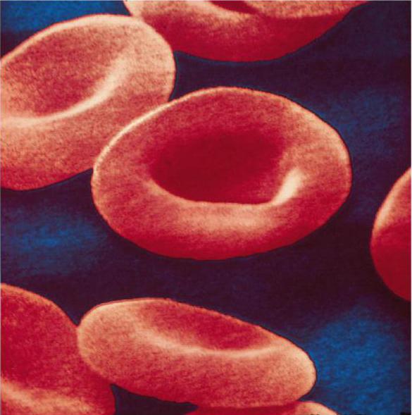
Red blood cells are highly specialized anucleate blood cells. Their core is lost during the maturation process. Red blood cells have the shape of a biconvex disk. On average, their diameter is about 7.5 microns, and the thickness at the periphery is 2.5 microns. Thanks to this shape, the surface of red blood cells for the diffusion of gases increases. In addition, their plasticity increases. Due to their high plasticity, they are deformed and easily pass through capillaries. Old and pathological red blood cells have low plasticity. Therefore, they are retained in the capillaries of the reticular tissue of the spleen and destroyed there.
The membrane of erythrocytes and the absence of a nucleus ensures their main function - the transport of oxygen and participation in the transfer of carbon dioxide. The erythrocyte membrane is impermeable to cations except potassium, and its permeability to chlorine anions, bicarbonate anions and hydroxyl anions is a million times greater. In addition, it allows oxygen and carbon dioxide molecules to pass through well. The membrane contains up to 52% protein. In particular, glycoproteins determine the blood group and provide its negative charge. It has a built-in Na–K–ATPase, which removes sodium from the cytoplasm and pumps in potassium ions. Chemoprotein makes up the bulk of red blood cells hemoglobin. In addition, the cytoplasm contains the enzymes carbonic anhydrase, phosphatases, cholinesterase and other enzymes.
Functions of red blood cells:
1. Transfer of oxygen from the lungs to the tissues.
2. Participation in the transport of CO 2 from tissues to the lungs.
3. Transport of water from tissues to the lungs, where it is released in the form of steam.
4. Participation in blood coagulation, releasing erythrocyte coagulation factors.
5. Transfer of amino acids on its surface.
6. Participate in the regulation of blood viscosity due to plasticity. As a result of their ability to deform, the viscosity of blood in small vessels is less than in large ones.
One microliter of a man’s blood contains 4.5-5.0 million red blood cells (4.5-5.0*10 12 /l). Women 3.7-4.7 million (3.7-4.7 * 10 12 / l).
The number of red blood cells is counted in Goryaev's cell. To do this, blood in a special capillary melanger (mixer) for red blood cells is mixed with a 3% sodium chloride solution in a ratio of 1:100 or 1:200. A drop of this mixture is then placed in a mesh chamber. It is created by the middle projection of the chamber and the cover glass. Chamber height 0.1 mm. On the middle protrusion there is a grid applied, forming large squares. Some of these squares are divided into 16 small ones. Each side of a small square has a size of 0.05 mm. Therefore, the volume of the mixture over the small square will be 1/10 mm * 1/20 mm * 1/20 mm = 1/4000 mm 3.
After filling the chamber, under a microscope, count the number of red blood cells in those 5 large squares that are divided into small ones, i.e. in 80 small ones. Then the number of red blood cells in one microliter of blood is calculated using the formula:
X = 4000*a*b/b.
Where a is the total number of red blood cells obtained during counting; b – the number of small squares in which the counting was carried out (b = 80); c – blood dilution (1:100, 1:200); 4000 is the reciprocal of the volume of liquid above a small square.
For quick calculations with a large number of tests, use photovoltaic erythrohemometers. The principle of their operation is based on determining the transparency of a suspension of red blood cells using a beam of light passing from a source to a light-sensitive sensor. Photoelectric calorimeters. An increase in the number of red blood cells in the blood is called erythrocytosis or erythremia ; decrease - erythropenia or anemia . These changes can be relative or absolute. For example, a relative decrease in their number occurs when water is retained in the body, and an increase occurs when dehydration occurs. An absolute decrease in the content of red blood cells, i.e. anemia, observed with blood loss, hematopoietic disorders, destruction of red blood cells by hemolytic poisons or transfusion of incompatible blood.
Hemolysis - This is the destruction of the red blood cell membrane and the release of hemoglobin into the plasma. As a result, the blood becomes clear.
The following types of hemolysis are distinguished:
1. By place of origin:
· Endogenous, i.e. in organism.
· Exogenous, outside of it. For example, in a bottle of blood, a heart-lung machine.
2. By character:
· Physiological. It ensures the destruction of old and pathological forms of red blood cells. There are two mechanisms. Intracellular hemolysis occurs in macrophages of the spleen, bone marrow, and liver cells. Intravascular– in small vessels from which hemoglobin is transferred to liver cells using the plasma protein haptoglobin. There, hemoglobin heme is converted into bilirubin. About 6-7 g of hemoglobin is destroyed per day.
· Pathological.
3. According to the mechanism of occurrence:
· Chemical. Occurs when red blood cells are exposed to substances that dissolve membrane lipids. These are alcohols, ether, chloroform, alkalis, acids, etc. In particular, when poisoned with a large dose of acetic acid, severe hemolysis occurs.
· Temperature. At low temperatures, ice crystals form in red blood cells, destroying their shell.
· Mechanical. Observed during mechanical ruptures of membranes. For example, when shaking a bottle of blood or pumping it with a heart-lung machine.
· Biological. Occurs under the influence of biological factors. These are hemolytic poisons of bacteria, insects, and snakes. As a result of transfusion of incompatible blood.
· Osmotic. Occurs when red blood cells enter an environment with an osmotic pressure lower than that of blood. Water enters the red blood cells, they swell and burst. The sodium chloride concentration at which 50% of all red blood cells are hemolyzed is a measure of their osmotic stability. It is determined in the clinic to diagnose liver diseases and anemia. Osmotic resistance must be at least 0.46% NaCl.
When red blood cells are placed in a medium with a higher osmotic pressure than blood, plasmolysis occurs. This is the shrinkage of red blood cells. It is used to count red blood cells.
Erythrocytes or red blood cells are one of the formed elements of blood that perform numerous functions that ensure the normal functioning of the body:
- the nutritional function is to transport amino acids and lipids;
- protective - in binding toxins with the help of antibodies;
- enzymatic is responsible for the transfer of various enzymes and hormones.
Red blood cells are also involved in regulating acid-base balance and maintaining blood isotonicity.
However, the main job of red blood cells is to deliver oxygen to the tissues and carbon dioxide to the lungs. Therefore, they are often called “respiratory” cells.
Features of the structure of red blood cells
The morphology of red blood cells differs from the structure, shape and size of other cells. In order for red blood cells to successfully cope with the gas transport function of blood, nature has endowed them with the following distinctive features:

The listed features are measures of adaptation to life on land, which began to develop in amphibians and fish, and reached their maximum optimization in higher mammals and humans.
This is interesting! In humans, the total surface area of all red blood cells in the blood is about 3,820 m2, which is 2,000 times more than the surface of the body.
Formation of red blood cells
The life of an individual red blood cell is relatively short - 100-120 days, and human red bone marrow reproduces about 2.5 million of these cells every day.
Full development of red blood cells (erythropoiesis) begins in the 5th month of intrauterine development of the fetus. Until this point, and in cases of oncological lesions of the main hematopoietic organ, red blood cells are produced in the liver, spleen and thymus.

The development of red blood cells is very similar to the process of human development. The birth and “intrauterine development” of red blood cells begins in the erythron - the red germ of the hematopoiesis of the red brain. It all starts with a pluripotent blood stem cell, which, changing 4 times, turns into an “embryo” - an erythroblast, and from this moment morphological changes in structure and size can already be observed.
Erythroblast. This is a round, large cell measuring from 20 to 25 microns with a nucleus that consists of 4 micronuclei and occupies almost 2/3 of the cell. The cytoplasm has a purple tint, which is clearly visible on a section of flat “hematopoietic” human bones. In almost all cells, so-called “ears” are visible, formed due to protrusion of the cytoplasm.
Pronormocyte. The dimensions of a pronormocyte cell are smaller than those of an erythroblast - already 10-20 microns, this occurs due to the disappearance of the nucleoli. The purple hue begins to lighten.
Basophilic normoblast. In almost the same cell size - 10-18 microns, the nucleus is still present. Chromantin, which gives the cell a light purple color, begins to gather into segments and the externally basophilic normoblast has a spotted color.
Polychromatophilic normoblast. The diameter of this cell is 9-12 microns. The core begins to change destructively. A high concentration of hemoglobin is observed.
Oxyphilic normoblast. The disappearing nucleus is displaced from the center of the cell to its periphery. The cell size continues to decrease - 7-10 microns. The cytoplasm becomes clearly pink with small remnants of chromantin (Joly bodies). Before entering the blood, normally the oxyphilic normoblast must squeeze out or dissolve its nucleus with the help of special enzymes.
Reticulocyte. The color of the reticulocyte is no different from the mature form of the erythrocyte. The red color provides the overall effect of the yellow-greenish cytoplasm and the violet-blue reticulum. The diameter of the reticulocyte ranges from 9 to 11 microns.
Normocyte. This is the name of a mature form of red blood cell with standard sizes, pinkish-red cytoplasm. The nucleus disappeared completely, and hemoglobin took its place. The process of increasing hemoglobin during red blood cell maturation occurs gradually, starting from the earliest forms, because it is quite toxic to the cell itself.
Another feature of red blood cells that causes a short lifespan is the absence of a nucleus does not allow them to divide and produce protein, and as a result, this leads to the accumulation of structural changes, rapid aging and death.
Degenerative forms of red blood cells

With various blood diseases and other pathologies, qualitative and quantitative changes in the normal levels of normocytes and reticulocytes in the blood, hemoglobin levels, as well as degenerative changes in their size, shape and color are possible. Below we will consider changes that affect the shape and size of red blood cells - poikilocytosis, as well as the main pathological forms of red blood cells and due to what diseases or conditions such changes occurred.
| Name | Changing shape | Pathologies |
| Spherocytes | A spherical shape of normal size with no characteristic clearing at the center. | Hemolytic disease of newborns (AB0 blood incompatibility), disseminated intravascular coagulation syndrome, specicymia, autoimmune pathologies, extensive burns, vascular and valve implants, other types of anemia. |
| Microspherocytes | Small balls from 4 to 6 microns. | Minkowski-Choffard disease (hereditary microspherocytosis). |
| Eliptocytes (ovalocytes) | Ovals or elongated shapes due to membrane abnormalities. There is no central clearing. | Hereditary ovalocytosis, thalassemia, liver cirrhosis, anemia: megablastic, iron deficiency, sickle cell. |
| Target-shaped red blood cells (codocytes) | Flat cells, reminiscent of a target in color - pale at the edges and a bright spot of hemoglobin in the center. The cell area is flattened and increased in size due to excess cholesterol. |
Thalassemia, hemoglobinopathies, iron deficiency anemia, lead poisoning, liver disease (accompanied by obstructive jaundice), removal of the spleen. |
| Echinocytes | Spikes of the same size are located at the same distance from each other. Looks like a sea urchin. | Uremia, stomach cancer, bleeding peptic ulcer complicated by bleeding, hereditary pathologies, lack of phosphates, magnesium, phosphoglycerol. |
| Acanthocytes | Spur-like protrusions of various sizes and sizes. Sometimes they resemble maple leaves. | Toxic hepatitis, cirrhosis, severe forms of spherocytosis, lipid metabolism disorders, splenectomy, with heparin therapy. |
| Sickle-shaped red blood cells (drepanocytes) | Looks like holly leaves or a sickle. Changes in the membrane occur under the influence of an increased amount of a special form of hemoglobin-s. | Sickle cell anemia, hemoglobinopathies. |
| Dental cells | Exceed the usual size and volume by 1/3. The central enlightenment is not round, but in the form of a strip. When sedimented, they become bowl-like. |
Hereditary spherocytosis and stomatocytosis, tumors of various etiologies, alcoholism, liver cirrhosis, cardiovascular pathology, taking certain medications. |
| Dacryocytes | They resemble a tear (drop) or a tadpole. | Myelofibrosis, myeloid metaplasia, tumor growth with granuloma, lymphoma and fibrosis, thalassemia, complicated iron deficiency, hepatitis (toxic). |
Let's add information about sickle erythrocytes and echinocytes.
Sickle cell anemia is most common in regions where malaria is endemic. Patients with such anemia have increased hereditary resistance to malaria infection, while sickle red blood cells are also resistant to infection. It is not possible to accurately describe the symptoms of sickle disease. Since sickle-shaped red blood cells are characterized by increased fragility of the membranes, this often causes capillary blockages, leading to a wide variety of symptoms in terms of severity and nature of manifestations. However, the most typical ones are obstructive jaundice, black urine and frequent fainting.

A certain number of echinocytes are always present in human blood. Aging and destruction of red blood cells is accompanied by a decrease in ATP synthesis. It is this factor that becomes the main reason for the natural transformation of disc-shaped normocytes into cells with characteristic protrusions. Before dying, the red blood cell goes through the following stages of transformation - first 3 classes of echinocytes, and then 2 classes of spheroechinocytes.
Red blood cells end their life in the spleen and liver. Such valuable hemoglobin will break down into two components - heme and globin. The heme will in turn be divided into bilirubin and iron ions. Bilirubin is excreted from the human body, along with other toxic and non-toxic remnants of red blood cells, through the gastrointestinal tract. But iron ions, as a building material, will be sent to the bone marrow for the synthesis of new hemoglobin and the birth of new red blood cells.
The red blood cell, the structure and functions of which we will consider in our article, is the most important component of blood. It is these cells that carry out gas exchange, ensuring respiration at the cellular and tissue level.
Red blood cell: structure and functions
The circulatory system of humans and mammals is characterized by the most perfect structure compared to other organisms. It consists of a four-chambered heart and a closed system of blood vessels through which blood continuously circulates. This tissue consists of a liquid component - plasma, and a number of cells: erythrocytes, leukocytes and platelets. Each cell plays its role. The structure of a human red blood cell is determined by the functions it performs. This refers to the size, shape and number of these blood cells.
Features of the structure of red blood cells
Red blood cells have the shape of a biconcave disc. They are not able to move independently in the bloodstream, like leukocytes. They reach tissues and internal organs thanks to the work of the heart. Red blood cells are prokaryotic cells. This means that they do not contain a formal core. Otherwise they would not be able to transport oxygen and carbon dioxide. This function is performed due to the presence of a special substance inside the cells - hemoglobin, which also determines the red color of human blood.

The structure of hemoglobin
The structure and functions of red blood cells are largely determined by the characteristics of this particular substance. Hemoglobin contains two components. These are an iron-containing component called heme and a protein called globin. For the first time, the English biochemist Max Ferdinand Perutz managed to decipher the spatial structure of this chemical compound. For this discovery he was awarded the Nobel Prize in 1962. Hemoglobin is a member of the group of chromoproteins. These include complex proteins consisting of a simple biopolymer and a prosthetic group. For hemoglobin, this group is heme. This group also includes plant chlorophyll, which ensures the process of photosynthesis.

How does gas exchange occur?
In humans and other chordates, hemoglobin is located inside red blood cells, and in invertebrates it is dissolved directly in the blood plasma. In any case, the chemical composition of this complex protein allows the formation of unstable compounds with oxygen and carbon dioxide. Blood saturated with oxygen is called arterial. It is enriched with this gas in the lungs.
From the aorta it goes to the arteries, and then to the capillaries. These small vessels are suitable for every cell of the body. Here, red blood cells give up oxygen and add the main product of respiration - carbon dioxide. With the flow of blood, which is already venous, they return to the lungs. In these organs, gas exchange occurs in the smallest bubbles - alveoli. Here hemoglobin detaches carbon dioxide, which is removed from the body through exhalation, and the blood is again saturated with oxygen.
Such chemical reactions are due to the presence of ferrous iron in heme. As a result of combination and decomposition, oxy- and carbhemoglobin are sequentially formed. But the complex protein of erythrocytes can also form stable compounds. For example, during incomplete combustion of fuel, carbon monoxide is released, which forms carboxyhemoglobin with hemoglobin. This process leads to the death of red blood cells and poisoning of the body, which can be fatal.

What is anemia
Shortness of breath, noticeable weakness, tinnitus, noticeable pallor of the skin and mucous membranes may indicate an insufficient amount of hemoglobin in the blood. The norm of its content varies depending on gender. In women, this figure is 120 - 140 g per 1000 ml of blood, and in men it reaches 180 g/l. The hemoglobin content in the blood of newborns is the highest. It exceeds this figure in adults, reaching 210 g/l.
Lack of hemoglobin is a serious disease called anemia or anemia. It can be caused by a lack of vitamins and iron salts in food, addiction to alcohol, the influence of radiation pollution and other negative environmental factors on the body.
A decrease in the amount of hemoglobin can also be due to natural factors. For example, in women, anemia can be caused by the menstrual cycle or pregnancy. Subsequently, the amount of hemoglobin normalizes. A temporary decrease in this indicator is also observed among active donors who often donate blood. But an increased number of red blood cells is also quite dangerous and undesirable for the body. It leads to an increase in blood density and the formation of blood clots. An increase in this indicator is often observed in people living in high mountain areas.
It is possible to normalize hemoglobin levels by consuming foods containing iron. These include liver, tongue, cattle, rabbit, fish, black and red caviar. Products of plant origin also contain the necessary microelement, but the iron they contain is much more difficult to absorb. These include legumes, buckwheat, apples, molasses, red peppers and herbs.
Shape and size
The structure of red blood cells is characterized primarily by their shape, which is quite unusual. It really resembles a disk, concave on both sides. This shape of red blood cells is not accidental. It increases the surface of red blood cells and ensures the most effective penetration of oxygen into them. This unusual shape also helps to increase the number of these cells. Thus, normally 1 cubic mm of human blood contains about 5 million red blood cells, which also contributes to the best gas exchange.

The structure of frog red blood cells
Scientists have long established that human red blood cells have structural features that ensure the most efficient gas exchange. This applies to form, quantity, and internal content. This is especially obvious when the structure of human and frog red blood cells is compared. In the latter, red blood cells are oval in shape and contain a nucleus. This significantly reduces the content of respiratory pigments. Frog red blood cells are much larger than human ones, and therefore their concentration is not so high. For comparison: if a person has more than 5 million of them per cubic mm, then in amphibians this figure reaches 0.38.
Evolution of red blood cells
The structure of human and frog erythrocytes allows us to draw conclusions about the evolutionary transformations of such structures. Respiratory pigments are also found in the simplest ciliates. In the blood of invertebrates they are contained directly in the plasma. But this significantly increases the thickness of the blood, which can lead to the formation of blood clots inside the vessels. Therefore, over time, evolutionary transformations went towards the appearance of specialized cells, the formation of their biconcave shape, the disappearance of the nucleus, a decrease in their size and an increase in concentration.
Ontogenesis of red blood cells
An erythrocyte, the structure of which has a number of characteristic features, remains viable for 120 days. Subsequently, they are destroyed in the liver and spleen. The main hematopoietic organ in humans is the red bone marrow. It continuously produces new red blood cells from stem cells. Initially they contain a nucleus, which as it matures is destroyed and replaced by hemoglobin.
Features of blood transfusion
There are often situations in a person's life that require a blood transfusion. For a long time, such operations led to the death of patients, and the real reasons for this remained a mystery. Only at the beginning of the 20th century was it established that the culprit was the erythrocyte. The structure of these cells determines human blood groups. There are four of them in total, and they are distinguished according to the AB0 system.
Each of them is distinguished by a special type of protein substances contained in red blood cells. They are called agglutinogens. People with the first blood group do not have them. From the second - they have agglutinogens A, from the third - B, from the fourth - AB. At the same time, the blood plasma contains agglutinin proteins: alpha, beta, or both at the same time. The combination of these substances determines the compatibility of blood groups. This means that the simultaneous presence of agglutinogen A and agglutinin alpha in the blood is impossible. In this case, red blood cells stick together, which can lead to the death of the body.

What is Rh factor
The structure of the human red blood cell determines the performance of another function - determining the Rh factor. This sign is also necessarily taken into account during blood transfusion. In Rh-positive people, a special protein is located on the red blood cell membrane. There are a majority of such people in the world - more than 80%. Rh negative people do not have this protein.
What is the danger of mixing blood with different types of red blood cells? During the pregnancy of an Rh-negative woman, fetal proteins may enter her blood. In response to this, the mother’s body will begin to produce protective antibodies that neutralize them. During this process, the red blood cells of the Rh-positive fetus are destroyed. Modern medicine has created special drugs that prevent this conflict.
Red blood cells are red blood cells whose main function is to transport oxygen from the lungs to cells and tissues and carbon dioxide in the opposite direction. This role is possible due to its biconcave shape, small size, high concentration and presence of hemoglobin in the cell.
Red blood cells as a concept appear in our lives most often in school during biology lessons in the process of becoming familiar with the principles of functioning of the human body. Those who did not pay attention to that material at that time may subsequently come into close contact with red blood cells (and these are erythrocytes) already in the clinic during an examination.
You will be sent to, and the results will be of interest in the level of red blood cells, since this indicator relates to the main indicators of health.
The main function of these cells is to supply oxygen to the tissues of the human body and remove carbon dioxide from them. Their normal quantity ensures the full functioning of the body and its organs. When the level of red cells fluctuates, various disorders and failures appear.
Erythrocytes are red blood cells of humans and animals containing hemoglobin.
They have a specific biconcave disk shape. Because of this special shape, the total surface area of these cells is up to 3000 m² and is 1500 times larger than the surface of the human body. For an ordinary person, this figure is interesting because a blood cell performs one of its main functions precisely with its surface.
For reference. The larger the total surface area of red blood cells, the better for the body.
If red blood cells had the usual spherical shape for cells, then their surface area would be 20% less than the existing one.
Due to their unusual shape, red cells can:
- Transport more oxygen and carbon dioxide.
- Pass through narrow and curved capillary vessels. Red blood cells lose their ability to travel to the most remote areas of the human body with age, as well as with pathologies associated with changes in shape and size.
One cubic millimeter of blood from a healthy person contains 3.9-5 million red blood cells.
The chemical composition of red blood cells looks like this:
- 60% – water;
- 40% – dry residue.
The dry residue of the bodies consists of:
- 90-95% – hemoglobin, red blood pigment;
- 5-10% - distributed between lipids, proteins, carbohydrates, salts and enzymes.
Blood cells lack cellular structures such as a nucleus and chromosomes. Red blood cells reach a nuclear-free state through successive transformations in the life cycle. That is, the hard component of the cells is reduced to a minimum. The question is, why?
For reference. Nature created red cells in such a way that, having a standard size of 7-8 microns, they pass through the smallest capillaries with a diameter of 2-3 microns. The absence of a hard core allows it to “squeeze” through the thinnest capillaries in order to bring oxygen to all cells.
Formation, life cycle and destruction of red cells
 Red blood cells are formed from previous cells that originate from stem cells. Red cells originate in the bone marrow of flat bones - the skull, spine, sternum, ribs and pelvic bones. In the case when, due to illness, the bone marrow is not able to synthesize red blood cells, they begin to be produced by other organs that were responsible for their synthesis in fetal development (liver and spleen).
Note that, having received the results of a general blood test, you may encounter the designation RBC - this is the English abbreviation for red blood cell count - the number of red blood cells.
For reference. Red blood cells (RBCs) are produced (erythropoiesis) in the bone marrow under the control of the hormone erythropoietin (EPO). Cells in the kidneys produce EPO in response to decreased oxygen delivery (as in anemia and hypoxia), as well as increased androgen levels. What is important here is that in addition to EPO, the production of red blood cells requires a supply of constituents, mainly iron, vitamin B 12 and folic acid, which are supplied either through food or as supplements.
Red blood cells live for about 3-3.5 months. Every second, from 2 to 10 million of them decay in the human body. Cell aging is accompanied by a change in their shape. Red blood cells are most often destroyed in the liver and spleen, forming breakdown products - bilirubin and iron.
Read also on the topic
What are reticulocytes in the blood and what can be learned from their analysis
In addition to natural aging and death, the breakdown of red blood cells (hemolysis) can occur for other reasons:
- due to internal defects - for example, with hereditary spherocytosis.
- under the influence of various unfavorable factors (for example, toxins).
When destroyed, the contents of the red cell are released into the plasma. Extensive hemolysis can lead to a decrease in the total number of red blood cells moving in the blood. This is called hemolytic anemia.
Tasks and functions of red blood cells
The main functions of blood cells are:
- Movement of oxygen from the lungs to tissues (with the participation of hemoglobin).
- Transfer of carbon dioxide in the opposite direction (with the participation of hemoglobin and enzymes).
- Participation in metabolic processes and regulation of water-salt balance.
- Transfer of fatty organic acids into tissues.
- Providing tissue nutrition (red blood cells absorb and transport amino acids).
- Directly involved in blood clotting.
- Protective function. Cells are able to absorb harmful substances and transfer antibodies - immunoglobulins.
- The ability to suppress high immunoreactivity, which can be used to treat various tumors and autoimmune diseases.
- Participation in the regulation of the synthesis of new cells - erythropoiesis.
- Blood cells help maintain acid-base balance and osmotic pressure, which are necessary for biological processes in the body.
By what parameters are red blood cells characterized?
Main parameters of a detailed blood test:
- Hemoglobin level
Hemoglobin is a pigment found in red blood cells that helps with gas exchange in the body. An increase and decrease in its level is most often associated with the number of blood cells, but it happens that these indicators change independently of each other.
The norm for men is from 130 to 160 g/l, for women – from 120 to 140 g/l and 180–240 g/l for infants. A lack of hemoglobin in the blood is called anemia. The reasons for an increase in hemoglobin levels are similar to the reasons for a decrease in the number of red cells. - ESR – erythrocyte sedimentation rate.
The ESR indicator can increase in the presence of inflammation in the body, and its decrease is due to chronic circulatory disorders.
In clinical studies, the ESR indicator gives an idea of the general condition of the human body. Normally, ESR should be 1-10 mm/hour in men, and 2-15 mm/hour in women.
With a reduced number of red cells in the blood, the ESR increases. A decrease in ESR occurs with various erythrocytosis.
Modern hematological analyzers, in addition to hemoglobin, red blood cells, hematocrit and other conventional blood tests, can also take other indicators called red blood cell indices.
- MCV– average volume of erythrocytes.
A very important indicator that determines the type of anemia based on the characteristics of red cells. High MCV levels indicate hypotonic plasma abnormalities. A low level indicates a hypertensive state.
- MSN– average hemoglobin content in an erythrocyte. The normal value of the indicator when studied in the analyzer should be 27 - 34 picograms (pg).
- ICSU– average concentration of hemoglobin in erythrocytes.
The indicator is interconnected with MCV and MCH.
- RDW- distribution of red blood cells by volume.
The indicator helps differentiate anemia depending on its values. The RDW indicator, together with the MCV calculation, decreases in microcytic anemias, but it must be studied simultaneously with the histogram.
Red blood cells in urine
An increased level of red cells is called hematuria (blood in the urine). This pathology is explained by the weakness of the capillaries of the kidneys, which allow red blood cells to pass into the urine, and failures in the filtration of the kidneys.Hematuria can also be caused by microtrauma to the mucous membrane of the ureters, urethra or bladder.
The maximum level of blood cells in the urine in women is no more than 3 units in the field of view, in men - 1-2 units.
When analyzing urine according to Nechiporenko, red blood cells in 1 ml of urine are counted. The norm is up to 1000 units/ml.
A reading of more than 1000 U/ml may indicate the presence of stones and polyps in the kidneys or bladder and other conditions.
Norms for the content of red blood cells in the blood
The total number of red blood cells contained in the human body as a whole and the number of red cells coursing through the system blood circulation are different concepts.
The total number includes 3 types of cells:
- those that have not yet left the bone marrow;
- located in the “depot” and waiting to be released;
- plying through blood channels.
