We convert a WEB camera into a small and remote USB microscope for pennies
Using the “scientific poke” method, it turned out that no extraneous lenses are needed to achieve the goal. The method turned out to be ridiculously simple.
And so, point by point:
- Unwind the web camera;
- Unscrew the lens (it is threaded);
- Turn the lens over to the other side;
- Gently glue it in a circle with tape or whatever is convenient for you;
- We slightly bore the hole in the housing for the lens;
- We twist the web camera.
Unscrew the camera body. 
Remove the plastic lens and unscrew it from the holder. 

The matrix itself. 
We put the lens on the back side and glue it. Then screw it into place. 
Then we bore a file or scratch out a hole in the front cover with scissors (whichever you prefer) so that our extended lens can fit through. Then we carefully twist everything into place. 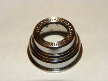


Congratulations, you are now the owner of a USB microscope.
Unfortunately, there are no many photographs, since I have not yet made a holder for it, and you cannot take photographs with a microscope. Even with not very high magnification, everything shakes and blurs. However, to visually assess its multiplicity, I will show you one photograph, which I managed to take with difficulty.
The photo shows the pixels of a laptop display. 
Unfortunately, I haven’t been able to get better quality yet, it requires more body movements, and the quality of the CMOS matrix leaves much to be desired, but what do you want from a microscope for $3.4.
To be continued…
Tags: usb microscope, web camera
Hello, habra users! This post will show you how to make one out of an old one. webcams qualitative microscope. It's really easy to do. If you are interested, continue under the hack.
Step 1: Required Materials
- Actually, the webcam itself
- Screwdriver
- Super glue
- Empty box
- Brain and some free time
Step 2: Opening the webcam
First, open up your camera. But be careful not to damage the CMOS sensor.
You need to extend the capture button wires to get still images. I also took out the LED on/off wires. They were gray and yellow (yours may vary). 
Step 3: Working with the Lens
Now we need to flip the lens over the CMOS sensor. Place it 2-3 mm from this sensor and secure it (for example, with superglue).

Step 4: Assembling the Camera
After turning the lens over, put the camera back together. It is now ready to use as a microscope.
Step 5: Final Stage
Now you need to attach the camera to the box, as shown in the photo. Now she is ready to receive images!You can also put a mirror so that the light spreads throughout the “object of study” and under it. Now our microscope is completely ready!
How to make a microscope from a webcam
If you take apart a suitable (adjustable focus) webcam, you can remove the lens and flip it over. In this case, the camera turns into... a microscope!
I used this camera (on chipset VC0345 with sensor OmniVision OV7670) with a two-lens lens:
Since wires for the microphone were added to the camera cable, which caused inconvenience in use, I unsoldered the standard cable and soldered another USB-cable: 
I use frosted glass as a stage for observing objects in the light: 
The glass is mounted on a plastic tube, and from below I illuminate it with white flashlight LEDs: 
Such a microscope is a transmitted light microscope and allows you to observe an object of interest in transmitted light in a bright field. The result is a shadow image of the object.
The main problem is keeping the webcam at the right distance from the object being watched, so I take many frames and choose the best one: 
For this I use a program I wrote :
Zooming in on my homemade digital microscope
Visual (geometric) magnification shows how many times the observed object on the computer screen is larger than its natural size. To estimate this parameter, you can use, for example, the distance between the strokes of a caliper. This magnification depends on the monitor used and is determined by the product of the lens magnification times the camera's own magnification.
The camera's own magnification is determined by the ratio of the size of the image on the screen (for example, diagonal) to the size of the light-receiving matrix.
For my microscope on a laptop screen, the distance between adjacent caliper strokes (1 millimeter) is 9 centimeters: 
Thus, the magnification of my homemade microscope is 90 times
.
Optical zoom microscope is determined by the aperture number of the lens. Aperture number $F$ (English) F-number, optical speed- optical speed) is directly proportional to the focal length of the lens $f$ and inversely proportional to the diameter $D$ of its entrance pupil: $F = ( f \over D )$. This value theoretically (due to the wave nature of light) cannot exceed 1500 once.
To determine the linear dimensions of objects in an enlarged form, I determined that the distance between the strokes of a caliper (1 mm) in the image is 365 pixels: 
LCD Pixels
Using this "modified" camera I got these pixel images LCD-laptop panels: ![]()
On the left it is shown that when you point the camera lens at the monitor area with white color, all three groups of subpixels glow - red ( R), green ( G) and blue ( B).
In this case, the pixel itself is square in shape, although the subpixels are rectangular, and the pixel side length is about 0.25 mm.
In the left image you can see that the gap width between red and blue pixels is larger than between blue and green and between green and red. But the image is upside down, i.e. true subpixel order RGB. This is confirmed by the test.
The right shows that to create a yellow pixel, only the red ones are lit ( R) and green ( G) subpixels.
And here is an image of the subpixels of another laptop’s monitor illuminated in white, along with a fragment of the symbol: 
And this is the picture I got for the white color on the phone screen Nokia 2710 Navigation Edition:![]()
Here is the interesting shape of the pixels of an LCD TV (blue color is reproduced): ![]()
Minerals
Salt

Sand

Clay

Biological objects
Human
Saliva

Saliva is one of the popular objects of observation under a microscope. It is claimed that saliva can be used to make diagnostics.
Hair

Animals
Mosquito

bird feather

The structure of the feather is visible - the shaft bearing the barbs that hold the barbs.
Plants
Bluebell seed

Bell seeds are very small - the weight of one seed is about 0.2 milligrams.
grape leaf
As you can see, a USB microscope from a web camera for soldering is quite easy to make from scrap materials within a few hours. For this will be needed:- Webcam;
- soldering iron with solder and flux;
- screwdrivers;
- tripod spare parts;
- LEDs, if they are not in the camera;
- glue or epoxy resin;
- program for broadcasting images to an LCD monitor.
This is the design of a homemade microscope from an SMD inspection chamber that can be obtained.
The following video is devoted to the principle of making a microscope from a webcam with your own hands. A tripod was used and a video of the soldering process of the USB connector is shown.
Microscope from a camera
To be honest, this “microscope” looks quite strange. The principle is the same as with a webcam - the optics are turned 180 degrees. There are even special ones for SLR cameras.
Below you can see the image obtained from such a homemade microscope for soldering. A large depth of field is visible - this is normal.
Disadvantages of a homemade microscope::
- short working distance;
- large dimensions;
- You need to come up with a way to mount the camera comfortably.
Advantages of a camera for soldering:
- can be made from an existing SLR camera;
- magnification is smoothly adjustable;
- there is autofocus.
Microscope from a mobile phone
The most popular way to make a microscope from a mobile phone with your own hands is to screw a lens from a CD or DVD player to the smartphone camera. This is the design of the microscope.
Lenses in this technique are used with a very short focal length. Therefore, using such a microscope, you can only monitor the state of soldering of SMD components and look in the solder. You simply cannot get a soldering iron between the board and the lens. Below is a video that shows what magnification such a homemade microscope gives.
Another option is a microscope for a mobile phone. This thing looks like this and costs just a penny.In more advanced cases, a mobile phone is hung on an existing stereo or mono microscope for small details. I got some good pictures this way. This method is important when photomicrographs need to be taken for training or consultation with other artists.
4th place - USB microscope for soldering
Chinese USB microscopes are now popular, essentially made from web cameras on and or even with a built-in monitor, for example USB microscopes and. Such electron microscopes are more intended for visual diagnostics of electronics, video inspection of soldering quality, or, for example, for checking the sharpness of knives.
Let me remind you that the video signal delay in such microscopes is significant. With a built-in monitor it is much easier to solder, but there is no depth of field and three-dimensional perception of micro-objects.
Disadvantages of a USB microscope:
- temporary lags that do not allow quick soldering;
- low optical resolution;
- lack of volumetric perception;
- As a rule, this is a stationary option, connected to a computer or outlet.
Advantages of a USB microscope:
- the ability to work at a comfortable eye distance;
- you can take videos and photos;
- relatively low cost;
- low weight and dimensions;
- You can easily look at the board at an angle.
Reviews about them are quite good. Both of them are certainly not role models, but they look impressive. The image quality is good, the working distance is 100 or 200 mm depending on the attachments. These microscopes can be used for soldering with proper setup and care.
See the mini-review in the video, the image through the lens is shown at the 9th minute.
2nd place - imported microscope for soldering
Among foreign brands, Carl Zeiss, Reichers, Tamron, Leica, Olympus, Nikon are famous for microscope equipment. Models such as Nikon SMZ-1, Olympus VMZ, Leica GZ6, Olympus SZ3060, Olympus SZ4045ESD, Nikon SMZ-645 have rightfully earned the title of folk binocular microscopes for soldering for their image quality. Below are approximate prices for popular foreign models:
- Leica s6e/s4e (7-40x) 110 mm - $1300;
- Leica GZ6 (7x-40x) 110 mm - $900;
- Olympus sz4045 (6.7x-40x) 110 mm - $500;
- Olympus VMZ 1-4x 10x 90 mm - $500;
- Nikon SMZ-645 (8x-50x) 115 mm - $800;
- Nikon SMZ-1 (7x-30x) 100 mm - $400;
- good Nikon SMZ-10a - $1500.
In principle, the prices are not astronomical, but these are used microscopes that can be bought on eBay or Amazon with paid delivery. The benefit here needs to be considered in each particular case separately.
1st place - domestic microscope for soldering
Among truly domestic microscopes, it is well known LOMO and they make applied microscopes under the SME brand. The most suitable new microscopes for soldering are MSP-1 option 23 or . True, their price tag is not childish.
I have to say that Altami, Biomed, Microhoney, Levenhuk- all these are domestic sellers of Chinese microscopes. Many people complain about the quality of workmanship. We do not consider them for professional use. True, there are tolerable specimens. This depends on the conditions of transportation and storage. The fact is that their optics are adjusted using silicone glue with appropriate reliability.
From old stocks or used, truly Soviet ones can be taken on Avito:
- BM-51-2 8.75x 140 mm - 5 thousand rubles. play around;
- MBS-1 (MBS-2) 3x-100x 65 mm - up to 20 thousand rubles;
- MBS-9 3x-100x 65 mm - up to 20 thousand rubles;
- OGME-P3 3x-100x 65/190mm - up to 20 thousand rubles. (I have one at work, I like it);
- MBS-10 3x-100x 95 mm— up to 30 thousand rubles;
- BMI-1Ts 45x 200 mm - more than 200 thousand rubles. - measuring.
Results of the microscope rating
If you are still thinking about which microscope to choose for soldering, then my winner is MBS-10- the people's choice for many years now.
Rating of microscopes by purpose
Microscope for mobile phone repair
The following microscopes for soldering and repairing smartphones are sorted by increasing image quality:
- MBS-10 (low contrast, unrealistic colors at high magnifications, discrete switching of magnifications, 90 mm distance);
- MBS-9 (65 mm distance and low contrast);
- Nikon SMZ-2b/2t 10cm (8x-50x)/(10-63x);
- Nikon SMZ-645 (8x-50x) 115 mm;
- Leica s6e/s4e (7-40x) 110 mm;
- Olympus sz61 (7-45x) 110 mm;
- Leica GZ6 (7x-40x) 110 mm;
- Olympus sz4045 (6.7x-40x) 110 mm;
- Olympus VMZ 1-4x 10x with a working distance of 90 mm;
- Olympus sz3060 (9x-40x) 110 mm;
- Nikon SMZ-1 (7x-30x) 100 mm;
- Bausch and Lomb StereoZoom 7 (working distance only 77 mm);
- Leica StereoZoom 7;
- Nikon SMZ-10a with Nikon Plan ED 1x lens and 10x/23 mm eyepieces;
- Nikon SMZ-U (7.5x-75x) working distance with Nikon Plan ED 1x 85 mm, with original 10x/24 mm eyepieces.
Microscope for repairing tablets and motherboards
For such applications, the issue of maximum resolution is not so important; magnifications of 7x-15x work there. They require a good universal tripod and a low minimum magnification. The following microscopes for soldering motherboards and tablets are sorted by degree of image quality magnification:
- Leica s4e/s6e (110mm) with 35mm field;
- Olympus sz4045/sz51/sz61 (110mm) with a field of 33 mm;
- Nikon SMZ-1 (100mm) with a field of 31.5 mm;
- Olympus sz4045;
- Olympus sz51/61;
- Leica s4e/s6e;
- Nikon SMZ-1.
Microscope for a jeweler or dental technician
The following microscopes for the dental technician or jeweler with a long working distance are sorted by degree of image quality improvement:
- Nikon SMZ-1 (7x-30x) with 10x/21 mm eyepieces;
- Leica GZ4 (7x-30x) 9 cm with 0.5x lens (19 cm);
- Olympus sz4045 150 mm;
- Nikon SMZ-10 150 mm.
Microscope for engraving
The following microscopes for engraving with a large depth of field are sorted in ascending order of image quality:
- Nikon SMZ-1;
- Olympus sz4045;
- Leica gz4.
How to check a used microscope when purchasing
Before purchasing a used microscope for soldering, it is easy to check (partially taken from this specialist):
- inspect frame microscope for scratches and impact marks. If there are signs of impact, the optics may be knocked off.
- check play of handles positioning - it should not exist.
- Mark a small dot on a piece of paper with a pencil or pen and check if the dot doubles at different magnifications.
- when turning the microscope adjustment knobs, listen for the presence crunch or slippage. If they are, the plastic gears may be broken and they are not sold separately.
- inspect the eyepieces for presence enlightenment. It is often scratched or erased due to improper care.
- rotate the eyepieces around their axis on a white background. If image artifacts are also spinning, then the problem is dirt on the eyepieces - that’s half the problem.
- if visible gray spots, faded image or dots, then the prism or auxiliary optics may be dirty. Sometimes a whitish coating, dust and even fungus are found on it.
- The most difficult thing in diagnosing a soldering microscope is to determine the weak ignorance vertically. If it is difficult for your eyes to adapt to the image in a couple of minutes, then it is better not to take such a microscope for soldering - it has severe misalignment. If, when soldering under a microscope, your eyes get tired within 30-60 minutes and your head starts to hurt, then this is weak ignorance. Slight differences in height between objects are difficult to determine when purchasing.
- inspect the spare parts, if available.
How to mount a microscope on your desktop
There are many ways to mount a soldering microscope to your workbench. Manufacturers solve these problems with the help of a barbell. They keep the microscope from falling and make it easy to position it relative to the board.
A homemade microscope stand or tripod is usually made from an old photographic enlarger or other available resources and parts.
But Master Sergei made a microscope stand for soldering microcircuits with his own hands from furniture tubes. It turned out well. See a video review of it below.
Master Sergei and Master Soldering worked on the material. In comments write what microscopes you use for soldering microcircuits and how good they are.
It's no secret that the world around us has subtle structures, the organization and structure of which cannot be discerned by the human eye. The entire universe remained inaccessible and unknown until the microscope was invented.
We all know this device from school. In it we looked at bacteria, living and dead cells, objects and objects that we all see every day. Through a narrow viewing lens, they miraculously turned into models of lattices and membranes, nerve plexuses and blood vessels. At such moments you realize how big and multifaceted this world is.
Recently, microscopes have begun to be made digital. They are much more convenient and efficient, because now you don’t have to look closely at the lens. Just look at the monitor screen, and we see an enlarged digital image of the object in question. Imagine that you can make such a miracle of technology with your own hands from an ordinary webcam. Don't believe me? We invite you to verify this with us.
Necessary resources for making a microscope
Materials:
- Perforated plate, corner and brackets for fastening wooden parts;
- A section of profile pipe 15x15 and 20x20 mm;
- Small fragment of glass;
- Webcam;
- LED flashlight;
- M8 bolt with four nuts;
- Screws, nuts.
Tools:
- Electric drill or screwdriver with a 3-4 mm drill bit;
- Pliers;
- Phillips screwdriver;
- Hot glue gun.
Assembling a microscope - step-by-step instructions
For the tripod base of the microscope we use perforated plates and metal corners. They are used to join wooden products. They are easily bolted together, and many holes allow this to be done at the required level.
Step one - install the base
We cover the flat perforated plate on the back side with soft furniture pads. We simply glue them on the corners of the rectangle.


The next element will be a bracket or corner with versatile shelves. We fasten the short shelf of the bracket and the base plate with a bolt and nut. We tighten them with pliers for reliability.


We mount two small brackets on the edge of the plate on both sides. We attach two more longer corners to them so that we form a small frame. This will be the base for the microscope viewing glass. It can be made from a small piece of thin glass.


Step two - make a tripod
We make a tripod from a piece of square profile pipe 15x15 mm. Its height should be about 200-250 mm. There is no point in doing more, since exceeding the distance from the viewing glass reduces the quality of the image, and less risks being overexposed and incorrect.
We attach the tripod to a perforated bracket, and on top of it we place a small piece of 20x20 pipe so that it moves freely along this stand.


We make an open frame from two brackets overlapped with each other. We choose longer bolts so that they are enough to tighten this frame around the moving section of the pipe. We place a plate with two holes on the sides on them and secure it with nuts.


To adjust the distance of the frame from the viewing glass, use an M8x100 mm bolt. We will need two nuts to fit the bolt size, and two larger ones. We take epoxy glue and glue the bolt nuts to the tripod in three places. A nut screwed onto the end of a bolt can also be secured with epoxy.


Step three - making the lens
In place of the tube with an eyepiece in our microscope there will be a regular webcam. The higher the resolution, the better; the connection to a computer can be either wired (USB 2.0, 3.0), or via Wi Fi or Bluetooth.
We free the camera from the body by unscrewing the motherboard with the matrix with a screwdriver.


We remove the protective cap and unscrew the lens with lenses and filter. All you need to do is place it in the same place, turning it 180 degrees.


We wrap the junction of the camera lens with the cylindrical body with electrical tape. If desired, it can be additionally glued with a hot glue gun. At this stage, the modified lens can already be tested in action.


Step four - final assembly of the microscope
We assemble the camera in reverse order, placing its body on the tripod frame with hot glue. The lens should be pointed downwards at the viewing glass of the microscope. The wiring harness can be secured with nylon ties to the tripod stand.
We adapt a low LED flashlight to the sight glass illuminator. It should fit freely under the microscope viewing panel. We connect the camera to the computer, and after a while the image will appear on the monitor screen.


The assembly is ready, it can be checked on any object, for example, by examining the crystal lattice of a pencil lead or the pixel structure of the screen of your smartphone. A popular trend today is the use of such homemade or inexpensive microscopes to control the soldering of small parts on electronic boards. Your child will undoubtedly like it, and perhaps awaken an interest in learning about the world around us.


