There are many diagnoses, the name of which says little to ordinary citizens. One of these diseases is chronic myeloid leukemia. Reviews of patients with this ailment are nevertheless able to attract attention, since this disease can not only cause significant damage to health, but also lead to a fatal outcome.
The essence of the disease
If one has to hear such a diagnosis as "chronic myeloid leukemia", then it is important to understand that we are talking about a serious tumor disease of the hematopoietic system, in which hematopoietic stem cells of the bone marrow are affected. It can be attributed to the group of leukemias, which are characterized by large formations of granulocytes in the blood.
At the very beginning of its development, myeloid leukemia manifests itself through an increase in the number of leukocytes, reaching almost 20,000 / μl. At the same time, in the progressive phase, this figure changes to 400,000 / μl. It is worth noting the fact that both in the hemogram and in the myelogram, the predominance of cells with different degrees of maturity is recorded. We are talking about promyelocytes, metamyelocytes, stab and myelocytes. In the case of the development of myeloid leukemia, changes in the 21st and 22nd chromosomes are detected.
This disease in most cases leads to a noticeable increase in the content of basophils and eosinophils in the blood. This fact is evidence that one has to deal with a severe form of the disease. In patients who suffer from such an oncological disease, splenomegaly develops, and a large number of myeloblasts are recorded in the bone marrow and blood.
How does the onset of the disease occur?
Chronic myeloid leukemia pathogenesis is quite interesting. Initially, a somatic mutation of a pluripotent hematopoietic blood stem cell can be determined as a triggering factor in the development of this disease. The main role in the mutation process is played by the cross translocation of chromosomal material between the 22nd and 9th chromosomes. In this case, the formation of the Ph-chromosome occurs.
There are cases (no more than 5%) when the Ph chromosome cannot be detected during a standard cytogenetic study. Although molecular genetic research reveals an oncogene.
Chronic myeloid leukemia can also develop due to exposure to various chemical compounds and radiation. Most often this disease is diagnosed in adulthood, extremely rarely in adolescents and children. As for gender, this type of tumor is recorded with the same frequency in both men and women aged 40 to 70 years.
Despite all the experience of doctors, the etiology of the development of myeloid leukemia is still not completely clear. Experts suggest that acute and chronic myeloid leukemia develops due to a violation of the chromosomal apparatus, which, in turn, is caused by the influence of mutagens or hereditary factors.
Speaking about the effect of chemical mutagens, it is worth paying attention to the fact that enough cases have been recorded when people exposed to benzene or using cytostatic drugs (Mustargen, Imuran, Sarkozolin, Leukeran, etc.) , myeloid leukemia developed.
Chronic myeloid leukemia: stages
With such a diagnosis as "myeloid leukemia", there are three stages in the development of this disease:
Initial. It is characterized by an enlargement of the spleen and a stable pumping of leukocytes in the blood. The patient's condition is examined in dynamics, without using radical treatment measures. The disease, as a rule, is diagnosed already at the stage of total generalization of the tumor in the bone marrow. At the same time, in the spleen, and in some cases in the liver, there is an extensive proliferation of tumor cells, which is characteristic of the expanded stage.

Expanded. Clinical signs at this stage begin to dominate, and the patient is prescribed treatment with specific drugs. At this stage, the myeloid tissue in the bone marrow, liver and spleen grows, the fat in flat bones is practically completely replaced. There is also a sharp predominance of the granulocytic lineage and three-growth proliferation. It is worth noting that in the expanded stage, the lymph nodes are extremely rarely affected by the leukemic process. In some cases, the development of myelofibrosis in the bone marrow is possible. There is a possibility of developing pneumosclerosis. As for the infiltration of the liver by tumor cells, in most cases it is expressed quite clearly.
Terminal. At this stage of the development of the disease, thrombocytopenia and anemia are progressing. The manifestations of various complications (infections, bleeding, etc.) become apparent. The development of a second tumor from immature stem cells is often observed.
What is the life expectancy to count on?
If we talk about people who have had to deal with chronic myeloid leukemia, then it is worth noting that modern methods of treatment have significantly increased the chances of such patients for a relatively long life. Due to the fact that discoveries were made in the field of pathogenetic mechanisms of the development of the disease, which made it possible to develop drugs that can affect the mutated gene, with such a diagnosis as chronic myeloid leukemia, the life expectancy of patients can be 30-40 years from the moment the disease is detected. But this is possible provided that the tumor was benign (slow swelling of the lymph nodes).
In the case of the development of a progressive or classical form, the average is from 6 to 8 years from the moment the disease was diagnosed. But in each individual case, the number of years that the patient can enjoy is significantly influenced by the measures that were taken in the course of treatment, as well as the form of the disease.
On average, according to statistics, during the first two years after the detection of the disease, up to 10% of patients die and 20% in subsequent years. Many patients with myeloid leukemia die within 4 years after being diagnosed.
Clinical picture
The development of a disease such as chronic myeloid leukemia is gradual. At first, the patient feels a deterioration in general health, rapid fatigue, weakness, and in some cases moderate pain in the left hypochondrium. After the study, an increase in the spleen is often recorded, and a blood test reveals a significant neutrophilic leukocytosis, characterized by the presence of a shift in the leukocyte formula to the left due to the action of myelocytes with an increased content of basophils, eosinophils and platelets. When the time comes for a detailed picture of the disease, then patients experience disability due to sleep disturbance, sweating, a stable increase in general weakness, a significant increase in temperature, pain in the spleen and bones. There is also a loss of mass and appetite. At this stage of the disease, the spleen and liver are significantly enlarged.

At the same time, chronic myeloid leukemia, the symptoms of which differ depending on the stage of development of the disease, already in the initial stage leads to the fact that the predominance of eosinophils, granular leukocytes and basophils is revealed in the bone marrow. This growth occurs due to a decrease in other leukocytes, normoblasts and red blood cells. If the process of the course of the disease begins to worsen, then the number of immature myeloblasts and granulocytes increases significantly, and hemocytoblasts begin to appear.
Blast crisis in chronic myeloid leukemia leads to total power metaplasia. In this case, there is a high fever, during which there are no signs of infection. Hemorrhagic syndrome develops (intestinal, uterine, mucous bleeding, etc.), leukemides in the skin, ossalgia, lymph nodes increase, complete resistance to cytostatic therapy and infectious complications are recorded.
If the process of the course of the disease could not be significantly influenced (or such attempts were not made at all), then the condition of the patients will progressively deteriorate, while thrombocytopenia will appear (the phenomena of hemorrhagic diathesis are felt) and severe anemia. Due to the fact that the size of the liver and spleen grows rapidly, the volume of the abdomen increases noticeably, the state of the diaphragm becomes high, the abdominal organs are compressed, and, as a result of the above factors, the respiratory excursion of the lungs begins to decrease. Moreover, the position of the heart changes.
When chronic myeloid leukemia develops to such a level, dizziness, shortness of breath, palpitations and headache appear against the background of pronounced anemia.
Monocytic crisis in myeloid leukemia
Concerning the topic of monocytic crisis, it should be noted that this is a rather rare phenomenon, during which young, atypical and mature monocytes appear and grow in the bone marrow and blood. Due to the fact that the bone marrow barriers are violated, at the terminal stage of the disease, fragments of the nuclei of megakaryocytes appear in the blood. One of the most important elements of the terminal stage in monocytic crisis is the suppression of normal hematopoiesis (regardless of the morphological picture). The disease process is aggravated by the development of thrombocytopenia, anemia and granulocytopenia.
In some patients, a rapid enlargement of the spleen may be recorded.
Diagnostics
The fact of the progression of a disease such as chronic myeloid leukemia, the prognosis of which may be rather uneasy, is determined by a whole complex of clinical data and specific changes in the process of hematopoiesis. In this case, histological studies, histograms and myelograms are taken into account without fail. If the clinical and hematological picture looks not clear enough and there is not enough data to confidently make a diagnosis, then doctors focus on the detection of the Ph chromosome in monocytes, megakaryocytes, erythrocytes and bone marrow granulocytes.

In some cases, it is necessary to differentiate chronic myeloid leukemia. Diagnostics, which can be defined as differential, is focused on identifying a typical picture of the disease with hyperleukocytosis and splenomegaly. If the option is atypical, then a histological examination of the spleen punctate is performed, as well as the study of the myelogram.
Certain difficulties can be observed when patients are admitted to the hospital in a state of blast crisis, the symptoms of which are very similar to myeloid leukemia. In such a situation, the data of a thoroughly collected anamnesis, cytochemical and cytogenetic studies, significantly help. Often, chronic myeloid leukemia has to be differentiated from osteomyelofibrosis, in which one can observe intense myeloid metaplasia in the lymph nodes, spleen, liver, as well as significant splenomegaly.
There are situations, and they are not uncommon, when a blood test helps to identify chronic myeloid leukemia in patients who underwent a routine examination (in the absence of complaints and an asymptomatic course of the disease).

Diffuse myelosclerosis can be ruled out by bone x-ray, which shows multiple sclerosis in the flat bones. Another disease, which, although rare, but still has to be differentiated from myeloid leukemia, is hemorrhagic thrombocythemia. It can be characterized by leukocytosis with a shift to the left and enlargement of the spleen.
Laboratory tests in the diagnosis of myeloid leukemia
In order to accurately determine the condition of a patient with suspected chronic myeloid leukemia, a blood test can be performed in several directions:
Blood chemistry. It is used to detect abnormalities in the functioning of the liver and kidneys, which are the result of the use of certain cytostatic agents or were triggered by the proliferation of leukemic cells.

- Clinical blood test (complete). It is necessary to measure the level of various cells: platelets, leukocytes and erythrocytes. In most patients who have had to deal with a disease such as chronic myeloid leukemia, the analysis reveals a large number of immature white cells. Sometimes there may be a low platelet or red blood cell count. Such results are not a basis for determining leukemia without an additional test, which is aimed at examining the bone marrow.
Microscopic examination of bone marrow and blood samples by a pathologist. In this case, the shape and size of the cells are studied. Immature cells are identified as blasts or myeloblasts. The number of hematopoietic cells in the bone marrow is also counted. The term "cellularity" is applicable to this process. In those with chronic myeloid leukemia, the bone marrow is usually hypercellular (a large accumulation of hematopoietic cells and a high content of malignant ones).
Treatment
With a disease such as chronic myeloid leukemia, treatment is determined depending on the stage of development of tumor cells. If we are talking about mild clinical and hematological manifestations in the chronic stage of the disease, then nutritious nutrition, enriched with vitamins, regular dispensary observation and general strengthening therapy should be considered as relevant therapeutic measures. The course of the disease can be favorably influenced by "Interferon".

In case of development of leukocytosis, doctors prescribe "Mielosan" (2-4 mg / day). If you have to deal with a higher leukocytosis, then the dose of "Mielosan" can rise to 6 or even 8 mg / day. The cytopenic effect should be expected no earlier than 10 days after the first dose of the drug. A decrease in the size of the spleen and the cytopenic effect occurs on average during the 3-6th week of treatment, if the total dose of the drug was from 200 to 300 mg. Further therapy involves taking once a week 2-4 mg of "Mielosan", which at this stage has a supporting effect. If the first signs of exacerbation make themselves felt, myelosanotherapy is performed.
It is possible to use a technique such as radiation therapy, but only if splenomegaly is determined as the main clinical symptom. For the treatment of patients whose disease is in a progressive stage, poly- and monochemotherapy is relevant. If a significant leukocytosis is recorded, with an insufficiently effective effect of "Mielosan", "Mielobromol" is prescribed (125-250 mg per day). At the same time, strict control of peripheral blood parameters is carried out.
In case of development of significant splenomegaly, "Dopan" is prescribed (once 6-10 g / day). Patients take the drug once for 4-10 days. The intervals between doses are determined depending on the degree and rate of decrease in the number of leukocytes, as well as the size of the spleen. As soon as the decrease in leukocytes reaches an acceptable level, the use of "Dopan" is discontinued.

If the patient develops resistance to "Dopan", "Mielosan", radiation therapy and "Myelobromol", "Hexaphosphamide" is prescribed for treatment. In order to effectively influence the course of the disease in a progressive stage, the CVAMP and AVAMP programs are used.
If resistance to cytostatic therapy develops in a disease such as chronic myeloid leukemia, treatment in the progression stage will be focused on the use of leukocytopharesis in combination with a specific chemotherapy regimen. As an urgent indication for leukocytopharesis, clinical signs of stasis in the vessels of the brain (a feeling of heaviness in the head, hearing loss, headaches), which are caused by hyperthrombocytosis and hyperleukocytosis, can be determined.
In the case of a blast crisis, various chemotherapy programs used for leukemia can be considered relevant. Indications for transfusions of erythrocyte mass, thrombocyte concentrate and antibacterial therapy are infectious complications, the development of anemia and thrombocytopenic hemorrhage.
Regarding the chronic stage of the disease, it should be noted that at this stage of the development of myeloid leukemia, bone marrow transplantation is quite effective. This technique is able to ensure the development of clinical and hematological remission in 70% of cases.
An urgent indication for the use of splenectomy in chronic myeloid leukemia is the threat of rupture or rupture of the spleen itself. Relative indications include severe abdominal discomfort.
Radiation therapy is indicated for those patients who have been diagnosed with extramedullary neoplastic formations that pose a threat to life.
Chronic myeloid leukemia: reviews
According to patients, such a diagnosis is too serious to be ignored. By examining the testimonies of various patients, it becomes obvious that there is a real possibility of defeating the disease. To do this, you need to timely undergo diagnostics and a course of subsequent treatment. Only with the participation of highly qualified specialists there is a chance to defeat chronic myeloid leukemia with minimal losses to health.

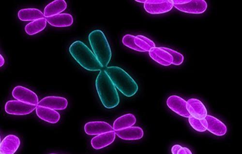
Blood smear of a patient with chronic myeloid leukemia
Chronic myeloid leukemia (CML) is a malignant neoplasm of hematopoietic tissue, accompanied by progressive proliferation of immature granulocytes. The disease initially has a sluggish character, gradually flowing into an exacerbation stage with severe symptoms and the formation of systemic disorders. It is one of the most dangerous and disabling diseases.
CML is the first oncological disease in which a link has been determined between the development of carcinogenesis and a mutation in a gene. The characteristic anomaly is based on the translocation of the 9th and 22nd chromosomes, that is, sections of these chromosomes change places, forming an aberrant chromosome. The mutated chromosome was identified by researchers from Philadelphia, so it was named Philadelphia or Ph-chromosome.
The study of the Ph chromosome and its influence allowed the development of a new means for suppressing oncological processes, thanks to which the life expectancy of patients has increased significantly. However, the disease still remains incurable. The number of primary CML is diagnosed in 1.5: 100,000 of the population per year, the peak incidence is at the age of 30-50 years, 30% of CML is detected in people over 60 years old, in children the disease is diagnosed in less than 5% of cases.
Reasons for development

Pesticides have a negative effect on hematopoiesis
The disease has been known to science since 1811, but until now the factors provoking a mutation in the gene have not been determined. There are a number of reasons that contribute to the development of pathology:
- radiation exposure, including radiation therapy;
- chemotherapy for other oncological diseases;
- a number of genetic diseases characterized by a chromosomal abnormality (for example, Down's syndrome);
- interaction with chemical compounds (petroleum products, pesticides).
Pathogenesis of chronic myeloid leukemia

Pathogenesis of chronic myeloid leukemia
The hybrid gene BCR-ABL 1, formed as a result of chromosome translocation, produces the synthesis of the BCR-ABL protein. This protein is a tyrosine kinase that normally promotes the transmission of signaling impulses for cell growth. The tyrosine kinase created by mutation becomes an active factor in cell proliferation; they begin to divide and spread independently of growth factors. The process of creating clones of the mutated cell takes place.
Uncontrolled division is accompanied by impaired apoptosis - programmed cell death. Also, hybrid tyrosine kinase inhibits the natural repair functions in DNA molecules, creating the prerequisites for subsequent mutations, which aggravates the pathological process.
Reproducing cells are immature, blast precursors of high-grade blood elements. Blast cells gradually displace functional erythrocytes, platelets and leukocytes. Abnormalities in other chromosomes are added, which triggers an accelerated process of destruction of the organism as a whole.
Stages of chronic myeloid leukemia

Blast crisis is one of the stages of myeloid leukemia
- Chronic -< 15% бластных клеток. Обычно стадия длится несколько лет. Признаки заболевания нередко обнаруживаются лишь в результатах общего анализа крови. Выявляется ХМЛ на этой стадии более чем у 80% пациентов. Мутировавшая клетка ещё контролируется геном BCR-ABL, способность к дифференцировке сохранена, а здоровые клетки функционируют в естественном режиме.
- Progressive (acceleration) - 15 - 29% of blast cells. The accelerated proliferation of immature cells reduces the median lifespan to one year. Thrombocytopenia develops, the number of leukocytes increases, and signs of resistance to therapy appear. At this stage, pathology is detected in 10-12% of patients. Tumor cells begin to suppress healthy ones, lose contact with the microenvironment, and actively move from the bone marrow into the bloodstream. Subsequent mutations in the chromosomes begin to emerge.
- Blast crisis -> 30% blast cells. The stage is characterized by the aggressive nature of the mutated cells, the patient's condition deteriorates sharply. Additional abnormalities both in the BCR-ABL gene and in the genome as a whole provoke a chain of pathological reactions that are no longer amenable to treatment. At this stage, tissues of internal organs, skin and mucous membranes can be affected, myeloid cells are transformed into sarcoma.
Symptoms and Signs

Hemorrhagic syndrome
The signs of CML become noticeable closer to the progressive stage.
- Symptoms of tumor intoxication: weight loss, fatigue, wave-like fever, pruritus, nausea, joint pain.
- Symptoms of tumor proliferation - enlargement of the spleen and liver, pain in the left hypochondrium, skin lesions.
- Anemic syndrome - dizziness, severe pallor, palpitations, feeling short of breath.
- Hemorrhagic syndrome - a tendency to bleeding of the mucous membranes, a rash in the form of red dots, prolonged bleeding with minor cuts.
Diagnosis of the disease

One of the methods for diagnosing the disease is X-ray
CML diagnostics include:
- Initial examination of the patient with the study of anamnesis, complaints, as well as examination by palpation of the size of the spleen and liver.
- A general blood test reveals the number and characteristics of the formed elements of the blood.
- Biochemical analysis is performed to determine the level of bilirubin, electrolytes, glucose, LDH, AST, ALT.
- A histological examination of the bone marrow determines the accumulation of blast cells.
- Cytogenetic analysis reveals the translocation of chromosomes.
- At the 3rd stage, immunophenotyping is performed to identify blast cells.
- Gene sequencing is used to detect gene mutations.
- An ultrasound scan of the internal organs, primarily the spleen and liver, is performed.
- Additionally, chest x-ray, ECG, echocardiography, ELISA for markers of various diseases, coagulogram and other studies are prescribed.
Treatment

The mainstay of treatment is tyrosine kinase inhibitors
CML therapy is currently based on the use of tyrosine kinase inhibitors. The I-generation agent imatinib blocks the activity of hybrid tyrosine kinase, penetrating into the "pocket" of the BCR-ABL protein. The development of imatinib has made a breakthrough in the treatment of CML due to its effectiveness. However, it is not uncommon for patients to develop resistance to the drug, which led to the creation of second-generation inhibitors. The combination with other methods of treatment allows to achieve high rates in improving the quality and duration of life.
The choice of drug and dose is determined depending on the stage of CML and the risk of side effects. Typically, treatment begins with 400 mg / day imatinib at the initial stage, 600 mg / day at the subsequent stages, then the dose can be increased or decreased. Various aberrations in genes cause low sensitivity to drugs, so the patient can change one inhibitor for another.
![]()
Bone marrow transplant
Interferon therapy is usually prescribed in the 1st stage of CML, since it is not effective in subsequent stages.
To reduce the mass of the tumor and if there is no result in the treatment with inhibitors, chemotherapy is carried out. In the stage of blast crisis, polychemotherapy is used similarly to the treatment of acute leukemia.
Radiation therapy may be prescribed for severe splenomegaly. At risk of rupture of the spleen, splenectomy is performed.
To date, research continues to create an even more perfect drug. With the help of the Skolkovo Foundation, Russian scientists are conducting clinical trials of a third-generation inhibitor, which should surpass the previous ones in its effectiveness.
Prevention and prognosis

The prognosis of the disease is determined by the doctor
The reason for the formation of CML has not been established, therefore, preventive measures are taken to avoid contact with carcinogenic substances, exposure to radiation.
The prognosis is determined by the stage and severity of the disease. One of the predictive models (Kantarjian H.M.) includes factors:
- advanced age of the patient at diagnosis;
- the concentration of blast cells in the blood ≥ 3%, in the bone marrow ≥ 5%;
- concentration of basophils ≥ 7%;
- platelet concentration ≥ 700 * 10 9 / l;
- severe splenomegaly.
This model is designed for the initial phase of CML, if there are ≥ 3 signs, the prognosis is poor, subsequent phases are considered “always unfavorable”. However, each case of CML is individual; there are known patients with a life expectancy of more than 30 years in the chronic stage. On average, with timely initiation of treatment with tyrosine kinase inhibitors, 70-80% of patients live for more than 10 years. With the transition of the disease into the progressive phase, the survival rate decreases by 3-4 times, with a blast crisis it is still up to 6 months.
Myeloid leukemia is not an independent disease, but means a condition characterized by increased and uncontrolled growth of myeloid cells in the red bone marrow and their accumulation in the bloodstream.
Popularly, leukemia is also called blood cancer, but the term is not correct. Nosologically, it is customary to distinguish two diseases associated with this condition - chronic (CML) and acute myeloid leukemia (AML).
In AML, there is a massive division of myelopoietic progenitor cells (blasts), which cannot differentiate into mature ones. According to WHO statistics, AML accounts for about 80% of all other types of leukemia. According to surveillance data, most often the disease affects patients under 15 and after 60 years. AML is less common in the gender ratio in women.
Unlike AML, in CML, malignant cells retain the ability to differentiate to mature forms. CML accounts for about 15% of all cases of leukemia. The annual incidence is approximately 1.6 per 100,000 population. Most often, the disease affects patients in the age group 20-50 years. In the gender ratio, men get sick more often than women, about 1.5: 1.
Classification
In addition to the classical ICD, there are several classifications that make it possible to obtain an accurate description of the pathological process. For acute myeloid leukemia, the most relevant is the French-American-British (FAB) classification based on the type and maturity of the cells from which the leukemia develops.
According to the hematological classification, chronic myeloid leukemia has about 5 main subtypes.
According to the international classification of diseases of the 10th revision (ICD-10), each subtype of the disease should be assigned a specific code:
C92.0 - Acute myeloid leukemia.
C92.1 - Chronic myeloid leukemia.
C92.2 - Atypical chronic myeloid leukemia.
C92.4 - Acute promyelocytic leukemia
C92.5 - Acute myelomonocytic leukemia.
C92.7 - Other myeloid leukemia.
C92.9 - Unspecified myeloid leukemia.
C93.1 - Chronic myelomonocytic leukemia
Causes and risk factors for AML
Acute myeloid leukemia is caused by damage to the DNA of the developing cells of the myeloid lineage of the bone marrow, which further provokes abnormal production of blood components. In AML, the bone marrow synthesizes immature cells called myeloblasts. These abnormal cells cannot function properly and, with abundant division and growth, begin to displace healthy elements of the bone marrow.
In most cases, it is unclear what causes the DNA mutation, but several factors have been identified as contributing to AML, including antecedent hematologic disorders, hereditary causes, environmental exposure, and drug effects. However, most patients with new-onset AML do not have an identifiable cause.
Antecedent hematological disorders. The most common cause of development is considered myelodysplastic syndrome (MDS). It is a bone marrow disorder of unknown etiology that most often occurs in elderly patients and is manifested by progressive cytopenia that develops over several months or years. There are also gradations of risk in patients with this syndrome. For example, in refractory anemia with annular sideroblasts, the risk of developing AML is significantly lower than in patients with MDS with an increased number of blast cells.
Congenital disorders. Congenital diseases that predispose patients to AML include: Bloom's syndrome, Down's syndrome, congenital neutropenia, Fanconi's anemia, and neurofibromatosis. Usually in these patients, acute myeloid leukemia develops from childhood, but it can appear at a more mature age.
In clinical studies, it has been noted that the risk of spreading AML is significantly increased with regular contact with benzene. This chemical is used as a solvent in various industries (chemical plants, refineries, rubber and footwear). Benzene is found in adhesives, cleaning products, paints, and cigarette smoke. Exposure to formaldehyde has also been associated with AML, but the exact effect is not yet known.

Chemotherapy. AML is more common in patients who have previously undergone chemotherapy. Some drugs have a close relationship with the development of secondary leukemia ("Mechlorethamine", "Procarbazine", "Chlorambucil", "Melphalan", "Etoposide", "Teniposide" and "Cyclophosphamide").
The risk increases if the patient receives radiation therapy at the same time as these chemotherapy drugs. Secondary leukemias occur about 10 years after treatment for Hodgkin's disease, non-Hodgkin's lymphoma, or childhood acute lymphocytic leukemia. Secondary leukemias can also occur after treatment for breast, ovarian, or other cancers.
Exposure to radiation. The effect of high radiation exposure is a known risk factor for AML as well as acute lymphoblastic leukemia. This was first noted among Japanese survivors after the atomic bombing of Hiroshima and Nagasaki. Within 6-8 years after the tragic events, many Japanese showed signs of acute myeloid leukemia.
Adverse radiation exposure can be observed during radiation therapy for cancer treatment, as well as with some types of diagnostic tests (X-ray, fluoroscopy, computed tomography).
The causes are unknown, but it has been noted that men suffer from AML more often than women. Also, the disease is more common in Caucasians. Unproven risk factors include living in an area of high electromagnetic radiation, exposure to pesticides, bleaches and hair dyes.
Causes and risk factors for the development of CML
In a healthy person, the cells of the body contain 23 pairs of chromosomes in their nucleus. In people suffering from CML, in the cells of the bone marrow, a violation of the structure of the chromosomes occurs, which consists in the movement of a site from the 22nd chromosome to the 9th. The ultrashort chromosome 22, also called Philadelphia (after the city where it was first discovered), is present in the blood of 90% of people with CML.
Against the background of these chromosomal changes, new genes are formed that begin to overproduce the enzyme tyrosine kinase. Subsequently, a large amount of tyrosine kinase leads to abnormal division of bone marrow cells, which contributes to the development of chronic myeloid leukemia. Abnormal white blood cells do not develop or die as normal, but they divide in large numbers, displace healthy blood cells and damage the bone marrow.
Until now, the exact reasons for the appearance of AML have not been clarified. It is now generally accepted that acute myeloid leukemia develops against the background of the accumulation of mutations in the progenitor cells of myelopoiesis. Except for a few differences, the factors that increase the risk of developing CML are similar to AML.
Weakened immunity. Clinical studies have shown that people with immunosuppression, such as AIDS, are 3 times more likely to develop CML compared to the general population. The adverse effect of cytostatic drugs in people forced to take them after organ transplantation has also been noted. In this case, the risk doubles.
The reasons are not fully understood, but after statistical analysis, it turned out that patients with inflammatory bowel diseases, such as ulcerative colitis or Crohn's disease, have a higher chance of developing CML compared to the general population.

Pesticides. Several studies have shown that men who are in daily contact with pesticides (farmers, agricultural workers) have an increased risk of developing chronic myeloid leukemia. Compared to the general population, the risk increases by about 40%.
Gender, age and other risk factors. As with AML, it is more common for CML to infect European men. There have been 4 studies that reported the adverse effects of obesity. Being overweight increases the likelihood of getting sick by about 25%.
Symptoms
Most of the clinical manifestations and signs of myeloid leukemia for both acute and chronic are associated with the displacement of healthy bone marrow growths by abnormal cells. For this reason, 4 main syndromes are distinguished during the course of the disease:
- Anemic. A decrease in the number of red blood cells causes fatigue, increased heart rate, pallor, and shortness of breath.
- Immunodeficient. The lack of normal production of white blood cells makes patients more susceptible to infection, since abnormal cells lack the mechanisms that contribute to a full immune response.
- Intoxicating. The early signs of myeloid leukemia are often nonspecific and may mimic those of the flu or other colds. Common symptoms include fever, fatigue, weight loss, poor appetite, shortness of breath, anemia, petechiae (bleeding spots on the skin), bone and joint pain.
- Hemorrhagic. A decrease in platelet synthesis leads to mild bruising or bleeding with minor trauma.
In addition, with CML, the spleen is enlarged in more than 50% of cases. It can reach such a large size that it begins to squeeze the abdominal organs. An enlarged spleen is sometimes associated with AML, but this process is usually slow and painless.
Due to leukocyte infiltration, some patients experience swelling of the gums. In rare cases, the primary symptom of AML is the formation of a dense leukemic mass or tumor (chloroma) outside the bone marrow. Lymph node enlargement and paraneoplastic inflammation of the skin are very rare in AML.
Stages
Dividing the course of chronic lymphocytic leukemia into phases allows doctors to better plan treatment and predict the outcome of the disease.
| Chronic phase | Blood and bone marrow contain less than 10% blast cells. The phase can last for several years, but without adequate treatment, the disease will progress and move to the next stages of development. In about 90% of patients, CML is diagnosed in the chronic phase. Clinical manifestations may be present. They are usually expressed as general weakness and slight weight loss, and the abdomen may enlarge due to splenomegaly. |
| Acceleration (acceleration) phase | A unified definition for this phase has not yet been developed, but an increase in the number of blasts from 10 to 19% or more than 20% of basophils in the peripheral blood is considered to be the main criterion for the transition. Basophils sometimes contain cytogenetic changes in addition to the Philadelphia chromosome. |
| Blast crisis | In its course, it resembles acute myeloid leukemia. In this phase, the number of blasts containing additional genetic changes increases to 20 percent or more. In 25% of cases, blasts may look like immature cells in acute lymphocytic leukemia or acute myeloid leukemia. Clinical manifestations in this phase are fever, enlargement of the spleen, and weight loss. |
Until now, standards have not been developed for determining the staging of acute myeloid leukemia, but it is customary to distinguish 3 key phases based on the general course of the disease.
| Newly diagnosed AML | The phase corresponds to newly diagnosed leukemia, which had not been purposefully treated before. It is possible that the patient was previously prescribed drugs for the symptoms of the disease (fever, bleeding), but not to suppress the growth of abnormal cells. At this stage of the course, up to 20% of blast cells are found. |
| Remission | Phase means that the patient received appropriate treatment, against which the blood count returned to normal. The main criterion for remission is the presence of less than 5% of blast cells in the aspirate and their absence in the peripheral blood and cerebrospinal fluid. |
| Relapse | Clinical manifestations and pathological changes in peripheral blood and aspirate returned after treatment. |
The most common types of myeloid leukemia
Acute myeloid leukemia with maturation (M2) accounts for about 25% of all AML cases. The subtype is characterized by the movement of part of the 8th chromosome to the 21st. On both sides of the splicing, a new set of DNA is formed from fragments that previously encoded the RUNX1 and ETO proteins. Then these two sequences combine and begin to encode one large protein called M2 AML, which allows the cell to divide unhindered.
Chronic granulocytic leukemia is most common in CML. That is, any pathological factor that provokes changes in the chromosome set affects blast cells, from which granulocytes are then formed. This form of CML occurs in about 95% of cases.
Diagnostics
Several studies may be ordered to confirm the diagnosis of leukemia. Diagnostics also allows you to determine the type of disease and, based on the data obtained, choose the best method of treatment. The basis of the diagnostic process when confirming the diagnosis of acute or chronic myeloid leukemia is made up of laboratory research methods.
Complete blood count (CBC). In most patients, a preliminary diagnosis of myeloid leukemia is made after a CBC. The essence of the test is to count the blood cells (erythrocytes, leukocytes, platelets). UAC is often performed as part of a regular medical check-up. People with CML will have a marked increase in leukocyte counts (usually due to granulocytes), combined with thrombocytosis and basophilia. In addition, elements of immature leukopoiesis are observed in the blood formula. When other bone marrow growths are inhibited in patients, the number of erythrocytes decreases. Due to an increase in the total number of leukocytes, leukemia is sometimes called leukemia.

Aspiration and biopsy. No specific tumor markers have been found to determine myeloid leukemia, so in most cases they are diagnosed by a combination of biopsy and aspiration. This is the only sure way to confirm the diagnosis. Aspiration is a procedure that allows a thin needle to remove the liquid portion of the bone marrow, and a biopsy takes a solid sample. These 2 procedures are very similar and are often performed at the same time to obtain more accurate information about the condition of the bone marrow.
A typical site for aspiration and biopsy is the iliac crest of the pelvic bone. After the collection of biological material, a specialist in the field of pathological anatomy conducts a detailed examination of the samples obtained. One of the main criteria indicating AML in a patient is the presence of more than 20% blasts in the blood and aspirate.
The analysis consists in testing leukemic cells for the content of certain genes, proteins and other factors that indicate their malignancy. Based on this research, individualized targeted therapy can be further developed.

Genetic research. Allows you to determine the genotype of AML and select the optimal treatment option for the patient. In addition, the test results can be used in the future to monitor the treatment process.
Cytogenetic research. A type of genetic testing that is used to analyze cell chromosomes. Sometimes this study can be performed on peripheral blood cells, but tissue samples obtained from the bone marrow are needed to establish an accurate diagnosis.
After starting treatment for CML, cytogenetic and / or molecular testing is repeated on another bone marrow sample to recount the number of cells containing the Philadelphia chromosome and evaluate the effectiveness of chemotherapy.
For most patients, the presence of the Philadelphia chromosome and the BCR-ABL fusion gene is the main marker for the presence of CML. In a small number of patients, the Philadelphia chromosome is undetectable on routine tests, despite the presence of the BCR-ABL fusion gene and an increase in blood cell counts. However, the tactics of treatment in this case will be the same as in patients with a detectable Philadelphia chromosome.
Imaging research methods. Are prescribed to assess the effect of leukemia on other parts of the body. For example, computed tomography and ultrasound are sometimes used to view and measure the size of the spleen in patients with leukemia.
How fast is it developing?
No specific methods have been developed to predict the duration of the chronic phase and the onset of a blast crisis in CML. However, it is customary to consider a sharp increase in the level of leukocytes, hepatosplenomegaly, and an increase in the percentage of blasts in the red bone marrow as unfavorable factors. The same goes for AML.
Features of the course and treatment in special categories of patients
The course of the disease, depending on age and gender, is not very different. The only factor that needs to be considered is the weight and age of the patients, since these characteristics affect the dosage of the drugs.
Pregnancy. During pregnancy, the diagnosis of myeloid leukemia is very rare, about 1 in 300,000 cases. Moreover, if you do not start timely treatment, then there is a high probability of developing a spontaneous abortion. In addition, an increased level of blast cells in the blood can cause intrauterine growth retardation, provoke premature birth, or lead to intrauterine fetal death.
Despite the presence of a hematoplacental barrier that protects the fetus from the effects of chemotherapy, termination of pregnancy may be recommended in the early stages. If the diagnosis was made in the 2-3rd trimester, then, as a rule, the remainder of the pregnancy is carried out under the guise of chemotherapy. In addition, breastfeeding should be avoided during the course of chemotherapy.
Treatment
In the treatment of myeloid leukemia, the cooperation of several specialists is required to create the optimal therapeutic tactics. It is especially important that the patient is under the supervision of an oncologist and / or hematologist.
Treatment options depend on several factors, including the phase of the illness, the expected side effects, the patient's preference, and the general condition of the body.
Targeted therapy. This is a type of treatment that targets the genes of malignant cells, their proteins and the tissue environment that promotes the growth and survival of leukemia. Targeted therapy blocks the growth and spread of malignant cells while limiting damage to healthy tissue.
The prescription of targeted drugs for AML directly depends on the specificity of mutations that have arisen in malignant cells. For example, "Midostaurin" (Rydapt) is indicated for patients with the FLT3 gene mutation (25-30% of cases). Enasidenib (IDHIFA) is recommended for people with recurrent or refractory AML with the IDH2 mutation.
In CML, the target for active substances is the enzyme tyrosine kinase BCR-ABL. There are 5 main drugs called tyrosine kinase inhibitors (TKIs): Imatinib (Gleevec), Dasatinib (Sprycel), Nilotinib (Tasigna), Bosutinib (Bosulif) and Pontinib (Iclusig). All 5 drugs can stop the BCR-ABL enzyme, causing CML cells to die quickly.
It is important to note that while taking TKI, men and women should avoid conceiving a child. Otherwise, there is a high risk of spontaneous abortion, intrauterine fetal death or the birth of a child with severe malformations. In addition, patients may develop idiopathic myelofibrosis as a side effect of CML therapy.
Chemotherapy. Drugs from this group are prescribed to destroy malignant cells by suppressing their ability to grow and divide. The form of administration of drugs can be in the form of intravenous, subcutaneous injection, or in the form of tablets. A chemotherapy regimen usually consists of a certain number of cycles given over a given period of time. The patient can take 1 drug or several at the same time.
It is the main treatment for AML. Due to the frequent development of complications, the treatment process is rather difficult, therefore, chemotherapy courses should be carried out on the basis of specialized hospitals. In the treatment of patients, it is customary to distinguish 4 phases:
- Induction of remission.
- Anchoring.
- Intensification.
- Supportive therapy (2-5 years).
The most commonly used combination is Cytarabin (Cytosar-U) and an anthracycline drug such as Daunorubicin (Cerubidine) or Idarubicin (idamycin). Some older people are unable to take these drugs, and Decitabine (Dacogen), Azacitidine (Vidaza) and / or low doses of Cytarabin can be used instead.
As a rule, to achieve remission, 2-5 courses of chemotherapy are needed, after which the patient enters the consolidation phase, and he is prescribed several more procedures. Supportive therapy begins approximately one week after the end of the hardening period. If modern protocols are followed, stable remission can be achieved in 60%, and recovery - in 30% of patients.

As a rule, in CML, hydroxyurea preparations (Droxia, Hydrea) are prescribed, which are good at reducing the number of leukocytes. Chemotherapy can help your blood counts return to normal within a few days or weeks while reducing the size of your spleen. However, hydroxyurea preparations do not reduce the content of cells with the Philadelphia chromosome and do not have such a pronounced effect in the blast crisis phase. Despite the fact that hydroxyurea has few side effects, most patients with newly diagnosed CML are advised to take Imatinib or another TKI. This means that patients do not need hydroxyurea or only use it for a short period.
Stem cell / bone marrow transplant. This is a medical procedure in which the affected bone marrow is replaced with hematopoietic stem cells from a healthy donor. The method is considered the most effective treatment for both types of leukemia. There are 2 types of stem cell transplants:
- allogeneic - transplantation from a compatible donor (usually a relative);
- autologous - own bone marrow transplant.
The success of transplantation is influenced by the phase of the disease, the results of previous treatment, the patient's age and general condition. Although transplantation is the only method that can guarantee complete recovery from CML, it is used less frequently than TKI due to the high risk of side effects.
![]()
Immunotherapy. The method increases the body's natural defense mechanisms to activate them to fight myeloid leukemia. Immunotherapy involves the use of drugs based on immunocomponents, made in laboratory or natural conditions. Interferon (Alferon, Infergen, Intron A, Roferon-A) is an effective group of drugs that can reduce the number of leukocytes, and in some cases even reduce the number of cells containing the Philadelphia chromosome.
Before Imatinib became available, interferon therapy was the mainstay of treatment for chronic phase CML. Currently, Interferon is not recommended as a first-line drug, as a number of studies have shown that TKIs work better and cause fewer side effects. At the same time, unlike ITK, "Interferon" is safe to take during pregnancy.
New treatments. Most major hematology and cancer centers are actively involved in clinical trials aimed at increasing the rate of successful recovery from myeloid leukemia. In consultation with a doctor, it is necessary to clarify the possibility of participating in research projects to obtain experimental treatment.
Promising techniques currently being tested include:
- combinations of "Imatinib" with other drugs;
- development of new schemes for the use of ITC;
- development of vaccines against BCR-ABL;
- development of new methods of stem cell transplantation aimed at reducing side effects.

Alternative treatment. Myeloid leukemias are very serious diseases characterized by high mortality and great difficulties in treatment. For this reason, the use of folk remedies will be ineffective or even harmful to the patient. Patients, if desired, can take decoctions made on pumpkin, blueberries or birch buds, but only in addition to the main treatment.
Rehabilitation
The protocols do not provide for a specific rehabilitation program, but physiotherapy courses, therapeutic baths, oxytherapy, psychological support and balanced nutrition can be recommended to improve the patient's well-being. It is important that the patient during the rehabilitation period was under the supervision of a specialist who would understand the patient's condition and be able to eliminate the side effects of therapy.
Relapse
In most cases, patients with acute myeloid leukemia develop a relapse after chemotherapy. In such cases, autologous stem cell transplantation is recommended. A number of hematological centers that adhere to this treatment tactic in the second remission or at the beginning of the first relapse, achieve recovery of patients in 25-50% of cases.
Such high results were achieved because many patients retained their stem cells during the first remission, after which they underwent successful transplantation. Harvesting stem cells after relapse is not as effective as less than half of patients receiving chemotherapy will achieve a second remission. The most optimal solution for patients who do not have previously preserved stem cells is allogeneic transplantation.
If the patient does not have the opportunity to carry out a stem cell transplant, then in such cases the main therapeutic tactic will be the appointment of high-dose chemotherapy.
Resistant flow
Most patients achieve remission (no signs and symptoms) after initial AML treatment. But in some patients, small sections of mutated cells remain in the body even after a full course of chemotherapy. Over time, the number of damaged cells will increase until they are found on tests or until symptoms return. This condition is called resistant leukemia.
After the end of treatment, the doctor must provide the patient with personal information about the possible risk of developing resistant myeloid leukemia.
Complications
Myeloid leukemia has a huge number of complications that develop both against the background of the course of the underlying disease and as a result of taking chemotherapy drugs. However, the greatest concern for doctors, due to the increased risk of death and reduced quality of life, are the following three:
- Due to a pathological increase in the number of immature blast cells, normal blood growths are displaced, which leads to a violation of the body's immune mechanisms.

- Bleeding. Against the background of pathological changes in the blood coagulation system, people with AML are more susceptible to sudden internal bleeding.
- Infertility. Many drugs used in the treatment of AML cause sterility as a side effect. As a rule, it is temporary, but in some cases it can be permanent.
Forecast (life expectancy)
In AML, the prognosis is determined by the type of cells involved in the pathological process, the patient's age and the adequacy of the treatment. Standard modern therapeutic techniques increase survival in adult patients (up to 60 years), but in older patients this figure is much lower.
The life expectancy of patients suffering from CML does not exceed 3.5 years from the date of diagnosis. The blast crisis phase is especially dangerous to life. It accounts for 85% of all CML deaths. Timely and appropriate treatment allows the patient to increase the survival rate by an average of 5-6 years from the moment the disease is detected.
Diet
Patients suffering from blood diseases are prescribed table number 11. The emphasis in nutrition should be on meat, chicken eggs, milk, cheese and kefir. Also, to replenish the loss of vitamins, regular consumption of vegetables and fruits is necessary. The total daily calorie content must reach at least 4500 kcal.

Prophylaxis
There is no specific prophylaxis for myeloid leukemia. One can only advise people at risk to exclude contact with benzene, pesticides and radioactive elements. One of the goals of follow-up prophylaxis after treatment is to check regularly for relapse. Therefore, it is recommended to undergo a preventive examination annually, which necessarily includes a general blood test.
Myeloid leukemia treatment in Israel
According to statistics on the treatment of acute myeloid leukemia in Israel, in 90% of cases, patients achieve stable remission, and more than half of them end in full recovery.
In Israeli clinics, the treatment of hematological diseases is based on advanced medical technologies, vast practical experience of specialists and modern protocols to increase patient survival.
Myeloid leukemia tests are performed in the hematology departments of clinics or specialized medical centers. Diagnostics include the following:
- Initial examination of the patient and collection of information about the history of the disease, the dynamics of its development and symptoms.
- Laboratory research methods, including a hemogram and a biochemical blood test. Cytogenetic testing is also performed to detect genetic changes and microscopically assess the state of chromosomes in blood cells, bone marrow and lymph nodes.
- A lumbar puncture involves taking samples of bone marrow and helping to detect the presence of abnormal cells. As a rule, the fence is made from the lumbar region under local anesthesia using a special puncture needle.
- Bone marrow biopsy is the main method for diagnosing leukemia. He confirms the diagnosis and determines the type of disease. The doctor collects tissue under local anesthesia, or intravenous sedation may be used if the patient wishes.
- Ultrasonography indicates enlarged lymph nodes in the abdominal region and also allows assessment of the structure and size of the liver, spleen, and kidneys.
In addition to this diagnostic standard, the doctor can prescribe additional research methods, as well as refer to other specialists for consultation.
Among the modern methods of treatment in Israel, the following are used:
- Chemotherapy aimed at suppressing the growth and division of malignant cells. The technique is based on the principles of increasing efficiency and reducing the risk of side effects.
- A method of monoclonal therapy based on the use of special antibodies that selectively attack atypical cells.
- Stem cell transplantation is the most radical method of treatment, in most cases it allows you to completely eliminate the disease.
- Targeted therapy based on the principle of targeted action directly on a malignant cell without damaging healthy tissues of the body.
![]()
An individual approach to each patient and the use of the latest technologies are the main principles of treatment used in Israeli clinics. Such tactics can significantly increase the patient's chances of recovery, as well as improve the prognosis for further quality of life.
Best hospitals in Israel
Medical center "Herzliya". Experienced hematologists guarantee their patients an effective treatment for leukemia. Herzliya Private Hospital is Israel's premier medical institution that provides its patients with first-class medical care and the best treatment standards that can be found. The treatment of hematological diseases at the Herzliya Medical Center is based on the latest scientific developments that allow you to achieve impressive results at all stages of the disease and meet the most stringent patient safety standards. The private hospital of the Herzliya Medical Center has all the conditions for diagnostics and treatment of any level of complexity.
Specialists offer their patients modern protocols of chemotherapy, bone marrow transplantation, as well as other therapeutic methods to achieve maximum results in the treatment of leukemia. The main goal of doctors is to improve the survival and quality of life of patients. At the Assuta Clinic, patients receive individualized treatment based on genetic information about the type of hematological pathology. The hospital has a team of experts who are constantly testing new ways to fight leukemia. This means that Assuta Hospital patients can participate in clinical trials of new treatment protocols that are not available in other hospitals.

Definition. Chronic myeloid leukemia is a myeloproliferative disease with the formation of a tumor bone marrow clone of progenitor cells capable of differentiating to mature granulocytes of predominantly neutrophilic series.
ICD10: C92.1 - Chronic myeloid leukemia.
Etiology. The etiological factor of the disease may be infection with a latent virus. The triggering factor that reveals the antigens of the latent virus can be ionizing radiation, toxic effects. A chromosomal aberration appears - the so-called Philadelphia chromosome. It is the result of a reciprocal translocation of part of the long arm of chromosome 22 to chromosome 9. Chromosome 9 contains the abl protooncogene, and chromosome 22 contains the c-sis protooncogene, which is a cellular homologue of the monkey sarcoma virus (virus-transforming gene), as well as the bcr gene. The Philadelphia chromosome appears in all blood cells with the exception of macrophages and T lymphocytes.
Pathogenesis. As a result of the influence of etiological and triggering factors, a tumor clone from a progenitor cell appears in the bone marrow, which is capable of differentiating to mature neutrophils. The tumor clone spreads in the bone marrow, displacing normal hematopoietic growths.
A huge number of neutrophils appears in the blood, comparable to the number of erythrocytes - leukemia. One of the causes of hyperleukocytosis is the shutdown of the bcr and abl genes related to the Philadelphia chromosome, which causes a delay in the final completion of the development of neutrophils with the expression of apoptosis (natural death) antigens on their membrane. Fixed macrophages of the spleen must recognize these antigens and remove old, worn-out cells from the blood.
The spleen cannot cope with the rate of destruction of neutrophils from the tumor clone, as a result of which compensatory splenomegaly is formed at first.
In connection with metastasis, foci of tumor hematopoiesis appear in the skin, other tissues and organs. Leukemic infiltration of the spleen contributes to its further increase. In the huge spleen, normal erythrocytes, leukocytes, platelets are also intensively destroyed. It is one of the leading causes of hemolytic anemia and thrombocytopenic purpura.
A myeloproliferative tumor, in the process of its development and metastasis, undergoes mutations and turns from monoclonal to multiclonal. This is evidenced by the appearance in the blood of cells with other, besides the Philadelphia chromosome, aberrations in the karyotype. As a result, an uncontrolled tumor clone of blast cells is formed. Acute leukemia occurs. Leukemic infiltration of the heart, lungs, liver, kidneys, progressive anemia, thrombocytopenia are incompatible with life, and the patient dies.
The clinical picture. Chronic myeloid leukemia passes through 3 stages in its clinical development: initial, advanced benign (monoclonal) and terminal malignant (polyclonal).
initial stage corresponds to myeloid hyperplasia of the bone marrow in combination with small changes in peripheral blood without signs of intoxication. The disease at this stage does not show any clinical symptoms and often goes unnoticed. Only in isolated cases can patients feel dull, aching pains in the bones, and sometimes in the left hypochondrium. Chronic myeloid leukemia at the initial stage can be recognized by accidental detection of "asymptomatic" leukocytosis, followed by a sternal puncture.
An objective examination at the initial stage may reveal a slight increase in the spleen.
Expanded stage corresponds to the period of monoclonal tumor proliferation with moderate metastasis (leukemic infiltration) outside the bone marrow. It is characterized by complaints of patients about progressive general weakness, sweating. Body weight is lost. There is a tendency to lingering colds. Disturbed by pain in the bones, in the left side in the area of the spleen, an increase in which patients notice themselves. In some cases, a prolonged subfebrile condition is possible.
An objective examination reveals severe splenomegaly. The organ can occupy up to half of the volume of the abdominal cavity. The spleen is dense, painless, and with extremely pronounced splenomegaly, it is sensitive. With a spleen infarction, intense pain suddenly appears in the left half of the abdomen, a rubbing noise of the peritoneum over the infarction zone, and the body temperature rises.
When pressing the hand on the sternum, the patient may experience severe pain.
In most cases, moderate hepatomegaly is found due to leukemic organ infiltration.
Symptoms of damage to other organs may appear: gastric ulcer and duodenal ulcer, myocardial dystrophy, pleurisy, pneumonia, leukemic infiltration and / or hemorrhage in the retina, menstrual irregularities in women.
Excessive production of uric acid during the disintegration of neutrophil nuclei often leads to the formation of urate stones in the urinary tract.
Terminal stage corresponds to the period of polyclonal hyperplasia of the bone marrow with multiple metastasis of various tumor clones to other organs and tissues. It is subdivided into the myeloproliferative acceleration phase and the blast crisis.
Phase myeloproliferative acceleration can be characterized as a pronounced exacerbation of chronic myeloid leukemia. All subjective and objective symptoms of the disease are aggravated. Constantly worried about severe pain in the bones, joints, in the spine.
In connection with leukemoid infiltration, severe damage to the heart, lungs, liver, kidneys occurs.
An enlarged spleen can occupy up to 2/3 of the volume of the abdominal cavity. Leukemides appear on the skin - spots of pink or brown color, slightly rising above the surface of the skin, dense, painless. These are tumor infiltrates consisting of blast cells and mature granulocytes.
Enlarged lymph nodes are revealed, in which solid tumors such as sarcomas develop. Focuses of sarcomatous growth can occur not only in the lymph nodes but also in any other organ, bones, which is accompanied by appropriate clinical symptoms.
There is a tendency to subcutaneous hemorrhage - thrombocytopenic purpura. Signs of hemolytic anemia appear.
In connection with a sharp increase in the content of leukocytes in the blood, often exceeding the level of 1000 * 10 9 / l (true "leukemia"), a clinical syndrome of hyperleukocytosis with shortness of breath, cyanosis, damage to the central nervous system, manifested by mental disorders, visual impairment as a result of edema optic nerve.
Blast crisis is the sharpest exacerbation of chronic myeloid leukemia and, according to clinical and laboratory data, is acute leukemia.
Patients are in serious condition, exhausted, with difficulty turning in bed. They are worried about the strongest pains in the bones, spine, exhausting fever, torrential sweats. The skin is pale bluish with multi-colored bruises (thrombocytopenic purpura), pink or brown leukemid lesions. The icterus of the sclera is noticeable. Sweet syndrome: acute neutrophilic dermatosis with high fever. Dermatosis is characterized by painful lumps, sometimes large nodes on the skin of the face, arms, trunk.
Peripheral lymph nodes are enlarged, stony density. The spleen and liver are enlarged to the maximum possible size.
As a result of leukemic infiltration, severe damage to the heart, kidneys, lungs with symptoms of cardiac, renal, pulmonary insufficiency occurs, which leads the patient to death.
Diagnostics.
In the initial stage of the disease:
Complete blood count: the number of erythrocytes and hemoglobin is normal or slightly reduced. Leukocytosis up to 15-30 * 10 9 / l with a shift of the leukocyte formula to the left to myelocytes and promyelocytes. Basophilia, eosinophilia, moderate thrombocytosis are noted.
Biochemical blood test: an increased level of uric acid.
Sternal punctate: increased content of cells of the granulocytic line with a predominance of young forms. The number of blasts does not exceed the upper limit of the norm. The number of megakaryocytes is increased.
In the expanded stage of the disease:
General blood test: the content of erythrocytes, hemoglobin is moderately reduced, the color indicator is about one. Reticulocytes, single erythrokaryocytes are detected. Leukocytosis from 30 to 300 * 10 9 / l and above. A sharp shift of the leukocyte formula to the left to myelocytes and myeloblasts. The number of eosinophils and basophils is increased (eosinophilic-basophilic association). Reduced the absolute content of lymphocytes. Thrombocytosis, reaching 600-1000 * 10 9 / l.
Histochemical study of leukocytes: in neutrophils, the content of alkaline phosphatase is sharply reduced.
Biochemical blood test: increased levels of uric acid, calcium, decreased cholesterol, increased LDH activity. The level of bilirubin may increase due to hemolysis of red blood cells in the spleen.
Sternal punctate: the brain is rich in cells. The number of cells of granulocyte lines is significantly increased. Blasts no more than 10%. Many megakaryocytes. The number of erythrokaryocytes is moderately reduced.
Cytogenetic analysis: the Philadelphia chromosome is detected in the myeloid cells of the blood, bone marrow, spleen. This marker is absent in T-lymphocytes and macrophages.
In the terminal stage of the disease in the phase of myeloproliferative acceleration:
Complete blood count: a significant decrease in the content of hemoglobin and erythrocytes in combination with anisochromia, anisocytosis, poikilocytosis. Single reticulocytes can be detected. Neutrophilic leukocytosis, reaching 500-1000 * 10 9 / l. A sharp shift of the leukocyte formula to the left to the blasts. The number of blasts can reach 15%, but there is no leukemic gap. The content of basophils (up to 20%) and eosinophils is sharply increased. Reduced platelet count. Functionally defective megatrombocytes, fragments of megakaryocyte nuclei are revealed.
Sternal punctate: the erythrocyte germ is suppressed more significantly than in the expanded stage, the content of myeloblastic cells, eosinophils and basophils is increased. Decreased the number of megakaryocytes.
Cytogenetic analysis: a specific marker of chronic myeloid leukemia, the Philadelphia chromosome, is detected in myeloid cells. Other chromosomal aberrations appear, which indicates the emergence of new clones of tumor cells.
The results of histochemical examination of granulocytes, the biochemical parameters of the blood are the same as in the advanced stage of the disease.
In the terminal stage of the disease in the phase of the blast crisis:
Complete blood count: a deep drop in the content of erythrocytes and hemoglobin with a complete absence of reticulocytes. Minor leukocytosis or leukopenia. Neutropenia. Sometimes basophilia. There are many blasts (over 30%). Leukemic failure: there are mature neutrophils and blasts in the smear, and there are no intermediate maturing forms. Thrombocytopenia.
Sternal punctate: reduced number of mature granulocytes, cells of erythrocyte and megakaryocytic lines. The number of blast cells is increased, including abnormal ones with enlarged, deformed nuclei.
Blast cells are detected in histological preparations of skin leukemides.
Generalized criteria for clinical and laboratory diagnosis of chronic myeloid leukemia:
Neutrophilic leukocytosis in the peripheral blood over 20 * 10 9 / l.
The presence in the leukocyte formula of proliferating (myelocytes, promyelocytes) and maturing (myelocytes, metamyelocytes) granulocytes.
Eosinophilic-basophilic association.
Myeloid hyperplasia of the bone marrow.
Decreased activity of alkaline phosphatase of neutrophils.
Detection of the Philadelphia chromosome in blood cells.
Splenomegaly.
Kaliniko-laboratory criteria for assessing risk groups necessary for choosing the optimal treatment tactics for the advanced stage of chronic myeloid leukemia.
In peripheral blood: leukocytosis over 200 * 10 9 / l, blasts less than 3%, the sum of blasts and promyelocytes is more than 20%, basophils more than 10%.
Thrombocytosis is more than 500 * 10 9 / l or thrombocytopenia is less than 100 * 10 9 / l.
Hemoglobin is less than 90 g / l.
Splenomegaly - the lower pole of the spleen 10 cm below the left costal arch.
Hepatomegaly - the anterior edge of the liver below the right costal arch by 5 cm or more.
Low risk - one of the signs. Intermediate risk - 2-3 signs. High risk - 4-5 signs.
Differential diagnosis. It is carried out with leukemoid reactions, acute leukemia. The fundamental difference between chronic myeloid leukemia and similar diseases is the detection in blood cells of the Philadelphia chromosome, a reduced content of alkaline phosphatase in neutrophils, and an eosinophilic-basophilic association.
Survey plan.
General blood analysis.
Histochemical study of the content of alkaline phosphatase in neutrophils.
Cytogenetic analysis of the karyotype of blood cells.
Biochemical blood test: uric acid, cholesterol, calcium, LDH, bilirubin.
Sternal puncture and / or trepanobiopsy of the iliac wing.
Treatment. In the treatment of patients with chronic myeloid leukemia, the following methods are used:
Cytostatic therapy.
The introduction of alpha-2-interferon.
Cytopheresis.
Radiation therapy.
Splenectomy.
Bone marrow transplantation.
Cytostatic therapy begins at the advanced stage of the disease. At low and medium risk, monotherapy with one cytostatic is used. At high risk and in the terminal stage of the disease, polychemotherapy with several cytostatics is prescribed.
The drug of first choice in the treatment of chronic myeloid leukemia is hydroxyurea, which has the ability to suppress mitosis in leukemic cells. Start with 20-30 mg / kg / day per os in one dose. The dose is adjusted weekly depending on changes in the blood picture.
In the absence of effect, mielosan is used at 2-4 mg per day. If the level of leukocytes in the peripheral blood is reduced by half, the dose of the drug is also halved. When leukocytosis drops to 20 * 10 ^ 9 / L, myelosan is temporarily canceled. Then they switch to a maintenance dose of 2 mg 1-2 times a week.
In addition to myelosan, myelobromol can be used at 0.125-0.25 once a day for 3 weeks, then maintenance treatment at 0.125-0.25 once every 5-7-10 days.
Polychemotherapy can be carried out according to the AVAMP program, which includes the introduction of cytosar, methotrexate, vincristine, 6-mercaptopurine, prednisolone. There are other schemes for multicomponent therapy with cytostatics.
The use of alpha-interferon (reaferon, intron A) is justified by its ability to stimulate antitumor and antiviral immunity. Although the drug does not have a cytostatic effect, it still contributes to leukopenia and thrombocytopenia. Alpha-interferon is prescribed in the form of subcutaneous injections of 3-4 million U / m 2 2 times a week for six months.
Cytopheresis allows you to reduce the content of leukocytes in the peripheral blood. A direct indication for this method is resistance to chemotherapy. Urgent cytopheresis is needed in patients with hyperleukocytosis and hyperthrombocytosis syndrome with predominant damage to the brain and retina. Cytopheresis sessions are carried out from 4-5 times a week to 4-5 times a month.
Indication for local radiation therapy is giant splenomegaly with perisplenitis, tumor-like leukemids. The dose of gamma-ray exposure to the spleen is about 1 Gray.
Splenectomy is used for threatening rupture of the spleen, deep thrombocytopenia, severe hemolysis of erythrocytes.
Bone marrow transplantation gives good results. Complete remission is achieved in 60% of patients undergoing this procedure.
Forecast. The average life expectancy of patients with chronic myeloid leukemia in the natural course without treatment is 2-3.5 years. The use of cytostatics increases life expectancy to 3.8-4.5 years. A more significant lengthening of the life expectancy of patients is possible after bone marrow transplantation.
- malignant myeloproliferative disease, characterized by a predominant lesion of the granulocytic lineage. It can be asymptomatic for a long time. It is manifested by a tendency to subfebrile condition, a feeling of fullness in the abdomen, frequent infections and an enlarged spleen. Anemia and changes in platelet levels are observed, accompanied by weakness, pallor and increased bleeding. In the final stage, fever, lymphadenopathy and skin rash develop. The diagnosis is established taking into account the history, clinical picture and laboratory data. Treatment - chemotherapy, radiotherapy, bone marrow transplant.

General information
Chronic myeloid leukemia is an oncological disease resulting from chromosomal mutation with damage to pluripotent stem cells and subsequent uncontrolled proliferation of mature granulocytes. It accounts for 15% of the total number of hematological malignancies in adults and 9% of the total number of leukemias in all age groups. Usually develops after 30 years, the peak incidence of chronic myeloid leukemia occurs at the age of 45-55 years. Children under 10 years old suffer extremely rarely.
Chronic myeloid leukemia is equally common in women and men. Due to the asymptomatic or asymptomatic course, it can become an accidental finding when examining a blood test taken in connection with another disease or during a routine examination. In some patients, chronic myeloid leukemia is detected at the final stages, which limits the possibilities of therapy and worsens survival rates. Treatment is carried out by specialists in the field of oncology and hematology.
Etiology and pathogenesis of chronic myeloid leukemia
Chronic myeloid leukemia is considered the first disease in which a connection between the development of pathology and a certain genetic disorder has been reliably established. In 95% of cases, the confirmed cause of chronic myeloid leukemia is a chromosomal translocation known as the "Philadelphia chromosome." The essence of translocation is the interchange of regions 9 and 22 chromosomes. As a result of this replacement, a stable open reading frame is formed. Frame formation accelerates cell division and inhibits DNA repair, which increases the likelihood of other genetic abnormalities.
Among the possible factors contributing to the appearance of the Philadelphia chromosome in patients with chronic myeloid leukemia, ionizing radiation and contact with certain chemical compounds are called. The result of the mutation is the increased proliferation of pluripotent stem cells. In chronic myeloid leukemia, mainly mature granulocytes proliferate, but the abnormal clone also includes other blood cells: erythrocytes, monocytes, megakaryocytes, less often B- and T-lymphocytes. In this case, normal hematopoietic cells do not disappear and, after suppression of the abnormal clone, can serve as the basis for normal proliferation of blood cells.
Chronic myeloid leukemia is characterized by a staged course. In the first, chronic (inactive) phase, there is a gradual aggravation of pathological changes while maintaining a satisfactory general condition. In the second phase of chronic myeloid leukemia - the acceleration phase, changes become apparent, progressive anemia and thrombocytopenia develop. The final stage of chronic myeloid leukemia is a blast crisis, accompanied by rapid extramedullary proliferation of blast cells. The source of blasts is the lymph nodes, bones, skin, central nervous system, etc. In the phase of the blast crisis, the state of the patient with chronic myeloid leukemia sharply deteriorates, severe complications develop, ending in the death of the patient. In some patients, the acceleration phase is absent, the chronic phase is immediately replaced by a blast crisis.
Chronic myeloid leukemia symptoms
The clinical picture is determined by the stage of the disease. The chronic phase lasts on average 2-3 years, in some cases - up to 10 years. This phase of chronic myeloid leukemia is characterized by an asymptomatic course or the gradual appearance of "mild" symptoms: weakness, some malaise, decreased ability to work and a feeling of fullness in the abdomen. An objective examination of a patient with chronic myeloid leukemia may reveal an enlargement of the spleen. According to blood tests, an increase in the number of granulocytes up to 50-200 thousand / μl with an asymptomatic course of the disease and up to 200-1000 thousand / μl with "mild" symptoms is revealed.
In the initial stages of chronic myeloid leukemia, a slight decrease in hemoglobin levels is possible. Subsequently, normochromic normocytic anemia develops. When examining a blood smear of patients with chronic myeloid leukemia, a predominance of young forms of granulocytes is noted: myelocytes, promyelocytes, myeloblasts. There are deviations from the normal level of grain in one direction or another (abundant or very scarce). The cytoplasm of cells is immature, basophilic. Anisocytosis is determined. If untreated, the chronic phase enters the acceleration phase.
The beginning of the phase of acceleration of chronic myeloid leukemia can be evidenced by both a change in laboratory parameters and a worsening of the patient's condition. Possibly an increase in weakness, an enlarged liver, and a progressive enlargement of the spleen. In patients with chronic myeloid leukemia, clinical signs of anemia and thrombocytopenia or trobocytosis are revealed: pallor, fatigue, dizziness, petechiae, hemorrhages, increased bleeding. Despite the ongoing treatment, the number of leukocytes in the blood of patients with chronic myeloid leukemia is gradually increasing. At the same time, there is an increase in the level of metamyelocytes and myelocytes, the appearance of single blast cells is possible.
The blast crisis is accompanied by a sharp deterioration in the patient's condition with chronic myeloid leukemia. New chromosomal abnormalities appear, a monoclonal neoplasm is transformed into a polyclonal one. An increase in cellular atypism is noted with the suppression of normal hematopoiesis germs. Pronounced anemia and thrombocytopenia are observed. The total number of blasts and promyelocytes in the peripheral blood is more than 30%, in the bone marrow - more than 50%. Patients with chronic myeloid leukemia lose weight and appetite. Extramedullary foci of immature cells (chloromas) appear. Bleeding and severe infectious complications develop.
Diagnostics of the chronic myeloid leukemia
The diagnosis is established on the basis of the clinical picture and the results of laboratory tests. The first suspicion of chronic myeloid leukemia often arises when the level of granulocytes in the general blood test is increased, prescribed as a routine examination or examination in connection with another disease. To clarify the diagnosis, the data of histological examination of the material obtained by sternal puncture of the bone marrow can be used, however, the final diagnosis of chronic myeloid leukemia is made when the Philadelphia chromosome is detected using PCR, fluorescent hybridization or cytogenetic research.
The question of the possibility of diagnosing chronic myeloid leukemia in the absence of the Philadelphia chromosome remains controversial. Many researchers believe that such cases can be explained by complex chromosomal abnormalities, due to which the identification of this translocation becomes difficult. In some cases, the Philadelphia chromosome can be detected using reverse transcription PCR. With negative research results and an atypical course of the disease, they usually speak not of chronic myeloid leukemia, but of undifferentiated myeloproliferative / myelodysplastic disorder.
Treatment of chronic myeloid leukemia
Treatment tactics are determined depending on the phase of the disease and the severity of clinical manifestations. In the chronic phase, with an asymptomatic course and poorly expressed laboratory changes, they are limited to general strengthening measures. Patients with chronic myeloid leukemia are advised to observe the regime of work and rest, eat food rich in vitamins, etc. When the level of leukocytes increases, busulfan is used. After normalization of laboratory parameters and reduction of the spleen, patients with chronic myeloid leukemia are prescribed supportive therapy or a course of treatment with busulfan. Radiation therapy is commonly used for leukocytosis in combination with splenomegaly. With a decrease in the level of leukocytes, a pause is made for at least a month, and then they switch to maintenance therapy with busulfan.
In the progressive phase of chronic myeloid leukemia, it is possible to use one chemotherapy drug or polychemotherapy. Apply mitobronitol, hexaphosphamide or chloroethylaminouracil. As in the chronic phase, intensive therapy is carried out until laboratory parameters are stabilized, and subsequently they are switched to maintenance doses. Polychemotherapy courses for chronic myeloid leukemia are repeated 3-4 times a year. With blast crises, treatment with hydroxycarbamide is carried out. If therapy is ineffective, leukocytapheresis is used. With severe thrombocytopenia, anemia, transfusions of thromboconcentrate and erythrocyte mass are performed. With chloromas, radiotherapy is prescribed.
Bone marrow transplant is performed in the first phase of chronic myeloid leukemia. Long-term remission can be achieved in 70% of patients. If indicated, splenectomy is performed. Emergency splenectomy is indicated in case of rupture or threat of rupture of the spleen, planned - with hemolytic crises, "wandering" spleen, recurrent perisplenitis and severe splenomegaly, accompanied by dysfunction of the abdominal organs.
Chronic myeloid leukemia prognosis
The prognosis for chronic myeloid leukemia depends on many factors, the determining one of which is the moment of initiation of treatment (in the chronic phase, the activation phase, or during the blast crisis). As unfavorable prognostic signs of chronic myeloid leukemia, consider a significant increase in the liver and spleen (the liver protrudes from under the edge of the costal arch by 6 cm or more, the spleen - by 15 cm or more), leukocytosis over 100x10 9 / l, thrombocytopenia less than 150x10 9 / l , thrombocytosis more than 500x10 9 / l, an increase in the level of blast cells in the peripheral blood up to 1% or more, an increase in the total level of promyelocytes and blast cells in the peripheral blood up to 30% or more.
The likelihood of an unfavorable outcome in chronic myeloid leukemia increases as the number of symptoms increases. Infectious complications or severe hemorrhages become the cause of death. The average life expectancy of patients with chronic myeloid leukemia is 2.5 years, however, with timely initiation of therapy and a favorable course of the disease, this indicator can increase to several decades.
