The regularity of the movement of blood in the blood circulation was discovered by Harvey (1628). Subsequently, the doctrine of the physiology and anatomy of blood vessels was enriched with numerous data that revealed the mechanism of general and regional blood supply to organs.
In goblin animals and humans, which have a four-chambered heart, there are large, small and cardiac circles of blood circulation (Fig. 367). The heart is central to the circulation.
367. The scheme of blood circulation (according to Kishsh, Sentagotai).
1 - general;
2 - aortic arch;
3 - pulmonary artery;
4 - pulmonary vein;
5 - left ventricle;
6 - right ventricle;
7 - celiac trunk;
8 - superior mesenteric artery;
9 - inferior mesenteric artery;
10 - inferior vena cava;
11 - aorta;
12 - common iliac artery;
13 - common iliac vein;
14 - femoral vein. 15 - portal vein;
16 - hepatic veins;
17 - subclavian vein;
18 - superior vena cava;
19 - internal jugular vein.
Small circle of blood circulation (pulmonary)
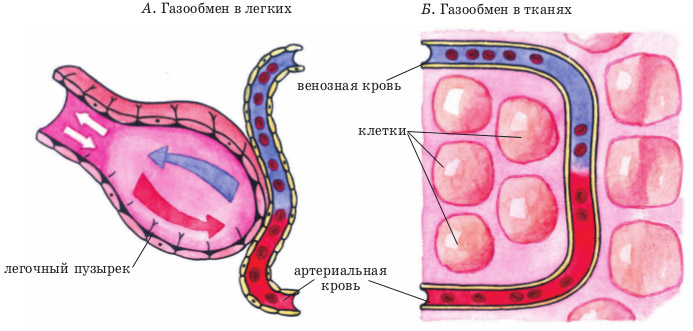
Venous blood from the right atrium passes through the right atrioventricular opening into the right ventricle, which, by contracting, pushes blood into the pulmonary trunk. It divides into the right and left pulmonary arteries, which enter the lungs. In the lung tissue, the pulmonary arteries divide into capillaries that surround each alveolus. After the release of carbon dioxide by erythrocytes and their enrichment with oxygen, venous blood turns into arterial. Arterial blood through four pulmonary veins (in each lung there are two veins) flows into the left atrium, then passes through the left atrioventricular opening into the left ventricle. The systemic circulation begins from the left ventricle.
A large circle of blood circulation
Arterial blood from the left ventricle during its contraction is thrown into the aorta. The aorta splits into arteries that supply blood to the limbs, trunk,. all internal organs and ending with capillaries. Nutrients, water, salts and oxygen are released from the blood of the capillaries into the tissues, metabolic products and carbon dioxide are resorbed. Capillaries collect in venules, where the venous vascular system begins, representing the roots of the superior and inferior vena cava. Venous blood through these veins enters the right atrium, where the systemic circulation ends.
Cardiac circulation
This circle of blood circulation starts from the aorta by two coronary cardiac arteries, through which blood enters all layers and parts of the heart, and then collects through small veins into the venous coronary sinus. This vessel opens with a wide mouth into the right atrium. Part of the small veins of the heart wall opens directly into the cavity of the right atrium and ventricle of the heart.
In mammals and humans, the circulatory system is the most complex. It is a closed system consisting of two circles of blood circulation. Providing warm-bloodedness, it is more energetically beneficial and allows a person to occupy the niche in which he is now located.
The circulatory system is a group of hollow muscular organs responsible for the circulation of blood through the vessels of the body. It is represented by a heart and vessels of various sizes. These are the muscle organs that form the circles of blood circulation. Their scheme is offered in all textbooks on anatomy and is described in this publication.
The concept of the circles of blood circulation
The circulatory system consists of two circles - bodily (large) and pulmonary (small). The circle of blood circulation is the vascular system of the arterial, capillary, lymphatic and venous type, which carries out the supply of blood from the heart to the vessels and its movement in the opposite direction. The heart is central, since in it, without mixing of arterial and venous blood, two circles of blood circulation intersect.
A large circle of blood circulation
The system of supplying peripheral tissues and its return to the heart is called the systemic circulation. It starts from the left ventricle, from where blood flows into the aorta through the aortic opening with a tricuspid valve. From the aorta, blood is directed to the smaller bodily arteries and reaches the capillaries. This is a set of organs that form a leading link.
Here oxygen enters the tissues, and carbon dioxide is captured from them by erythrocytes. Also in the tissue, the blood transports amino acids, lipoproteins, glucose, the metabolic products of which are carried out from the capillaries into the venules and further into the larger veins. They drain into the vena cava, which return blood directly to the heart in the right atrium.
The right atrium ends with the systemic circulation. The diagram looks like this (along the blood circulation): the left ventricle, aorta, elastic arteries, muscular-elastic arteries, muscle arteries, arterioles, capillaries, venules, veins and hollow veins that return blood to the heart in the right atrium. The brain, all skin and bones are nourished from the systemic circulation. In general, all human tissues are fed from the vessels of the systemic circulation, and the small one is only the place of blood oxygenation.
Small circle of blood circulation
The pulmonary (small) circle of blood circulation, the diagram of which is presented below, originates from the right ventricle. Blood enters it from the right atrium through the atrioventricular opening. From the cavity of the right ventricle, oxygen-depleted (venous) blood enters the pulmonary trunk through the outlet (pulmonary) tract. This artery is thinner than the aorta. It divides into two branches that go to both lungs.
The lungs are the central organ that forms the pulmonary circulation. The human diagram described in anatomy textbooks explains that pulmonary blood flow is needed to oxygenate the blood. Here she gives off carbon dioxide and takes in oxygen. In the sinusoidal capillaries of the lungs with a diameter of about 30 microns atypical for the body, gas exchange takes place.
Subsequently, oxygenated blood is directed through the intrapulmonary vein system and collected in 4 pulmonary veins. They are all attached to the left atrium and carry oxygen-rich blood there. This is where the circles of blood circulation end. The diagram of the pulmonary circle looks like this (along the blood flow): right ventricle, pulmonary artery, intrapulmonary arteries, pulmonary arterioles, pulmonary sinusoids, venules, pulmonary veins, left atrium.
Features of the circulatory system

A key feature of the circulatory system, which consists of two circles, is the need for a heart with two or more chambers. In fish, the circle of blood circulation is the same, because they do not have lungs, and all gas exchange takes place in the vessels of the gills. As a result, a single-chambered fish heart is a pump that pushes blood in only one direction.
Amphibians and reptiles have respiratory organs and, accordingly, circulatory circles. The scheme of their work is simple: from the ventricle, blood is sent to the vessels of the great circle, from the arteries - to the capillaries and veins. Venous return to the heart is also realized, however, blood from the right atrium enters the ventricle, which is common to the two circles of blood circulation. Since the heart of these animals is three-chambered, the blood from both circles (venous and arterial) is mixed.
In humans (and mammals), the heart has a 4-chambered structure. In it, two ventricles and two atria are separated by partitions. The lack of mixing of the two types of blood (arterial and venous) has become a gigantic evolutionary invention that ensured warm-bloodedness in mammals.

Blood supply to the lungs and heart
In the circulatory system, which consists of two circles, the nutrition of the lung and heart is of particular importance. These are the most important organs that ensure the closure of the bloodstream and the integrity of the respiratory and circulatory systems. So, the lungs have two circles of blood circulation in their thickness. But their tissue is nourished by the vessels of the great circle: bronchial and pulmonary vessels branch off from the aorta and from the intrathoracic arteries, carrying blood to the parenchyma of the lung. And the organ cannot feed from the right sections, although part of the oxygen diffuses from there. This means that the large and small circles of blood circulation, the scheme of which is described above, perform different functions (one enriches the blood with oxygen, and the second sends it to the organs, taking deoxygenated blood from them).
The heart also feeds on the vessels of the great circle, but the blood in its cavities is capable of providing oxygen to the endocardium. In this case, part of the myocardial veins, mainly small ones, flows directly into it. It is noteworthy that the pulse wave does not propagate into the cardiac diastole. Therefore, the organ is supplied with blood only when it is "resting".

The human circulation circles, the diagram of which is presented above in the relevant sections, provide both warm-bloodedness and high endurance. Let a person not be the animal that often uses its strength for survival, but this allowed the rest of the mammals to populate certain habitats. Previously, they were inaccessible to amphibians and reptiles, and even more so to fish.
In phylogeny, a large circle appeared earlier and was characteristic of fish. And the small circle supplemented it only in those animals that wholly or completely went out onto the land and inhabited it. Since its inception, the respiratory and circulatory systems are considered together. They are functionally and structurally related.
This is an important and already indestructible evolutionary mechanism for leaving and settling the land. Therefore, the continuing complication of mammalian organisms will now be directed not towards the complication of the respiratory and circulatory systems, but towards an increase in the oxygen-binding system and an increase in the area of the lungs.
Heart is the central organ of blood circulation. It is a hollow muscular organ, consisting of two halves: left - arterial and right - venous. Each half consists of communicating atrium and ventricle of the heart.
The central organ of blood circulation is heart... It is a hollow muscular organ, consisting of two halves: left - arterial and right - venous. Each half consists of communicating atrium and ventricle of the heart.
Venous blood flows through the veins into the right atrium and further into the right ventricle of the heart, from the latter into the pulmonary trunk, from where it follows through the pulmonary arteries into the right and left lungs. Here the branches of the pulmonary arteries branch out to the smallest vessels - capillaries.
In the lungs, venous blood is saturated with oxygen, becomes arterial and through four pulmonary veins is sent to the left atrium, then enters the left ventricle of the heart. From the left ventricle of the heart, blood enters the largest arterial highway - the aorta and through its branches, which decay in the tissues of the body to the capillaries, is carried throughout the body. By giving oxygen to the tissues and taking carbon dioxide from them, the blood becomes venous. Capillaries, once again connecting with each other, form veins.
All veins of the body are connected in two large trunks - the superior vena cava and the inferior vena cava. V superior vena cava blood is collected from areas and organs of the head and neck, upper extremities and some sections of the walls of the body. The inferior vena cava fills with blood from the lower extremities, walls and organs of the pelvic and abdominal cavities.
Systemic circulation video.
Both hollow veins bring blood to the right atrium, which also receives venous blood from the heart itself. So the circle of blood circulation is closed. This blood pathway is divided into a small and a large circle of blood circulation.

Small circle of blood circulation video
Small circle of blood circulation(pulmonary) begins from the right ventricle of the heart with the pulmonary trunk, includes the branching of the pulmonary trunk to the capillary network of the lungs and the pulmonary veins flowing into the left atrium.
A large circle of blood circulation(corporal) begins from the left ventricle of the heart with the aorta, includes all its branches, the capillary network and veins of organs and tissues of the whole body and ends in the right atrium.
Consequently, blood circulation takes place in two interconnected circles of blood circulation.
When the human circulatory system is divided into two circles of blood circulation, the heart is exposed to less stress than if the body had a common blood supply system. In the pulmonary circulation, blood travels from the heart to the lungs and then back thanks to a closed arterial and venous system that connects the heart and lungs. Its path begins in the right ventricle and ends in the left atrium. In the pulmonary circulation, blood with carbon dioxide is carried by arteries, and blood with oxygen is carried by veins.
From the right atrium, blood enters the right ventricle and is then pumped into the lungs through the pulmonary artery. From the right ventricle, venous blood enters the arteries and capillaries of the lungs, where it gets rid of carbon dioxide, and then is saturated with oxygen. Through the pulmonary veins, blood flows into the left atrium, then it enters the systemic circulation and then goes to all organs. Since it flows slowly in the capillaries, carbon dioxide has time to enter it, and oxygen has time to penetrate into the cells. Because blood enters the lungs at low pressure, the pulmonary circulation is also called a low pressure system. The time for blood to pass through the pulmonary circulation is 4-5 seconds.
With an increased demand for oxygen, for example, during intense sports, the pressure generated by the heart increases and the blood flow accelerates.
A large circle of blood circulation
The systemic circulation begins from the left ventricle of the heart. Oxygenated blood flows from the lungs into the left atrium and then into the left ventricle. From there, arterial blood enters the arteries and capillaries. Through the walls of the capillaries, the blood transfers oxygen and nutrients into the tissue fluid, taking carbon dioxide and metabolic products. From the capillaries, it enters the small veins that form larger veins. Then, through two venous trunks (superior vena cava and inferior vena cava), it enters the right atrium, ending the systemic circulation. The blood circulation in the systemic circulation is 23-27 seconds.
Blood flows through the superior vena cava from the upper parts of the body, and along the lower - from the lower parts.
The heart has two pairs of valves. One of them is located between the ventricles and the atria. The second pair is located between the ventricles and arteries. These valves provide direction for blood flow and prevent blood from flowing back. Blood is pumped into the lungs under great pressure, and it enters the left atrium under negative pressure. The human heart has an asymmetrical shape: since its left half does more heavy work, it is somewhat thicker than
Blood provides normal human activity, saturating the body with oxygen and energy, while removing carbon dioxide and toxins.
The central organ of the circulatory system is the heart, which consists of four chambers separated by valves and partitions, which act as the main channels for blood circulation.
Today, it is customary to divide everything into two circles - large and small. They are united into one system and are closed on each other. The circulation is made up of arteries - the vessels that carry blood from the heart, and veins - the vessels that carry blood back to the heart.
Blood in the human body can be arterial and venous. The first carries oxygen into the cells and has the highest pressure and, accordingly, speed. The second removes carbon dioxide and delivers it to the lungs (low pressure and low speed).
Both circles of blood circulation are two loops connected in series. The main organs of blood circulation can be called the heart, which acts as a pump, the lungs, which exchange oxygen, and which cleanses the blood of harmful substances and toxins.
In the medical literature, you can often find a wider list, where the human circulation circles are presented in this form:
- Big
- Small
- Cordial
- Placental
- Willisiev
A large circle of human circulation
The large circle originates from the left ventricle of the heart.
Its main function is to deliver oxygen and nutrients to organs and tissues through capillaries, the total area of which reaches 1500 square meters. m.
In the process of passing through the arteries, the blood takes carbon dioxide and returns to the heart, through the vessels, closing the blood flow in the right atrium with two vena cava - the lower and upper.
The whole cycle of passage takes from 23 to 27 seconds.
Sometimes the name of the corporal circle is found.
Small circle of blood circulation
The small circle originates from the right ventricle, then passing through the pulmonary arteries, delivers venous blood to the lungs.
Through the capillaries, carbon dioxide is displaced (gas exchange) and the blood, having become arterial, returns to the left atrium.

The main task of the small circle of blood circulation is heat exchange and blood circulation
The main task of the small circle is heat exchange and circulation. The average blood circulation time is no more than 5 seconds.
It can also be called the pulmonary circulation.
"Additional" circles of blood circulation in humans
Through the placental circle, oxygen is supplied to the fetus in the womb. It has a displaced system and does not belong to any of the main circles. At the same time, arterial-venous blood flows through the umbilical cord with a ratio of oxygen and carbon dioxide of 60/40%.
The heart circle is part of the body (great) circle, but due to the importance of the heart muscle, it is often distinguished into a separate subcategory. At rest, up to 4% of the total cardiac output (0.8 - 0.9 mg / min) is involved in the bloodstream, with an increase in the load, the value increases up to 5 times. It is in this part of a person's blood circulation that there is a blockage of blood vessels by a thrombus and a lack of blood in the heart muscle.
The circle of Willis provides blood supply to the human brain, it also stands out separately from the large circle due to the importance of functions. With blockage of individual vessels, it provides additional oxygen delivery through other arteries. Often atrophied and has hypoplasia of individual arteries. A full-fledged circle of Willis is observed only in 25-50% of people.
Features of the blood circulation of individual human organs
Although the entire body is supplied with oxygen thanks to the large circle of blood circulation, some individual organs have their own unique oxygen exchange system.
The lungs have a double capillary network. The first belongs to the body circle and feeds the organ with energy and oxygen, while taking away metabolic products. The second to the pulmonary - here there is a displacement (oxygenation) of carbon dioxide from the blood and its enrichment with oxygen.

The heart is one of the main organs of the circulatory system
Venous blood flows from the unpaired abdominal organs in a different way; it preliminarily passes through the portal vein. Vienna is so named because of its connection with the gate of the liver. Passing through them, it is cleared of toxins and only after that it returns through the hepatic veins to the general circulation.
The lower third of the rectum in women does not pass through the portal vein and is connected directly to the vagina, bypassing hepatic filtration, which is used to administer some drugs.
Heart and brain. Their features were revealed in the section on additional circles.
Few facts
Up to 10,000 liters of blood pass through the heart per day, besides, it is the strongest muscle in the human body, contracting up to 2.5 billion times in a lifetime.
The total length of the vessels in the body reaches 100 thousand kilometers. This may be enough to get to the moon or wrap the earth around the equator several times.
The average amount of blood is 8% of the total body weight. With a weight of 80 kg, about 6 liters of blood flows in a person.
Capillaries have such "narrow" (no more than 10 microns) passages that blood cells can only pass through them one at a time.
Watch an informative video about the circles of blood circulation:
Have you noticed a mistake? Highlight it and press Ctrl + Enter to let us know.
The continuous movement of blood through a closed system of cavities of the heart and blood vessels is called blood circulation. The circulatory system contributes to the maintenance of all vital functions of the body.
The movement of blood through the blood vessels occurs due to the contractions of the heart. In humans, a large and small circles of blood circulation are distinguished.
Large and small circles of blood circulation
A large circle of blood circulation begins with the largest artery - the aorta. Due to the contraction of the left ventricle of the heart, blood is thrown into the aorta, which then breaks down into arteries, arterioles that supply blood to the upper and lower extremities, head, trunk, all internal organs and ending in capillaries.
Passing through the capillaries, the blood gives the tissues oxygen, nutrients and takes away the products of dissimilation. From the capillaries, blood is collected in small veins, which, merging and increasing their cross section, form the superior and inferior vena cava.
Ends with a large circle of blood circulation in the right atrium. Arterial blood flows in all arteries of the systemic circulation, venous blood flows in the veins.
Small circle of blood circulation begins in the right ventricle, where venous blood flows from the right atrium. The right ventricle contracts and pushes blood into the pulmonary trunk, which divides into two pulmonary arteries that carry blood to the right and left lungs. In the lungs, they divide into capillaries that surround each alveolus. In the alveoli, the blood gives off carbon dioxide and is saturated with oxygen.
Through four pulmonary veins (each lung has two veins), oxygenated blood enters the left atrium (where the pulmonary circulation ends), and then into the left ventricle. Thus, venous blood flows in the arteries of the pulmonary circulation, and arterial blood flows in its veins.
The regularity of the movement of blood in the circles of blood circulation was discovered by the English anatomist and physician W. Harvey in 1628.
Blood vessels: arteries, capillaries and veins

There are three types of blood vessels in humans: arteries, veins, and capillaries.
Arteries- a cylindrical tube through which blood moves from the heart to organs and tissues. The walls of the arteries are made up of three layers that give them strength and elasticity:
- Outer connective tissue membrane;
- middle layer formed by smooth muscle fibers, between which elastic fibers lie
- inner endothelial membrane. Due to the elasticity of the arteries, the periodic expulsion of blood from the heart to the aorta turns into a continuous movement of blood through the vessels.
Capillaries are microscopic vessels, the walls of which consist of one layer of endothelial cells. Their thickness is about 1 micron, the length is 0.2-0.7 mm.
Due to the structural features, it is in the capillaries that the blood performs its main functions: it gives oxygen and nutrients to the tissues and carries away carbon dioxide and other dissimilation products to be released from them.
Due to the fact that the blood in the capillaries is under pressure and moves slowly, in the arterial part of it, water and nutrients dissolved in it seep into the intercellular fluid. At the venous end of the capillary, the blood pressure decreases and the intercellular fluid flows back into the capillaries.
Veins- the vessels that carry blood from the capillaries to the heart. Their walls consist of the same membranes as the walls of the aorta, but much weaker than arterial ones and have fewer smooth muscle and elastic fibers.
The blood in the veins flows under slight pressure, so the surrounding tissues, especially the skeletal muscles, have a greater influence on the movement of blood through the veins. Unlike arteries, veins (with the exception of hollow veins) have pocketed valves that prevent blood from flowing back.
By analogy with the root system of plants, the blood inside a person transports nutrients through various sized vessels.
In addition to the nutritional function, work is carried out to transport oxygen in the air - cellular gas exchange is carried out.
Circulatory system
If you look at the scheme of blood distribution throughout the body, then its cyclical path is striking. If we do not take into account the placental blood flow, then among the isolated there is a small cycle that provides respiration and gas exchange of tissues and organs and affects the lungs of a person, as well as a second, large cycle that carries nutrients and enzymes.

The task of the circulatory system, which became known thanks to the scientific experiments of the scientist Harvey (in the 16th century, he discovered the blood circles), in general, is to organize the movement of blood and lymphatic cells through the vessels.
Small circle of blood circulation
From above, venous blood from the right atrial chamber enters the right heart ventricle. Veins are medium-sized vessels. Blood flows in portions and is pushed out of the cavity of the heart ventricle through a valve that opens towards the pulmonary trunk.

From it, the blood goes into the pulmonary artery, and, as the distance from the main muscle of the human body, the veins flow into the arteries of the pulmonary tissue, transforming and breaking up into a multiple network of capillaries. Their role and primary function is to carry out gas exchange processes in which alveolocytes take carbon dioxide.
As oxygen is distributed through the veins, arterial features become characteristic of the blood flow. So, through the venules, the blood goes to the pulmonary veins, which open into the left atrium.
A large circle of blood circulation
Let's trace the big blood cycle. The systemic circulation begins from the left cardiac ventricle, where the arterial flow enriched in O 2 and depleted in CO 2 enters, which is supplied from the pulmonary circulation. Where does the blood from the left ventricle of the heart go?

After the left ventricle, the trailing aortic valve pushes arterial blood into the aorta. It distributes O 2 in high concentration to all arteries. Moving away from the heart, the diameter of the artery tube changes - it decreases.
All CO 2 is collected from the capillary vessels, and the great circle flows enter the vena cava. From them, the blood again enters the right atrium, then into the right ventricle and pulmonary trunk.
Thus, the systemic circulation in the right atrium ends. And to the question - where does the blood from the right ventricle of the heart get, the answer is to the pulmonary artery.
Diagram of the human circulatory system
The diagram with arrows of the blood flow process described below briefly and clearly demonstrates the sequence of the blood flow path in the body, indicating the organs involved in the process.

Human circulatory organs
These include the heart and blood vessels (veins, arteries, and capillaries). Consider the most important organ in the human body.

The heart is a self-governing, self-regulating, self-correcting muscle. The size of the heart depends on the development of skeletal muscles - the higher their development, the larger the heart. In structure, the heart has 4 chambers - 2 ventricles and 2 atria, and is placed in the pericardium. The ventricles are separated from each other and between the atria by special heart valves.
Responsible for replenishing and saturating the heart with oxygen are the coronary arteries, or “coronary vessels” as they are called.
The main function of the heart is to carry out the work of a pump in the body. Failures are due to several reasons:
- Insufficient / excessive blood supply.
- Cardiac muscle injury.
- External squeezing.
The blood vessels are the second most important in the circulatory system.
Linear and volumetric blood flow velocity
 When considering the velocity parameters of blood, the concepts of linear and volumetric velocities are used. There is a mathematical relationship between these concepts.
Where does blood move at the fastest rate? The linear blood flow velocity is in direct proportion to the volumetric one, which varies depending on the type of vessels.
The highest velocity of blood flow in the aorta.
Where is the blood moving at the slowest speed? The lowest speed is in the vena cava.
Time of complete blood circulation
For an adult, whose heart produces about 80 beats per minute, blood completes all the way in 23 seconds, distributing 4.5-5 seconds for a small circle and 18-18.5 seconds for a large one.

The data are confirmed empirically. The essence of all research methods lies in the principle of marking. A traceable substance that is not typical for the human body is injected into the vein and its location is dynamically established.
So it is noted how long the substance will appear in the vein of the same name, located on the other side. This is the time of complete blood circulation.
Conclusion
The human body is a complex mechanism with various kinds of systems. The circulatory system plays the main role in its proper functioning and life support. Therefore, it is very important to understand its structure and keep the heart and blood vessels in perfect order.
In the human body, the circulatory system is designed to fully meet its internal needs. An important role in the advancement of blood is played by the presence of a closed system in which arterial and venous blood flows are separated. And this is done with the help of the presence of blood circulation circles.
History reference
In the past, when scientists did not yet have informative devices at hand capable of studying physiological processes in a living organism, the greatest scientists were forced to search for anatomical features in corpses. Naturally, the heart of a deceased person does not contract, so some of the nuances had to be conjectured on their own, and sometimes simply fantasized. So, back in the second century AD Claudius Galen, student on the works of himself Hippocrates, assumed that the arteries contain air in their lumen instead of blood. Over the next centuries, many attempts have been made to combine and link together the available anatomical data from the standpoint of physiology. All scientists knew and understood how the circulatory system works, but how does it work?
Scientists have made a colossal contribution to the systematization of data on the work of the heart Miguel Servet and William Harvey in the 16th century. Harvey, the scientist who first described the large and small circles of blood circulation , in 1616 determined the presence of two circles, but how the arterial and venous channels are connected, he could not explain in his writings. And only later, in the 17th century, Marcello Malpighi, one of the first who began to use the microscope in his practice, discovered and described the presence of the smallest capillaries invisible to the naked eye, which serve as a connecting link in the blood circulation.
Phylogenesis, or the evolution of the circulation
Due to the fact that, with evolution, animals of the class of vertebrates became more and more progressive in anatomical and physiological terms, they required a complex structure of the cardiovascular system. So, for a more rapid movement of the liquid internal environment in the body of a vertebrate animal, it became necessary to have a closed blood circulation system. In comparison with other classes of the animal kingdom (for example, with arthropods or with worms), the rudiments of a closed vascular system appear in chordates. And if a lancelet, for example, lacks a heart, but there is an abdominal and dorsal aorta, then fish, amphibians (amphibians), reptiles (reptiles) have a two- and three-chambered heart, respectively, and birds and mammals have a four-chambered heart, a feature of which is the focus in it of two circles of blood circulation, not mixing with each other.

Thus, the presence in birds, mammals and humans, in particular, of two separated circles of blood circulation is nothing more than the evolution of the circulatory system necessary for better adaptation to environmental conditions.
Anatomical features of the circulatory system
The circulatory system is a collection of blood vessels, which is a closed system for the supply of oxygen and nutrients to the internal organs through gas exchange and the exchange of nutrients, as well as for the removal of carbon dioxide and other metabolic products from cells. The human body is characterized by two circles - the systemic, or large circle, as well as the pulmonary, also called the small circle.

Video: circulatory circles, mini-lecture and animation
A large circle of blood circulation
The main function of the great circle is to ensure gas exchange in all internal organs, except for the lungs. It starts in the left ventricular cavity; represented by the aorta and its branches, the arterial bed of the liver, kidneys, brain, skeletal muscles and other organs. Further, this circle continues with the capillary network and the venous bed of the listed organs; and by the confluence of the vena cava into the cavity of the right atrium ends in the latter.

So, as already mentioned, the beginning of the great circle is the cavity of the left ventricle. This is where the arterial blood stream is directed, which contains more oxygen than carbon dioxide. This flow enters the left ventricle directly from the circulatory system of the lungs, that is, from the small circle. The arterial flow from the left ventricle through the aortic valve is pushed into the largest great vessel - the aorta. The aorta can be figuratively compared to a kind of tree, which has many branches, because arteries extend from it to internal organs (to the liver, kidneys, gastrointestinal tract, to the brain - through the carotid artery system, to skeletal muscles, to subcutaneous fat fiber, etc.). Organ arteries, also having numerous ramifications and bearing the names corresponding to the anatomy, carry oxygen to each organ.
In the tissues of internal organs, arterial vessels are subdivided into vessels of smaller and smaller diameters, and as a result, a capillary network is formed. Capillaries are the smallest vessels that practically do not have a middle muscle layer, but are represented by an inner membrane - an intima lined with endothelial cells. The gaps between these cells at the microscopic level are so large in comparison with other vessels that they allow proteins, gases and even formed elements to freely penetrate into the intercellular fluid of the surrounding tissues. Thus, between the capillary with arterial blood and the liquid intercellular medium in one organ or another, there is an intense gas exchange and the exchange of other substances. Oxygen penetrates from the capillary, and carbon dioxide, as a product of cell metabolism, enters the capillary. The cellular stage of respiration is carried out.
After more oxygen has passed into the tissues, and all carbon dioxide has been removed from the tissues, the blood becomes venous. All gas exchange is carried out with each new inflow of blood, and during the period of time while it moves along the capillary towards the venule - a vessel that collects venous blood. That is, with each cardiac cycle in one or another part of the body, oxygen is supplied to the tissues and carbon dioxide is removed from them.
These venules are combined into larger veins, and a venous bed is formed. Veins, similar to arteries, bear the names in which organ they are located (renal, cerebral, etc.). From the large venous trunks, tributaries of the superior and inferior vena cava are formed, and the latter then flow into the right atrium.
Features of blood flow in the organs of a large circle

Some of the internal organs have their own characteristics. So, for example, in the liver there is not only the hepatic vein, which "carries" the venous flow from it, but also the portal vein, which, on the contrary, brings blood to the hepatic tissue, where the blood is purified, and only then the blood is collected in the tributaries of the hepatic vein to get to the big circle. The portal vein brings blood from the stomach and intestines, so everything that a person has eaten or drunk must undergo a kind of "cleaning" in the liver.
In addition to the liver, certain nuances exist in other organs, for example, in the tissues of the pituitary gland and kidneys. So, in the pituitary gland, the presence of the so-called "miraculous" capillary network is noted, because the arteries that bring blood to the pituitary gland from the hypothalamus are divided into capillaries, which are then collected in venules. Venules, after the blood with molecules of releasing hormones is collected, are again divided into capillaries, and then veins are formed that carry the blood from the pituitary gland. In the kidneys, the arterial network is divided twice into capillaries, which is associated with the processes of excretion and reabsorption in the kidney cells - in the nephrons.
Small circle of blood circulation
Its function is to carry out gas exchange processes in the lung tissue in order to saturate the "spent" venous blood with oxygen molecules. It begins in the cavity of the right ventricle, where from the right atrial chamber (from the "end point" of the great circle) venous blood flow enters with an extremely small amount of oxygen and a high content of carbon dioxide. This blood moves through the valve of the pulmonary artery into one of the large vessels called the pulmonary trunk. Further, the venous flow moves along the arterial bed in the lung tissue, which also breaks down into a network of capillaries. By analogy with capillaries in other tissues, gas exchange takes place in them, only oxygen molecules enter the lumen of the capillary, and carbon dioxide penetrates into the alveolocytes (alveolar cells). Air from the environment enters the alveoli with each act of breathing, from which oxygen penetrates through the cell membranes into the blood plasma. With the exhaled air during exhalation, the carbon dioxide that has entered the alveoli is removed to the outside.

After saturation with O 2 molecules, the blood acquires arterial properties, flows through the venules and eventually reaches the pulmonary veins. The latter, consisting of four or five pieces, open into the left atrial cavity. As a result, venous blood flow flows through the right half of the heart, and arterial blood flows through the left half; and normally these streams should not mix.
The lung tissue has a double network of capillaries. With the help of the first, gas exchange processes are carried out in order to enrich the venous flow with oxygen molecules (interconnection directly with the small circle), and in the second, the lung tissue itself is nourished with oxygen and nutrients (interrelation with the large circle).
Additional circles of blood circulation
With these concepts, it is customary to distinguish the blood supply to individual organs. So, for example, to the heart, which needs oxygen more than others, arterial inflow is carried out from the branches of the aorta at its very beginning, which are called the right and left coronary (coronary) arteries. Intensive gas exchange occurs in the capillaries of the myocardium, and venous outflow is carried out into the coronary veins. The latter are collected in the coronary sinus, which opens directly into the right atrial chamber. In this way, cardiac, or coronary circulation.

coronary (coronary) circle of blood circulation in the heart
Circle of willis is a closed arterial network of cerebral arteries. The brain circle provides additional blood supply to the brain in case of disturbance of cerebral blood flow through other arteries. This protects such an important organ from a lack of oxygen, or hypoxia. The cerebral circulation is represented by the initial segment of the anterior cerebral artery, the initial segment of the posterior cerebral artery, the anterior and posterior communicating arteries, and the internal carotid arteries.

circle of Willis in the brain (classic version of the structure)
Placental circulation functions only during gestation by a woman and performs the function of "breathing" in a child. The placenta forms from 3-6 weeks of gestation and begins to function at full strength from the 12th week. Due to the fact that the lungs of the fetus do not work, the flow of oxygen into its blood is carried out through the flow of arterial blood into the umbilical vein of the child.

Fetal circulation before birth
Thus, the entire human circulatory system can be conditionally divided into separate interconnected areas that perform their functions. The correct functioning of such areas, or circles of blood circulation, is the key to the healthy functioning of the heart, blood vessels and the whole organism as a whole.
Circulation- This is the movement of blood through the vascular system, providing gas exchange between the body and the external environment, the exchange of substances between organs and tissues and humoral regulation of various functions of the body.
Circulatory system includes and - aorta, arteries, arterioles, capillaries, venules, veins, etc. Blood moves through the vessels due to the contraction of the heart muscle.
Blood circulation takes place in a closed system consisting of small and large circles:
- The systemic circulation provides all organs and tissues with blood containing nutrients.
- The small, or pulmonary, circle of blood circulation is designed to enrich the blood with oxygen.
Circles of blood circulation were first described by the English scientist William Harvey in 1628 in the work "Anatomical studies of the movement of the heart and blood vessels."
Small circle of blood circulation starts from the right ventricle, during the contraction of which venous blood enters the pulmonary trunk and, flowing through the lungs, gives off carbon dioxide and is saturated with oxygen. Oxygenated blood from the lungs through the pulmonary veins enters the left atrium, where the small circle ends.
A large circle of blood circulation starts from the left ventricle, with the contraction of which oxygen-enriched blood is pumped into the aorta, arteries, arterioles and capillaries of all organs and tissues, and from there flows through the venules and veins into the right atrium, where the great circle ends.
The largest vessel in the systemic circulation is the aorta, which exits the left ventricle of the heart. The aorta forms an arch from which arteries branch off to carry blood to the head (carotid arteries) and to the upper limbs (vertebral arteries). The aorta runs down the spine, where branches extend from it, carrying blood to the abdominal organs, to the muscles of the trunk and lower extremities.
Arterial blood, rich in oxygen, passes throughout the body, supplying the cells of organs and tissues with the nutrients and oxygen necessary for their activity, and in the capillary system it turns into venous blood. Venous blood, saturated with carbon dioxide and cellular metabolic products, returns to the heart and from it enters the lungs for gas exchange. The largest veins of the systemic circulation are the superior and inferior vena cava, which flow into the right atrium.
Rice. The scheme of small and large circles of blood circulation
It should be noted how the circulatory systems of the liver and kidneys are included in the systemic circulation. All blood from the capillaries and veins of the stomach, intestines, pancreas and spleen enters the portal vein and passes through the liver. In the liver, the portal vein branches into small veins and capillaries, which then rejoin into the common trunk of the hepatic vein, which flows into the inferior vena cava. All the blood of the abdominal organs before entering the systemic circulation flows through two capillary networks: the capillaries of these organs and the capillaries of the liver. The portal system of the liver plays an important role. It ensures the neutralization of toxic substances that are formed in the large intestine during the breakdown of amino acids not absorbed in the small intestine and are absorbed by the colon mucosa into the blood. The liver, like all other organs, also receives arterial blood through the hepatic artery, which branches out from the abdominal artery.
The kidneys also have two capillary networks: there is a capillary network in each Malpighian glomerulus, then these capillaries are connected to an arterial vessel, which again disintegrates into capillaries, entwining convoluted tubules.

Rice. Circulation diagram
A feature of blood circulation in the liver and kidneys is a slowdown in blood flow due to the function of these organs.
Table 1. Difference between blood flow in the systemic and pulmonary circulation
|
Blood flow in the body |
A large circle of blood circulation |
Small circle of blood circulation |
|
In which part of the heart does the circle begin? |
In the left ventricle |
In the right ventricle |
|
In which part of the heart does the circle end? |
In the right atrium |
In the left atrium |
|
Where does the gas exchange take place? |
In the capillaries located in the organs of the chest and abdominal cavities, the brain, upper and lower extremities |
In the capillaries located in the alveoli of the lungs |
|
What kind of blood moves through the arteries? |
Arterial |
Venous |
|
What kind of blood moves through the veins? |
Venous |
Arterial |
|
Time of blood circulation in a circle |
||
|
Circle function |
Oxygen supply to organs and tissues and carbon dioxide transport |
Saturation of blood with oxygen and removal of carbon dioxide from the body |
Blood circulation time - the time of a single passage of a blood particle through the large and small circles of the vascular system. More details in the next section of the article.
Regularities of the movement of blood through the vessels
Basic principles of hemodynamics
Hemodynamics- This is a section of physiology that studies the patterns and mechanisms of blood flow through the vessels of the human body. When studying it, the terminology is used and the laws of hydrodynamics - the science of the movement of fluids - are taken into account.
The speed at which blood flows through the vessels depends on two factors:
- from the difference in blood pressure at the beginning and end of the vessel;
- from the resistance that liquid meets on its way.
The pressure difference facilitates the movement of the liquid: the larger it is, the more intense this movement. The resistance in the vascular system, which reduces the speed of blood movement, depends on a number of factors:
- the length of the vessel and its radius (the greater the length and the smaller the radius, the greater the resistance);
- the viscosity of blood (it is 5 times more than the viscosity of water);
- friction of blood particles against the walls of blood vessels and among themselves.
Hemodynamic indicators
The blood flow velocity in the vessels is carried out according to the laws of hemodynamics, in common with the laws of hydrodynamics. Blood flow velocity is characterized by three parameters: volumetric blood flow velocity, linear blood flow velocity and blood circulation time.
Volumetric blood flow velocity - the amount of blood flowing through the cross section of all vessels of a given caliber per unit of time.
Linear blood flow velocity - the speed of movement of an individual blood particle along the vessel per unit of time. In the center of the vessel, the linear velocity is maximum, and near the vessel wall, it is minimum due to increased friction.
Blood circulation time - the time during which the blood passes through the large and small circles of blood circulation. Normally, it is 17-25 seconds. It takes about 1/5 to go through the small circle, and 4/5 of this time to go through the big one.
The driving force of blood flow through the vascular system of each of the circles of blood circulation is the difference in blood pressure ( ΔР) in the initial section of the arterial bed (aorta for the great circle) and the final section of the venous bed (vena cava and right atrium). Difference in blood pressure ( ΔР) at the beginning of the vessel ( Р1) and at the end of it ( P2) is the driving force of blood flow through any vessel of the circulatory system. The force of the blood pressure gradient is spent on overcoming the resistance to blood flow ( R) in the vascular system and in each individual vessel. The higher the gradient of blood pressure in the circle of blood circulation or in an individual vessel, the greater the volumetric blood flow in them.
The most important indicator of the movement of blood through the vessels is volumetric blood flow rate, or volumetric blood flow (Q), which is understood as the volume of blood flowing through the total cross-section of the vascular bed or the section of an individual vessel per unit of time. The volumetric blood flow rate is expressed in liters per minute (l / min) or milliliters per minute (ml / min). To assess the volumetric blood flow through the aorta or the total cross section of any other level of the vessels of the systemic circulation, use the concept volumetric systemic blood flow. Since the entire volume of blood ejected by the left ventricle during this time flows through the aorta and other vessels of the systemic circulation per unit of time (minute), the concept of systemic volumetric blood flow is synonymous with the concept of systemic volumetric blood flow (MOC). The IOC of an adult at rest is 4-5 l / min.
There are also volumetric blood flow in the organ. In this case, they mean the total blood flow flowing per unit of time through all the arterial or outflowing venous vessels of the organ.
Thus, the volumetric blood flow Q = (P1 - P2) / R.
This formula expresses the essence of the basic law of hemodynamics, which states that the amount of blood flowing through the total cross-section of the vascular system or an individual vessel per unit of time is directly proportional to the difference in blood pressure at the beginning and at the end of the vascular system (or vessel) and inversely proportional to the resistance to current blood.
The total (systemic) minute blood flow in the great circle is calculated taking into account the values of the mean hydrodynamic blood pressure at the beginning of the aorta P1, and at the mouth of the vena cava P2. Since in this part of the veins the blood pressure is close to 0 , then in the expression for calculating Q or the IOC is substituted with the value R, equal to the average hydrodynamic arterial blood pressure at the beginning of the aorta: Q(IOC) = P/ R.
One of the consequences of the basic law of hemodynamics - the driving force of blood flow in the vascular system - is due to the blood pressure created by the work of the heart. Confirmation of the decisive value of the blood pressure value for blood flow is the pulsating nature of the blood flow throughout the cardiac cycle. During systole, when blood pressure reaches its maximum level, blood flow increases, and during diastole, when blood pressure is at its lowest, blood flow decreases.
As blood moves through the vessels from the aorta to the veins, blood pressure decreases and the rate of its decrease is proportional to the resistance to blood flow in the vessels. The pressure in the arterioles and capillaries decreases especially quickly, since they have a high resistance to blood flow, having a small radius, large total length and numerous branches, which create an additional obstacle to blood flow.

The resistance to blood flow created in the entire vascular bed of the systemic circulation is called total peripheral resistance(OPS). Therefore, in the formula for calculating the volumetric blood flow, the symbol R you can replace it with an analogue - OPS:
Q = P / OPS.
A number of important consequences are derived from this expression, which are necessary for understanding the processes of blood circulation in the body, assessing the results of measuring blood pressure and its deviations. The factors affecting the resistance of the vessel for the fluid flow are described by Poiseuille's law, according to which
![]()
where R- resistance; L- the length of the vessel; η - blood viscosity; Π - number 3.14; r Is the radius of the vessel.
It follows from the above expression that since the numbers 8 and Π are permanent, L in an adult, little changes, the value of peripheral resistance to blood flow is determined by the changing values of the radius of the vessels r and blood viscosity η ).
It has already been mentioned that the radius of muscle-type vessels can change rapidly and have a significant effect on the amount of resistance to blood flow (hence their name - resistive vessels) and the amount of blood flow through organs and tissues. Since the resistance depends on the magnitude of the radius to the 4th power, then even small fluctuations in the radius of the vessels have a strong effect on the values of resistance to blood flow and blood flow. So, for example, if the radius of the vessel decreases from 2 to 1 mm, then its resistance will increase 16 times, and with a constant pressure gradient, the blood flow in this vessel will also decrease 16 times. Reverse changes in resistance will be observed when the radius of the vessel is doubled. With a constant average hemodynamic pressure, blood flow in one organ can increase, in another it can decrease, depending on the contraction or relaxation of the smooth muscles of the arterial vessels and veins of this organ.
The viscosity of blood depends on the content in the blood of the number of erythrocytes (hematocrit), protein, lipoproteins in the blood plasma, as well as on the state of aggregation of the blood. Under normal conditions, the viscosity of the blood does not change as quickly as the lumen of the vessels. After blood loss, with erythropenia, hypoproteinemia, blood viscosity decreases. With significant erythrocytosis, leukemia, increased aggregation of erythrocytes and hypercoagulation, blood viscosity can increase significantly, which entails an increase in resistance to blood flow, an increase in the load on the myocardium and may be accompanied by impaired blood flow in the vessels of the microvasculature.
In the established circulatory regime, the volume of blood expelled by the left ventricle and flowing through the cross section of the aorta is equal to the volume of blood flowing through the total cross section of the vessels of any other part of the systemic circulation. This volume of blood returns to the right atrium and enters the right ventricle. From it, the blood is expelled into the pulmonary circulation and then through the pulmonary veins returns to the left heart. Since the MVC of the left and right ventricles are the same, and the large and small circles of blood circulation are connected in series, the volumetric blood flow velocity in the vascular system remains the same.
However, during a change in blood flow conditions, for example, when moving from a horizontal to a vertical position, when gravity causes a temporary accumulation of blood in the veins of the lower trunk and legs, for a short time the MVC of the left and right ventricles can become different. Soon, intracardiac and extracardiac mechanisms of regulation of the work of the heart equalize the volumes of blood flow through the small and large circles of blood circulation.
With a sharp decrease in venous return of blood to the heart, causing a decrease in stroke volume, blood pressure may decrease. With a pronounced decrease in it, blood flow to the brain may decrease. This explains the feeling of dizziness that can occur with a sharp transition of a person from a horizontal to a vertical position.
Volume and linear velocity of blood currents in the vessels
The total blood volume in the vascular system is an important homeostatic indicator. Its average value is 6-7% for women, 7-8% of body weight for men and is in the range of 4-6 liters; 80-85% of the blood from this volume is in the vessels of the systemic circulation, about 10% is in the vessels of the pulmonary circulation and about 7% is in the cavities of the heart.
Most of the blood is contained in the veins (about 75%) - this indicates their role in the deposition of blood in both the large and pulmonary circulation.
The movement of blood in the vessels is characterized not only by volumetric, but also linear blood flow velocity. It is understood as the distance that a blood particle moves per unit of time.
There is a relationship between volumetric and linear blood flow velocity, described by the following expression:
V = Q / Pr 2
where V- linear blood flow velocity, mm / s, cm / s; Q - volumetric blood flow velocity; P- a number equal to 3.14; r Is the radius of the vessel. The magnitude Pr 2 reflects the cross-sectional area of the vessel.

Rice. 1. Changes in blood pressure, linear blood flow velocity and cross-sectional area in different parts of the vascular system

Rice. 2. Hydrodynamic characteristics of the vascular bed
From the expression of the dependence of the linear velocity on the volumetric velocity in the vessels of the circulatory system, it can be seen that the linear blood flow velocity (Fig. 1) is proportional to the volumetric blood flow through the vessel (s) and is inversely proportional to the cross-sectional area of this vessel (s). For example, in the aorta with the smallest cross-sectional area in the systemic circulation (3-4 cm 2), linear blood velocity the largest and is alone about 20-30 cm / s... With physical exertion, it can increase by 4-5 times.
Towards the capillaries, the total transverse lumen of the vessels increases and, therefore, the linear velocity of blood flow in the arteries and arterioles decreases. In capillary vessels, the total cross-sectional area of which is greater than in any other part of the great circle vessels (500-600 times the cross-section of the aorta), the linear blood flow velocity becomes minimal (less than 1 mm / s). The slow flow of blood in the capillaries creates the best conditions for metabolic processes between blood and tissues. In veins, the linear blood flow velocity increases due to a decrease in the area of their total cross-section as they approach the heart. At the mouth of the hollow veins, it is 10-20 cm / s, and under loads it increases to 50 cm / s.
The linear velocity of plasma movement depends not only on the type of vessel, but also on their location in the blood stream. There is a laminar type of blood flow, in which the notes of blood can be conditionally divided into layers. In this case, the linear velocity of movement of blood layers (mainly plasma), close or adjacent to the vessel wall, is the lowest, and the layers in the center of the flow are the highest. Friction forces arise between the vascular endothelium and the parietal blood layers, creating shear stresses on the vascular endothelium. These stresses play a role in the production of vasoactive factors by the endothelium that regulate vascular lumen and blood flow velocity.
Erythrocytes in vessels (with the exception of capillaries) are located mainly in the central part of the blood stream and move in it at a relatively high speed. Leukocytes, on the contrary, are located mainly in the parietal layers of the blood flow and make rolling movements at a low speed. This allows them to bind to adhesion receptors in places of mechanical or inflammatory damage to the endothelium, adhere to the vessel wall, and migrate into tissues to perform protective functions.
With a significant increase in the linear velocity of blood movement in the narrowed part of the vessels, in the places where its branches leave the vessel, the laminar nature of blood movement can change to turbulent. In this case, in the blood flow, the layer-by-layer movement of its particles may be disturbed; between the vessel wall and the blood, greater forces of friction and shear stresses may arise than with laminar motion. Vortex blood flows develop, the likelihood of damage to the endothelium and the deposition of cholesterol and other substances into the intima of the vessel wall increases. This can lead to mechanical disruption of the structure of the vascular wall and the initiation of the development of parietal thrombi.
Time of complete blood circulation, i.e. The return of a blood particle to the left ventricle after its ejection and passing through the large and small circles of blood circulation is 20-25 s in mowing, or after about 27 systoles of the ventricles of the heart. Approximately a quarter of this time is spent on moving blood through the vessels of the small circle and three quarters - along the vessels of the systemic circulation.
