Angiography of the cerebral vessels is an instrumental research method that literally allows one to "see" the cerebral vessels. To conduct the study, it is necessary to introduce a contrast agent into the corresponding vessel of the brain and the presence of an X-ray apparatus, with the help of which the image of the vessels filled with this contrast will be recorded. Angiography of cerebral vessels is not a routine diagnostic method; it has its own indications and contraindications, as well as, unfortunately, complications. What is this diagnostic method, in what cases it is used, how exactly it is carried out, and you can learn about other nuances of angiography of cerebral vessels from this article.
Angiography in a broad sense is the acquisition of an image of any vessel in the body using X-rays. Angiography of cerebral vessels is just one of the varieties of this extensive research method.
Angiography has been known in medicine for almost 100 years. It was first suggested by the Portuguese neurologist E. Moniz back in 1927. In 1936, angiography was used in clinical practice, and in Russia the method began to be used since 1954 thanks to the Rostov neurosurgeons V.A. Nikolsky and E.S. Temirov. Despite such a long period of use, angiography of cerebral vessels continues to improve to the present.
What is cerebral angiography?
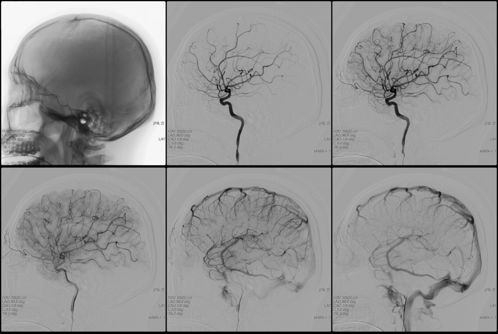
The essence of this research method is as follows. A radiopaque substance, usually based on iodine (Urografin, Triyodtrust, Omnipak, Ultravist and others), is injected into a specific artery of the brain (or the entire network of cerebral arteries) to the patient. This is done so that it is possible to fix the image of the vessel on an X-ray film, since the vessels are poorly visualized in a conventional image. The introduction of a radiopaque substance is possible by puncture of the corresponding vessel (if technically feasible) or through a catheter brought to the required vessel from the periphery (usually from the femoral artery). When the contrast agent enters the vascular bed, a series of X-rays are taken in two projections (frontal and lateral). The obtained images are evaluated by a radiologist, he draws conclusions about the presence or absence of a certain pathology of the cerebral vessels.
Varieties
Depending on the method of administration of the drug, this research method may be:
- puncture (when the contrast is introduced by puncturing the corresponding vessel);
- catheterization (when the contrast is delivered through a catheter inserted through the femoral artery and advanced along the vascular bed to the desired location).
According to the vastness of the study area, angiography of cerebral vessels is:
- general (all vessels of the brain are visualized);
- selective (one basin is considered, carotid or vertebrobasilar);
- superselective (a smaller-caliber vessel in one of the blood vessels is being investigated).
Superselective angiography is used not only as a research method, but also as a method of endovascular treatment, when, after identifying a “problem” in a particular vessel, this problem is “eliminated” using microsurgical techniques (for example, embolization or thrombosis of an arteriovenous malformation).
Due to the widespread introduction of modern diagnostic methods such as computed tomography (CT) and magnetic resonance imaging (MRI), CT angiography and MR angiography are increasingly being performed recently. These studies are carried out in the presence of appropriate tomographs, they are less traumatic and safer than just angiography. But more on that later.
Indications for conducting
Angiography of cerebral vessels is a specialized diagnostic method, which should only be prescribed by a doctor. It is not performed at the request of the patient. The main indications are:
- suspicion of arterial or arteriovenous;
- suspicion of arteriovenous malformation;
- determination of the degree of stenosis (narrowing) or occlusion (blockage) of the vessels of the brain, that is, the establishment of the lumen of the corresponding vessels. In this case, the severity of atherosclerotic changes in the vessels and the need for subsequent surgical intervention are established;
- establishing the relationship of the cerebral vessels with the adjacent one for planning the surgical access;
- control of the location of the clips applied to the vessels of the brain.
I would like to note that simply complaints of dizziness, headache, tinnitus and the like are not in themselves an indication for angiography. Patients with such symptoms should be examined by a neurologist, and based on the results of the examination, as well as other research methods, the need for angiography is determined. This necessity is established by the doctor!
Contraindications
The main contraindications are:
- an allergic reaction (intolerance) to iodine preparations and other x-ray contrast agents;
- pregnancy (due to ionizing radiation during the procedure). In this case, MR angiography is possible;
- mental illness that does not allow you to comply with all the conditions for the procedure (for example, a person will not be able to move while taking a picture);
- acute infectious and inflammatory diseases (as the risk of complications increases);
- violation of the indicators of the blood coagulation system (both downward and upward);
- the general condition of the patient, regarded as severe (this may be grade III heart failure, end-stage renal and hepatic failure, coma, and so on). As such, this subgroup of contraindications is relative.
Preparing for angiography
To obtain accurate results and reduce the risk of complications from the procedure, it is recommended:
- to pass general and biochemical blood tests, including - to determine the indicators of the coagulation system (the test period should not exceed 5 days). The blood group and Rh factor are also determined in case of possible complications;
- make an ECG and FG (FG, if one has not been performed within the last year);
- do not drink alcoholic beverages for 14 days;
- during the last week, do not take drugs that affect blood clotting;
- perform an allergic test with a contrast agent. To do this, within 1-2 days, 0.1 ml of the appropriate drug is administered intravenously to the patient and the reaction is assessed (the appearance of itching, rash, difficulty breathing, etc.). In case of a reaction, the procedure is contraindicated!
- the day before, take antihistamines (antiallergic) drugs and tranquilizers (if necessary and only as prescribed by a doctor!);
- do not eat for 8 hours and do not drink water 4 hours before the study;
- swim and shave (if necessary) the site of puncture or catheterization of the vessel;
- remove all metal objects (hairpins, jewelry) before the examination.
Research technique
At the very beginning, the patient signs a consent to conduct this type of research. The patient is placed with an intravenous peripheral catheter to have immediate access to the circulatory system. Then premedication is carried out (approximately 20-30 minutes before the procedure): antihistamines, tranquilizers, pain relievers are administered to minimize discomfort during the procedure and the risk of complications.
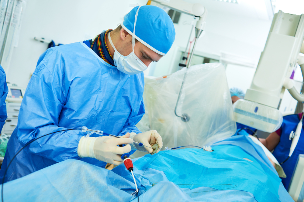
The patient is placed on the table and connected to devices (cardiac monitor, pulse oximeter). After the skin is treated with a local anesthetic and anesthesia, the corresponding vessel (carotid or vertebral artery) is punctured. Since it is not always possible to accurately get into these arteries, most often a small skin incision and puncture of the femoral artery is made, followed by immersion of the catheter and passing it through the vessels to the study site. The advancement of the catheter along the arterial bed is not accompanied by pain, since the inner wall of the vessels is devoid of pain receptors. Control of the progress of the catheter is carried out using X-ray. When the catheter is brought to the mouth of the required vessel, a contrast agent preheated to body temperature in a volume of 8-10 ml is injected through it. The introduction of contrast can be accompanied by the appearance of a metallic taste in the mouth, a feeling of heat, and a rush of blood to the face. These sensations go away on their own within a few minutes. After the contrast is injected, X-rays are taken in frontal and lateral projections almost every second several times (which allows you to see both arteries, and the capillary phase, and veins). The pictures are developed and evaluated immediately. If something remains incomprehensible to the doctor, an additional portion of the contrast agent is injected, and the images are repeated. Then the catheter is removed, a sterile pressure bandage is applied to the puncture site of the vessel. The patient should be monitored by medical personnel for at least 6-10 hours.
Complications
According to statistics, complications during this diagnostic method occur in 0.4-3% of cases, that is, not so often. Their occurrence can be associated both with the procedure itself (for example, the outflow of blood from the puncture site of the vessel), and with the use of a contrast agent. It should be borne in mind that compliance with all conditions in the preparation and conduct of angiography is the prevention of possible complications. The use of the latest generation of iodine-containing drugs (Omnipak and Ultravist) is characterized by lower statistics of complications.
So, possible complications of cerebral angiography are:
- vomit;
- an allergic reaction to an iodine-containing drug: itching, swelling and redness at the injection site, and then the appearance of shortness of breath (reflex breathing disorder), a drop in blood pressure, an irregular heart rhythm. In severe cases, anaphylactic shock may develop, which is a life-threatening condition;
- spasm of cerebral vessels and, as a result, acute cerebrovascular accident (up to);
- seizures;
- penetration of the contrast agent into the soft tissues in the puncture zone of the vessel (outside the vascular bed). If the volume of the drug poured into the tissue is up to 10 ml, then the consequences are minimal, if more, then inflammation of the skin and subcutaneous fat develops;
- the outflow of blood from the puncture site of the vessel.
CT and MR angiography: what are the features?
CT and MR-angiography of cerebral vessels in their essence represent a similar study as angiography. But there are a number of certain features of these procedures that distinguish them from cerebral angiography. Let's talk about this.
- it is done with a tomograph, not a conventional X-ray machine. The study is also based on X-rays. However, its dose is significantly less than with conventional angiography of cerebral vessels, which is safer for the patient;
- computer processing of information allows you to obtain a three-dimensional image of blood vessels absolutely at any point of the study (this applies to the so-called spiral CT angiography performed on a special spiral tomograph);
- the contrast agent is injected into a vein in the elbow rather than into the arterial network (which significantly reduces the risk of complications, since the administration of the drug becomes a routine intravenous injection through a peripheral catheter).
- for CT angiography, there is a limitation on the weight of a person. Most tomographs can support body weight up to 200 kg;
- the procedure is performed on an outpatient basis and does not require observation of the patient at the end of the procedure.
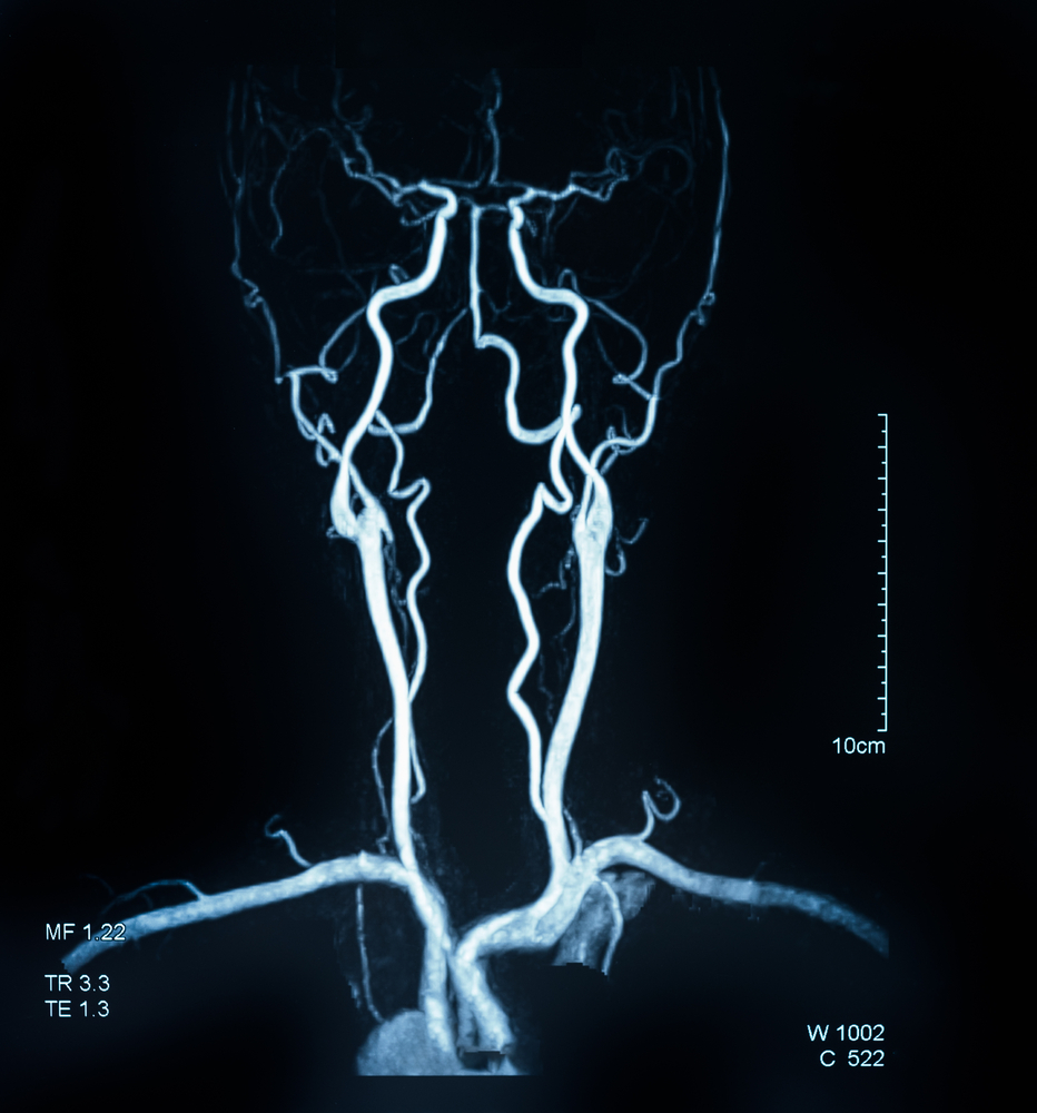
MR angiography is characterized by the following features:
- it is carried out using a magnetic resonance imager, that is, the method is based on the phenomenon of nuclear magnetic resonance. This means the complete absence of X-rays during the procedure (and therefore MR-angiography is allowed during pregnancy);
- can be performed both with the use of a contrast agent (for better visualization), and without it (for example, with intolerance to iodine preparations in patients). This nuance is undeniable
an advantage over other types of angiography. If it is necessary to use contrast, the substance is also injected into the vein of the ulnar flexure through a peripheral catheter; - the image of the vessels is obtained in three-dimensional due to computer processing;
- a series of images takes a slightly longer period of time compared to other types of angiography, while a person needs to lie in the tomograph tube all the time. For persons suffering from claustrophobia (fear of enclosed spaces), this is not feasible;
- the procedure is contraindicated in the presence of an artificial pacemaker, metal clips on the vessels, artificial joints, electronic implants of the inner ear);
- performed on an outpatient basis, and the patient is immediately released home.
In general, we can say that CT and MR angiography are modern, less dangerous and more informative research methods than conventional angiography of cerebral vessels. However, they are not always feasible, therefore, conventional angiography of cerebral vessels is still a relevant method for studying vascular pathology of the brain.
Thus, angiography of cerebral vessels is a very informative method for diagnosing, mainly, vascular diseases of the brain, including stenoses and occlusions, which are the cause of strokes. The method itself is quite affordable, it only requires an X-ray apparatus and a contrast agent. Subject to all the conditions for the preparation and conduct of the study, angiography of the cerebral vessels gives an accurate answer to the question posed to it with a minimum number of complications. In addition, modern medicine has such innovative methods as CT and MR-angiography, which are more dramatic, less harmful and traumatic for the patient. CT and MR angiography allow you to obtain a three-dimensional image of the vessels, which means, with a greater degree of probability, not to miss the existing pathology.
Medical animation on the topic "Cerebral Angiography":
