The brain is the main control center of the body. It is he who receives and analyzes information. But there are situations when he begins to behave incorrectly and an encephalogram of the brain is required. These can be tumors, diseases, traumatic brain injuries, which did not give a complete description of the picture on ultrasound or tomography. Also, such a procedure can be carried out for young children in case of their mental retardation or impaired speech motility. This unique opportunity gives you a chance to learn about brain activity in critical situations.
What it is
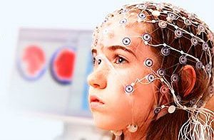
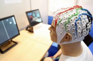
People calmly perceive x-rays, ultrasounds and are wary of an encephalogram. But this is a simple and quick process. As already scientifically proven, our brain emits electrical signals. The principle of an encephalogram is based on the fixation of signals. Sensors are placed on various parts of the head, the data is transmitted to a computer, which displays the tone of neuronal cells.
A bit of history
In the middle of the 19th century, it was found that muscle tissue and the brain emit weak electrical signals. For the first time the activity of electric waves was investigated by the Englishman Caton and the Russian Danilevsky. Only in 1913 was the first brain encephalography recorded by the scientist Pravdich-Nemytsky, and the German scientist Berger introduced the concept of an encephalogram. This method was applied in practice in 1934. Currently, doctors refer to the name in abbreviated form as the abbreviation EEG.
What is the EEG for?
Such a diagnosis allows doctors to assess the damage to brain activity, to establish the location of the affected area. EEG information can be compared with other diagnostics and the effectiveness of drug treatment can be determined. The examination also makes it possible to study the tone of the nervous system, prevent convulsive seizures, and determine the vital activity of paralyzed patients. With this procedure, conclusions about a tumor or cystic formations are checked.
In what cases is EEG prescribed?
An encephalogram is prescribed for cranial injuries and concussions, in order to establish the consequences of an operation that affected the activity of the brain.
If an accurate diagnosis is required: vegetative-vascular dystonia, headaches, osteochondrosis of the cervical spine, unstable or high blood pressure.
EEG is prescribed for seizures of epilepsy or epilepsy, fainting, numbness of the extremities. A study is carried out if the child has a delay in speech motor skills or mental thinking.
Examinations are made to confirm the death of a part of the neurons, when it is seen how a person turns into a vegetable, and to carry out operations of a neural nature. With violations of blood circulation in the head, lesions of the central nervous system, various types of neurosis. EEG is also taken by patients who underwent MRI and they did not have any abnormalities or deviations of the endocrine system. The direction on the EEG is received by sick people with oncology, which affects the activity of the central nervous system.
EEG is provided for in some types of medical examination and for obtaining a driver's license, assessing the activity of neurotoxic poisons, doubts about Parkinson's and Alzheimer's diseases.
Conducting an EEG in case of doubt about falling sickness
Falling disease, also called epilepsy, is not very easy to recognize. EEG is the most reliable way to measure. In addition, you can identify the type of seizures and their changes. So the doctor can individually select the treatment that will be most beneficial for the patient. In the absence of seizures, patients undergo an EEG twice a year. If there are seizures and it is necessary to change the treatment, then the EEG is done often.
Features of the EEG procedure for children
Adults, having received a prescription for an EEG examination of a child, begin to doubt its safety. There is nothing wrong with this procedure, but the process itself can be difficult. Children under the age of 1 year undergo the procedure in a sleeping mode. The baby's hair is washed, fed and the EEG is adjusted for sleep.
It is useful to know: Modern technologies - how MRI of the brain is done
If it is not a problem for a baby under 1 year old to make an encephalogram of the brain, then it is more difficult for children under 3 years old. It is necessary to persuade them. If the children are contact, then the procedure can be carried out in a state of wakefulness, and in other situations, preference is given to the sleeping mode. At this age, it is difficult to persuade a child to sit quietly even for 15 minutes. At this time they are interested in everything, they will turn their heads. If the mother can persuade or captivate the baby for this time for peace of mind, then the EEG will work.
EEG
Before the procedure, it is necessary to exclude coffee and avoid the presence of varnishes, lotions, gels on the hair. Get rid of metal objects, earrings, hairpins, piercings. You should not eat heavily. EEG is not performed during acute colds.
The process is absolutely harmless and not painful. The patient sits down, closes the eyelids and relaxes. Special sensors are covered with a gel, attached to the skin, a cap is put on, and a process has begun that registers the biological tone of the brain.
The electrodes are connected to the amplifier because the activity signals are weak. The readings are recorded by a device, an electroencephalograph. The indicators are displayed, according to which the deviation from the norm is determined. They can be captured either to tape or to a file on a computer. The procedure takes no more than 30 minutes.
During the EEG, you can use provocative moments to close and open your eyes, breathe deeply and often through your mouth. At certain points, the doctor says when it is necessary for the patient to do this. An encephalogram is performed in a calm position, but it can also be done in sleep mode.
Video monitoring
This brain encephalogram takes much longer and can last up to one day. This is a more detailed picture of the activity of the brain. It has a feature that several video cameras are watching the patient. Special sensors record work from calm to active state. A specialist can see a picture of brain activity in a calm state, mental, emotional and physical stress. This method helps to identify the most subtle violations.
Decoding EEG data
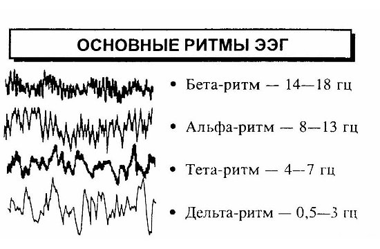
When determining the results, the number of years of the patient and his condition are taken into account. During wakefulness, an encephalogram of the brain is considered good, when the registered Alpha waves with a frequency of pulsations from 8-12 MG and a swing of 50 μV, and Beta waves with a frequency of pulsations of more than 12 Hz and a swing of no more than 20 μV. The appearance of peaks of alpha and beta waves indicate disturbances in the body.
When decoding, they focus on the main indicators:
- the alpha rhythm is characteristic of everyone during relaxation;
- beta rhythm - for an active thought process;
- theta rhythm - a state of drowsiness, half-sleep, superficial sleep;
- delta rhythm - deep co;
- besides these waves, other oscillations can be registered, but they are not of interest. Therefore, they do not focus on them when decoding.
