Medicine constantly presents to society new methods for diagnosing serious pathologies. The success of the treatment of various diseases depends on their timely detection, the appointment of the necessary therapy. Duplex scanning of the vessels of the head and neck is an innovative research method that allows you to see in a two-dimensional projection the smallest tubular hollow formations of the human body. The non-invasive nature of the technique facilitates the procedure, does not require recovery after the manipulation.
What is Duplex Vascular Scan
How to check your head non-invasively? The unique properties of ultrasound help it pass through the tissues of the human body and, reflecting from the blood cells, send a signal in the form of an image of the area under study to the screen of the diagnostician's monitor. By means of duplex scanning of the vessels of the head and neck, the specialist can assess the parameters of blood hemodynamics, obtain information about the anatomical features of the veins and arteries. Different Doppler technologies use the properties of the sound wave in the same way, but have different functionalities:
- Doppler ultrasound (ultrasound dopplerography). This study helps to assess the patency of the vessels of the brain, neck, and other organs. USDG carries only one functional load - the determination of hemodynamics.
- Duplex ultrasound scanning. Using this method, it is possible to diagnose the presence in the arteries and veins of atherosclerotic plaques, blood clots, which contribute to the narrowing of the vascular lumen. During monitoring, a tubular formation with surrounding tissues is visualized. Duplex scanning is divided into the following subtypes:
- extracranial - examines the great vessels;
- intracranial - checks the intracerebral "pools";
- transcranial - Provides color duplex scanning of the brain.
- Triplex scanning. Doppler sonography of the vessels of the head and neck, during which, in addition to information about the intensity of blood movement, the diagnostician receives a color image of the tubular formation with the surrounding tissues.
- Ultrasound procedure. Shows the "big picture" of the structure of the arteries and veins. Doppler ultrasound helps to find out the characteristics of blood flow movement, to conduct an examination for the presence of pathologies.
Indications for the purpose of the study

The study of vessels of a planned nature should be carried out without fail once a year. Detection of an anomaly at an early stage of development helps to avoid the negative consequences associated with a progressive form of the disease, and to take measures to prescribe the necessary therapy. Duplex scanning of the patency of the vessels of the head and neck is often prescribed to verify the results obtained during MRI, USDG of the vessels of the neck and head. The indications for duplex are the following symptoms:
- headache;
- dizziness;
- fainting;
- numbness of the hands;
- lack of coordination;
- memory loss;
- smoking;
- a history of strokes;
- cervical osteochondrosis;
- arterial hypertension;
- previously identified vascular dystonia;
- family ties with a hypertensive or diabetic;
- vasculitis (vascular inflammation).
How to prepare
Examination of the head and neck does not require special training from the patient. On the day of the procedure, it is necessary to abandon the use of drugs that increase the tone of blood vessels: coffee, nicotine, tea, energy. Cancellation of drugs that can distort the results of the ultrasound scan - "Betaserc", "Cinnazirin" - requires consultation with a neurologist. Before scanning, the patient will need to remove all foreign objects from the study area in the form of chains, hairpins, etc.
How is the procedure carried out
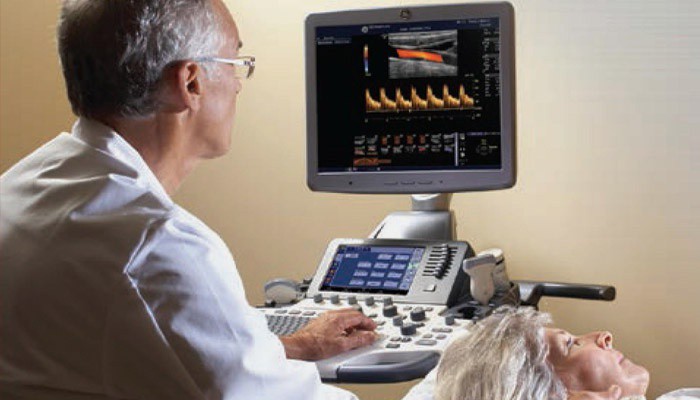
Duplex scanning can be done on the referral of the attending physician in the neurological departments of large city hospitals or go to the clinic according to the area of residence. The manipulation is carried out according to the general rule. The patient is placed on a couch, a firm pillow or roller is placed under the head, the head is moved to the side opposite to the sensor.
Before starting the procedure, the doctor applies a little special gel to the area under study, with which you can easily "drive" the transducer over the skin surface, analyzing the arterial and venous channels. The cerebral vessels are checked through the bones of the skull. The skin is preliminarily treated with a water-soluble gel, then the doctor places the sensors on the following areas:
- temples;
- above the eye sockets;
- alignment of the occipital bone with the spine;
- occipital bone.
Decoding the results

At the end of the examination, the doctor receives comprehensive information about the condition of the arteries and veins. The analysis of the venous bed contains practically no digital data, but includes the parameters:
- anatomy;
- patency;
- blood speed;
- the presence of abnormal formations inside the lumen.
Doppler ultrasonography of arterial vessels collects digital data, which are compared with normal values. The presence of the following indicators can be considered a satisfactory condition of the common and carotid arteries:
- the limiting speed of blood movement in the artery is less than 0.9;
- percentage of stenosis - 0;
- peak velocity in diastole - less than 0.5;
- absence of formations inside the lumen;
- wall thickness - 0.9-1.1.
Are there any contraindications

The advantage of duplex scanning is the absence of a negative effect on the human body. The non-invasive nature of the study helps to diagnose blood vessels in an adult and a child without any restrictions. Relative contraindications can be considered a serious condition of the patient or the presence of diseases that prevent the patient from moving to a horizontal position.
