Downtrend headaches, bad sleep, fast fatigue, irritability - all this may be a consequence of bad blood circulation in the brain or abnormalities in the nervous system. For timely diagnosis of negative disorders in vessels, EEG is used - the electroencephalogram of the brain. This is the most informative and affordable method of examination, which does not harm the patient and can be safely used in childhood.
Electroencephalogram is used to examine the brain vessels
EEG brain - what is it?
The enencephalogram of the head is a study of a vital organ by exposure to electrical impulses.
The method determines the bioelectric activity of the brain, is very informative and most accurate, as it shows a complete clinical picture:
- level and distribution of inflammatory processes;
- the presence of pathological changes in vessels;
- early signs of epilepsy;
- tumor processes;
- the degree of brain functioning due to the pathologies of the nervous system;
- the consequences of stroke or operational intervention.
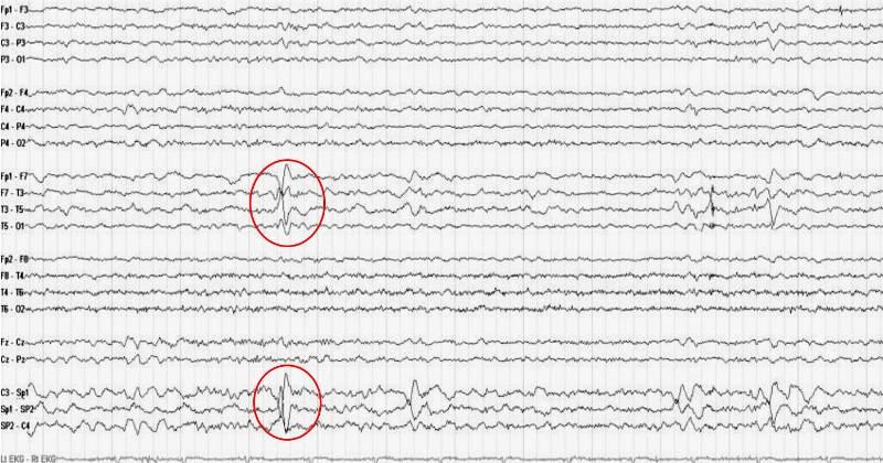
EEG helps to identify signs of epilepsy
EEG helps to monitor changes in the brain, both structural and reversible. This allows you to monitor the activity of a vital organ during therapy, and adjust the treatment of identified diseases.
Where you can do the survey price
Electricencephalography can be made in any specialized medical center. Institutions can be both public and private. Depending on the ownership form, the level of qualifications of the clinic, as well as the equipment used, the prices of the procedure differ significantly.
In addition, the cost of the encephalogram is influenced by the following factors:
- duration of the diagnostic procedure;
- conducting functional samples;
- use of special programs (for mapping, studying epileptic impulse, comparison of zones of symmetric brain zones).
Indications for the electroencephalogram
Before prescribing a patient with encephalography, a specialist inspects a person and analyzes his complaints.
The following states may become a reason for EEG:
- problems with sleep - insomnia, frequent awakening, walking in a dream;
- regular dizziness, faint;
- fast fatigue and constant feeling of fatigue;
- downtry headaches.

With frequent pains in the head you need to go through EEG
Minor, at first glance, changes in well-being, may be a consequence of irreversible processes in the brain.
Therefore, doctors can assign a encephalogram when identifying or suspicions of such pathology as:
- diseases of the neck and head vessels;
- vegeta dystonia, certificates in cardiac activity;
- after stroke condition;
- speech delay, stuttering, autism;
- inflammatory processes (meningitis, encephalitis);
- endocrine disorders or suspicion of tumor foci.
A mandatory study of EEG is considered for people who have underwent head injuries, neurosurgical surgery, or epilepsy suffering from seizures.
How to prepare for research
Monitoring the electrical activity of the brain requires simple preparation. For the accuracy of the results, it is important to fulfill the basic recommendations of the doctor.
- Do not use anticonvulsant, sedative medicines, as well as tranquilizers in 3 days before the procedure.
- 24 hours before the study does not drink any carbonated drinks, tea, coffee and energy. To eliminate chocolate. Do not smoke.
- On the eve of the procedure, thoroughly wash the hair part of the head. To eliminate the use of cosmetics (gels, varnishes, foams, mousse).
- Before starting the study, you need to remove all metal decorations (earrings, chain, clips, hairpins)
- Hair must be loose - all kinds of weaving must be broken.
- It is necessary to maintain peace of mind to the procedure (do not allow stress and nervous disruptions in 2-3 days) and during its holding (not afraid of noise and flashes of light).
An hour before the examination, it is necessary to eat well - the study is not carried out on an empty stomach.

During the day, it is impossible to eat chocolate
How the electroencephalogram is carried out
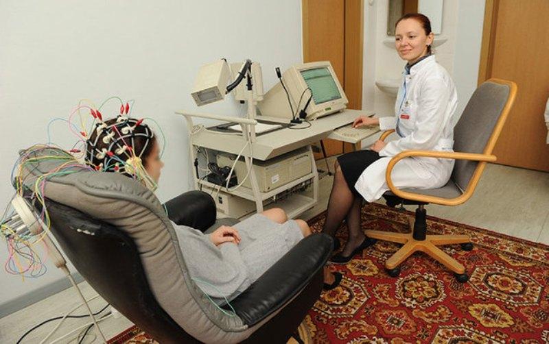
Evaluation of the electrical activity of cerebral cells is carried out with the help of encephalographer. It consists of sensors (electrodes) that resemble a hat for the pool, block and monitor, where monitoring results are transmitted. The study is carried out in a small room, which is isolated from light and sound.
The EEG method takes a little time and includes several stages:
- Preparation. The patient takes a convenient pose - sitting on a chair or go to the couch. Then there is an overlap of electrodes. On the head of a person, a specialist puts on a "cap" with sensors whose wiring is connected to the device, which fixes the bioelectric imaging of the brain.
- Study. After turning on the encephalographer, the device begins to read the information by passing it to the monitor in the form of a graph. At this time, the power of electric fields and its distribution of various parts of the brain can be recorded.
- Use of functional samples. This is the implementation of simple exercises - to frort, look at the light outbreaks, rarely or breathe deeply, listen to sharp sounds.
- Completion of the procedure. The specialist removes the electrodes and prints the results obtained.
During the EEG, the patient accept a convenient position and relax
If the study requires a deeper study (day monitoring), interruptions are possible. Sensors are disconnected from the wires, and the patient can go to the toilet, eat, chat with relatives.
Features of the EEG in children
Monitoring brain activity in children has its own nuances. If the child is up to a year, then the study is carried out in a state of sleep. To do this, the baby should be fed and then specified. After a year, children examine in wakefulness.
So that the procedure has passed successfully, it is important to prepare the child:
- On the eve of the examination with the child, it is recommended to talk, talk about the upcoming procedure. You can come up with the game so that the baby is faster to adapt, calling it a superhero or astronaut.
- Take with you your favorite toys. This will help distract fume and calm it at the right moment.
- Feed the child before starting the study.
- To specify with the doctor the time of manipulation and pick up a comfortable clock when the child is awake and it is not clone into sleep.
- On the eve of the survey, wash your baby's head well. If this is a girl, hair break, remove all the decorations (immediately before monitoring).
How much the procedure lasts
The usual encephalogram is a routine EEG or the diagnosis of paroxysmal state. The duration of such a method depends on the studied area and application in monitoring functional samples. On average, the procedure does not take more than 20-30 minutes.
During this time, a specialist has time to spend:
- rhythmic photostimulation of different frequency;
- hyperventilation (breaths deep and rare);
- load in the form of a slow blink (to open and close the eyes at the right moments);
- detect a number of functional changes in a hidden character.
In case of insufficiency of the information received, specialists can resort to a deeper survey.
There are several options:
- Night sleep enencephalogram. A long section is being studied - wakefulness before bedtime, nap, destruction to sleep and morning awakening.
- EEG with deprivation. The method is that the patient is deprived of night sleep. He must wake up for 2-3 hours earlier than usually and not to sleep the next night.
- Continuous electroencephalogram. Monitoring of bioelectric activity of the brain occurs during daytime sleep. The method is very effective in suspected paroxysm (seizure) or identifying the causes of sleep disorders.
Based on the EEG method, the duration of such a study may vary from 20 minutes to 8-15 hours.
Decoding EEG indicators
The interpretation of the results of the encephalogram is engaged in qualified diagnostics.
When deciphering, clinical symptoms of the patient and the main EEG indicators are taken into account:
- the state of rhythms;
- symmetry of hemispheres;
- changes in gray matter when using functional samples.
The results obtained are compared with the established norms, and deviations (dieselmia) are recorded in the conclusion.
Table "Decoding EEG"
| Indicators | Norm | Deviation | Possible pathological processes | |
| In adults | The child has | |||
| Alpha Rhythm | 8-15 Hz - rhythm is distinguished by regularity, observed at rest or with closed eyes. Maximum concentration of pulses in the back zone of the skull and temke | The appearance of alpha waves in the frontal part of the brain. Rhythm becomes paroxysmal. Violation of frequency stability and symmetry of hemispheres (above 30%) | Development of tumor processes, the appearance of a cyst. Structure of stroke or heart attack. Availability of serious damage to the skull of injuries | Neurosis of varying degrees Psychopathy Psychomotor Development Delay - Neurophysiological immaturity of cerebral cells |
| Beta rhythm | 12-30 Hz - reflects the excitement, alarming nervousness and depression. Sensitive to sedatives. Localizes in obscurity | Diffuse Beta Waves Increase amplitude Violations of symmetry hemisphey Paroxysmal discharges |
Concussion of the brain Encephalitis |
|
| Delta Rhythm | 0.5-3 Hz - fixes the state of natural sleep. Does not exceed 15% of all rhythms. Amplitude not higher than 40 μV | High amplitude The appearance of the delta and theta waves outside of sleep, localization in all parts of the brain High frequency rhythms |
Irritation of the structural centers of the gray substance (irritation) Dementia |
|
| Theta rhythm | 3.5-8 Hz - reflects the normal state during adults in adults. In children, such an indicator is dominant | |||
Based on the study of rhythms, a conclusion is made on the bioelectric activity of the brain. In normal condition, it must be without seizures (paroxysis), have regular rhythm and synchronization. Diffuse (moderate) changes are admissible if other pathological disorders (irritation of parts of the brain, dysfunction of regulatory systems, rhythm disorganization) are not detected. In this case, the specialist can prescribe corrective treatment and observe patients.
It is important to take into account that moderate changes in rhythms (Delta and Teta), the emergence of paroxysmal discharges and epileptic activity on EEG in children and people under 21 is the norm and does not apply to deviations in the structures of a vital organ.
Duration of electroencephalography
The results of the encephalogram are valid from 1 to 6 months.
Terms may differ depending on:
- diseases;
- therapies (re-EEG are needed when adjusting the treatment or evaluation of the effectiveness of prescribed drugs);
- informativeness of the selected EEG method.
If a person is healthy or electroencephalogram has light changes, the conclusion is valid for half a year. In the case of serious deviations or the need for regular monitoring of cerebral activity (especially in children), the EEG period may be a month or week.
The use of electroencephalography to estimate the state of brain activity allows you to identify a number of pathologies in the early stages. The EEG method makes it possible to determine the development delay in children before the first manifestations. In addition, the procedure is completely harmless, it can be made an unlimited number of times, even in early childhood. Encephalogram is used not only to detect deviations, but also as a tool for monitoring the effectiveness of treatment.
