EEG, or electroencephalography, is a diagnostic method for studying the functional activity of neurons in the brain. It is based on the registration of impulses emanating from certain brain centers, with their subsequent decoding. It is used to identify pathological processes occurring in the central nervous system (for example, if epilepsy, cancer, and others are suspected). What does the EEG of the brain show? Why is this examination prescribed? More on this later in the article.
What is the essence of the procedure?
EEG can be performed on patients of different ages
Nerve cells in the head, while performing their functions, generate an electrical impulse with a certain frequency and amplitude. This neuronal activity can be captured and recorded by placing electrodes on the surface of the head. As a result, everything that the EEG detects will be reflected on paper or on a monitor in the form of waves.
Different people have their own indicators of the electrical activity of nerve cells in the brain.
Despite the fact that the average indicators still exist, during the deciphering of the electroencephalogram, specialists must take into account such parameters as: age characteristics, the presence or absence of neurological diseases, the therapy being carried out at the time of the study (or its absence), and others.
What rhythms of the brain are recorded by an electroencephalogram
As mentioned above, electrical oscillations arising in the neurons of the cerebral hemispheres are displayed on the monitor screen in the form of waves. Changes in the following rhythms are of diagnostic value: alpha, beta, theta, delta. There are other rhythms of functional activity of neurons (gamma, kappa, mu). However, they are not of particular interest in relation to diagnosis, since their occurrence is associated with a sufficiently high mental and mental stress. And what the electroencephalogram of the brain shows is revealed in a state of complete rest, sometimes during sleep.

Types of brain waves
Brain rhythms
So, what does the EEG of the brain reveal? Below are the main rhythms of electrical activity of neurons in the cerebral hemispheres and their brief characteristics.
- Alpha rhythm. It is characterized by a frequency of 8-13 Hz and an amplitude of about 50 μV. Such indicators are normally recorded by the device at a time when a person is awake, but does not show physical and mental activity. In addition, his eyes must be closed. When the eyes are opened, the visual analyzer is switched on, as a result of which the activity of nerve cells increases; in this case, alpha waves pass into waves with a higher frequency - beta. The same happens with sounds, any activity, feelings of fear, anxiety and other conditions.
- Beta rhythm. The frequency range of these waves is in the range of 14-30 Hz, and the amplitude is approximately 25 μV. In a calm state, they are expressed to an insignificant degree. An increase in the beta rhythm is due to a stress factor, as well as high mental activity.
- Theta rhythm. These waves are characterized by a frequency of 4-7 Hz and an amplitude of about 100 μV. Theta waves occur when a person begins to doze. In addition, they are enhanced by various neurological pathologies, concussion, prolonged stress, emotional and mental overload, mental disorders.
- Delta rhythm. It occurs during deep sleep (including under the influence of anesthesia), as well as during various pathological processes in the brain. It is characterized by a frequency of 0.5-3.5 Hz and an amplitude of 100-300 μV.
What does an electroencephalogram show?
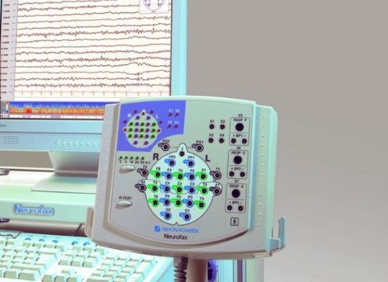
An EEG device is called an electroencephalograph.
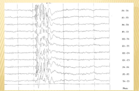
The curve that appears on the monitor screen during the EEG makes it possible to diagnose various changes in the functioning of the nerve cells of the brain. A specialist, evaluating the activity of neurons, reflected in the encephalogram, can determine the following points:
- find out the presence or absence of violations from the cerebral cortex;
- assess the severity of brain damage, if detected;
- pinpoint the location of the injury;
- identify those parts of the brain that are the source of epileptic seizures;
- to study the features of the periodization of sleep and wakefulness;
- detect a neoplasm;
- determine how effectively drug therapy was carried out;
- find out how the brain works in the periods between attacks;
- find the cause of fainting and other crisis moments and much more.
Features of decoding an electroencephalogram
Based on what the EEG shows, the specialist makes a decoding and draws up a conclusion. The following are considered good results:
- the alpha rhythm is fixed in the occipital and parietal regions, and its frequency and amplitude are within normal limits;
- indicators of frequency and amplitude of beta waves also have normal values and are fixed in the forehead area (they can alternate with theta waves, which are at their normal frequency).
When decoding, it is important to take into account that by themselves, taken separately, the rhythms do not yet indicate the presence of any specific disease or disorder. For example, healthy people may have waves that are characteristic of epilepsy. In addition, EEG readings taken in the intervals between epileptic seizures do not record changes in all patients. Thus, if, as a result of the examination, no neuronal activity corresponding to epilepsy was found, this does not mean that there is no disease (provided that there are pronounced clinical symptoms). In this case, the doctor chooses other diagnostic methods.
Juvenile absence epilepsy
What else does the EEG of the brain show in an adult? Other diseases (except epilepsy) can be detected by electroencephalography in the form of lesions. So, if an increase in delta and theta rhythms is recorded, then a specialist can assume the presence of a tumor, edema, stroke.
Diffuse changes in the brain can be evidence of diseases and conditions such as:
- concussion, head trauma;
- meningitis;
- encephalopathy.
In some cases, an EEG study can show changes in the functional activity of neurons in people who do not complain about well-being.
If this happens, do not panic, especially since there are no clinical symptoms. EEG analysis at the next diagnosis is likely to show normal results.
EEG diagnostics in childhood
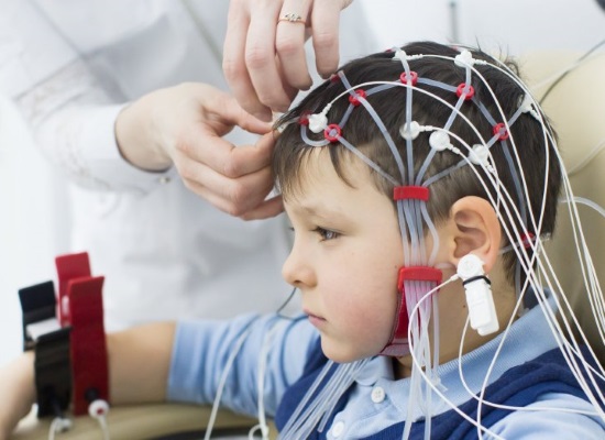
Before the procedure, a helmet with electrodes is put on the child's head
In some cases, the doctor may prescribe a diagnosis using electroencephalography in a child. Most often, the indications are:
- trauma, concussion of the head;
- frequent crying for no apparent reason;
- fluctuations in blood pressure indicators;
- sleep disorders;
- nervous cramps, seizures;
- fainting;
- headache complaints;
- irritability, erratic behavior;
- dizziness and other symptoms and conditions.
An analysis of what the electroencephalogram shows makes it possible to assess the development of the child in the first year of life, shows whether the formation of the central nervous system in the baby is proceeding correctly, and makes it possible to identify ischemic areas at an early stage. If any pathologies are detected, the doctor has the opportunity to stop their further development and eliminate the disease through timely therapy.
Usually EEG in children is carried out in a state of sleep. The procedure is absolutely safe for the health of the child, so parents should not worry, even if it is assigned to a newborn.
Despite the fact that the electroencephalogram is considered an outdated diagnostic method, which is actively supplanted by more modern CT and MRI, it is still relevant. This is due to its sufficient information content, low cost and availability. Therefore, if the doctor directed you to undergo an EEG procedure, you should not neglect it.
