The brain, regardless of whether a person is sleeping or doing mental work, exhibits bioelectrical activity. The method that allows you to register this activity is called electroencephalography, and the image obtained during the study is called an electroencephalogram (EEG).
Electroencephalography is widely used in both pediatric and adult neurology. With the help of an EEG, an experienced doctor can assess the state of the brain, detect areas of ischemic, traumatic or any other lesion in it, as well as identify foci of epileptic activity in the brain tissue. You can undergo this examination in specialized neuropsychiatric medical institutions and multidisciplinary diagnostic centers. Adult and pediatric neuropathologists, neurosurgeons and psychiatrists give directions to the EEG.
What does an electroencephalogram show?
Electroencephalography is one of the main instrumental methods for examining patients with neurological, mental and speech disorders. Indications for an EEG are the following pathological conditions:
How to prepare for research
The EEG does not require any special preparation. Before the procedure, it is advisable not to drink alcohol and strong coffee, not to smoke, not to engage in heavy physical labor and not to be very nervous, as this may distort the result of the study.
There is no need to starve or follow a diet, on the contrary, it is impossible to go to the study without eating. Hunger and a drop in blood sugar (hypoglycemia) provoke a change in brain activity, which can be regarded by a doctor as a sign of pathology, although it is actually not.
 For people taking psychotropic drugs, preparation for an EEG may involve temporarily withdrawing one or another drug. However, it is not worth making any adjustments to the course of treatment on your own. All appointments should be made by a doctor who gives direction to the EEG.
In patients with suspected epilepsy, electroencephalography is often performed not according to the standard method, but during sleep, or vice versa after a long abstinence from sleep. With the help of such techniques, doctors can register abnormal brain activity that is not always noticeable during a routine examination. If you plan to conduct an EEG using one of these methods, special preparation may be required: abstinence from sleep for 24-36 hours or taking sleeping pills) drugs.
How is electroencephalography performed?
Electroencephalography is a non-invasive study, not associated with the impact on the human body of any radiation. It does not cause harm to health, it is usually easily tolerated by the examined, therefore it can be carried out repeatedly. The only "minus" of the EEG is the need for prolonged immobility (at least 20 minutes). When examining a small child, this “minus” can become a serious problem, but doctors will definitely offer various options for solving it. From the mother, it is required to feed the baby, take him to the toilet and change the diaper before the procedure.
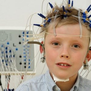
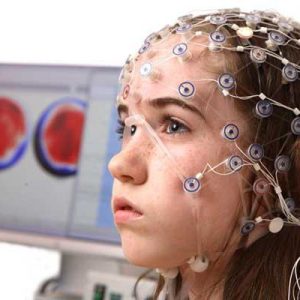
To conduct an electroencephalographic study, the patient is placed on a special chair or couch (usually in a reclining position), a special gel is applied to the head and a cap with electrodes is attached, each of which should be located clearly above its own parts of the brain. When the device is turned on, the subject should be as calm and motionless as possible. If there is a need to change the position, the registration of the electroencephalogram is suspended.
During the study, the doctor usually asks the patient to do some manipulations: open and close the eyes, breathe deeply and often, follow the flashing light, etc. it stress tests, which allow the doctor to assess the reaction of the brain to stimuli. With their help, they can reveal what remains hidden when a person remains absolutely calm.
Deciphering the electroencephalogram
The bioelectrical activity of the brain, registered by an electroencephalograph, is displayed on paper in the form of curved lines - waves (rhythms). There are as many of them as there are electrodes on the subject's head. Each such wave has its own amplitude and frequency of oscillations. Depending on the magnitude of these indicators, the following EEG rhythms are distinguished:
- alpha rhythm(8-13 vibrations per second). It is typical for an adult and quite healthy person who is in a state of calm wakefulness. This rhythm is best expressed in the leads of the occipital and parietal regions of the brain.
- beta rhythm, its frequency is higher than that of the alpha rhythm. There is a predominance of this rhythm during active wakefulness, mental stress, emotional arousal, REM sleep. The beta rhythm is generated by the frontal lobes of the cerebral hemispheres.
- Gamma rhythm. It has a frequency even greater than that of the beta rhythm. There is such brain activity in a state of maximum concentration.
- Theta rhythm- lower in frequency than the alpha rhythm. It is most pronounced in children 2-8 years old, in adults it can be during sleep.
- delta rhythm- the rhythm of the lowest frequency. It is typical for healthy babies of the first year of life, it can also be considered a variant of the norm for children under 6 years old (it all depends on clinical data). In adults, the delta rhythm appears during very deep natural sleep, general anesthesia, coma. In the waking state, this rhythm occurs when EEG is recorded from brain regions bordering on pathological foci and tumors.
- Straight line - there is no rhythm. Such an EEG pattern indicates the absence of electrical activity in the brain, that is, its possible death.
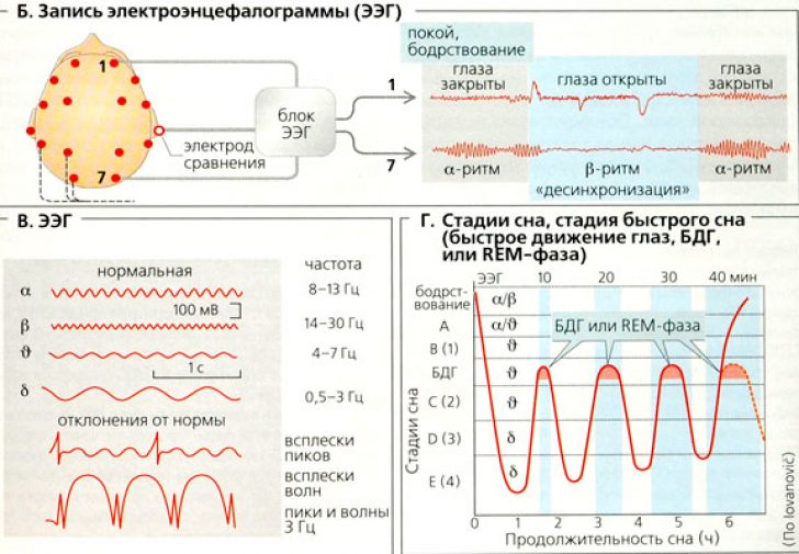
Since each of the described rhythms corresponds to a certain state of the brain, the replacement of one rhythm by another may indicate the presence of pathology. In addition, the appearance of waves that are uncharacteristic for some kind of abstraction or a significant increase or decrease in the amplitude of their oscillations is also regarded as a deviation from the norm.
In order to decipher the electroencephalogram as correctly as possible, the doctor must take into account the age of the patient (for children, adults and the elderly, their own norms for the bioelectrical activity of the brain tissue) and separately evaluate the data obtained at rest and with stimulation.
Thus, having examined the EEG, a specialist (a doctor involved in functional diagnostics) can determine whether there is a deviation from the norm, which part of the brain generates “wrong” waves, distinguish diffuse damage to the brain tissue from a local one, a superficial pathological focus from a deep one, identify an epileptic activity, recognize coma and establish the degree of its severity. These data are indispensable for neuropathologists and neurosurgeons, thanks to which doctors are able to "look" into the patient's brain, understand what is happening there, and, based on the information received, choose the most correct treatment tactics.
Zubkova Olga Sergeevna, medical commentator, epidemiologist
