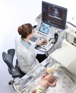
- Ultrasound examination of the brain. This is a common diagnostic procedure given to newborns to rule out possible head pathologies due to birth injuries, infections, or congenital diseases.
Ultrasound is a painless procedure and does not harm a fragile child's body, therefore, having received a referral for it, you should not panic, and even more so ignore it.
Why do research on newborns?
Neurosonography is designed to promptly identify or exclude suspicions of brain diseases in infants. Therefore, the list of indications for its implementation is very wide:
- lack of oxygen in the prenatal period or during childbirth;
- uncharacteristic head shape in a newborn;
- protracted or, conversely, too fast delivery;
- the absence of the first cry of a newborn;
- premature birth;
- trauma during childbirth;
- the child's weight is less than normal (2.8 kilograms);
- low Apgar score (less than 7);
- the woman in labor was given a caesarean section;
- during childbirth, obstetric manipulations were performed (for example, turning the fetus);
- the child often spit up;
- seizures for unknown reasons;
- intrauterine infection;
- predisposition to genetic diseases;
- developmental delay;
- malformations at the stage of pregnancy;
- also, the examination is carried out at the request of the parents without any medical indications.
Preparing for NSG in infants
in infants - a simple procedure that does not require prior preparation. It can be carried out during sleep or wakefulness of the baby, in some cases, the study is done even in intensive care, without taking the child out of the box.
It is not necessary to follow a diet or a special daily regimen before an ultrasound, but so that the baby does not worry and allows himself to be examined, it is better to feed him in advance.
The only strict prohibition is the use of creams or ointments on the child's head, they impair the performance of the sensors, which, in turn, affects the quality of the study.
Reference! Neurosonography is done without anesthesia or any additional drugs, the total procedure time will not take more than twenty minutes.
How is an ultrasound of the head done for a child up to a year old?
The peculiarity is that it is carried out only through the holes in the skull, since the bones do not transmit ultrasound. Therefore, ultrasound is more often done for children under one year old, preferably until the large fontanel closes.
The sensors of the equipment are installed in the region of the fontanel, less often in the temporal region or the occipital foramen, pre-treated with a conductive gel.
The doctor observes a dynamic image on the screen, noting the state of the structures, sulci and convolutions, the ventricles of the brain, the presence of cysts or formations, the condition of the membranes.
The examination ends with a snapshot and the derivation of digital indicators, according to which a transcript of the results is given.
Norm indicators
The absence of violations is indicated by:
- symmetry of the location of brain structures;
- ventricles of normal shape and size with clear and even boundaries;
- immutability of the membranes of the brain;
- the absence of foreign entities.
Digital indicators for newborns:
- the depth of the anterior horn of the lateral ventricle is from 1 to 2 millimeters;
- the depth of the body of the lateral ventricle does not exceed 4 millimeters;
- the size of the third ventricle is not more than 6 millimeters;
- the width of the gap between the hemispheres is not more than 2 millimeters;
- the size of a large tank is from 3 to 6 millimeters;
- the width of the subarachnoid space is up to 3 millimeters.
Important! Only a specialist should decipher the results of an ultrasound scan. To avoid unnecessary panic, do not self-diagnose.
The results of ultrasound are only part of the information, they are evaluated in conjunction with the external signs of pathologies and the indications of other studies by a neuropathologist. In addition, minor deviations from the norm in infants, both at 1 month and at 6 months, can be age-related and disappear with time. Only a professional can judge this.
What does the examination show and what pathologies does it reveal?
As a result of the study, pathologies can be identified. A number of them do not require additional intervention, but some need to be treated, so for a detailed decoding of ultrasound, you must definitely contact a neurologist.
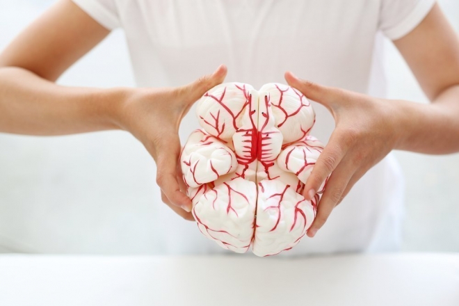
According to the results of neurosonography, diseases such as:
- Cysts are fluid-filled vesicles of various shapes and sizes. By itself, the cyst does not harm the health of the child and may resolve over time, but when it is detected, it is important to observe the dynamics of the development of the anomaly to prevent complications.
- Tumors are a serious lesion. I must say that they are not often observed in infants, but depending on the size, nature and location, they can be very dangerous.
- Dropsy of the brain is a dangerous disease in which fluid accumulates in the ventricles of the brain. With untimely treatment, hydrocephalus seriously affects the mental development of the child, can lead to impaired speech. With such a disease, ultrasound is prescribed repeatedly and consultation of narrow specialists is required.
- Ischemic lesions are observed in premature infants. The dangerous consequences of ischemia are the death of brain cells, and in difficult cases, even entire areas.
- Hematomas and hemorrhages are common in infants born before 36 weeks or who have received birth injuries. Such phenomena should be under the constant supervision of physicians, tk. may be symptoms of other diseases.
- An increase in intracranial pressure may be due to the displacement of any hemisphere of the brain or an excess of cerebrospinal fluid (liquid).
- Aneurysms - expansion of the walls of various areas.
- Meningitis is a very dangerous infectious disease of the brain, for successful treatment it is important to diagnose it as early as possible.
Where can I get an ultrasound of the brain?
If there are indications, neurosonography is done in the perinatal center. Subsequently, for examination, you need to contact specialized diagnostic centers or any multidisciplinary hospital.
In private and public clinics, the cost of the procedure varies from five hundred to one and a half thousand rubles.
Harm of neurosonography for children's health
 Ultrasound examination of the brain has been used in medicine since the 1970s.
And, although some doctors insist on carrying out the procedure only as prescribed by a doctor, referring to the negative thermal effects of ultrasonic waves, for almost fifty years of practice, no real dangers to the child's body have been identified.
The procedure is absolutely painless, has no contraindications and gives fast results for analysis. To date, this is the most affordable way to diagnose deviations. With timely neurosonography, you can see deviations, avoid complications, save the child's health and life.
For the most accurate results, doctors advise conduct neurosonography of the baby at least three times: in the first two days, a month and three months. This is due to the rapid growth of the child and the impossibility of earlier (for example, intrauterine) diagnosis of certain diseases.
