Angiography is a method of examining blood vessels, which is carried out in order to clarify their condition and functioning. It is performed using radiography, during which a special substance is used. When there is a suspicion of vascular cerebral pathologies or their diagnosis is necessary, angiography of the cerebral vessels is performed ( cerebral angiography).
When is angiography done?
This procedure is performed if it is necessary to identify the source of hemorrhage, if there is a suspicion of a brain tumor, the presence of narrowing, blood clots or aneurysm of blood vessels, which lead to impaired cerebral circulation. With the help of angiography, it is possible to reveal excessive tortuosity of blood vessels or abnormalities in their structure.
Indications for
- Complaints of unreasonable headaches and dizziness,
- The appearance of regular or intermittent nausea,
- Fainting
- Vegeto-vascular dystonia,
- The need to research problems after traumatic brain injury,
- Patient complaints about neck pain,
- The presence of coronary artery disease,
- Postponed stroke or.
With the help of the procedure, it is possible not only to detect vascular disorders, but also to determine how pronounced and widespread they are.
This type of diagnosis makes it possible to assess the venous outflow of blood, the state of blood circulation in the bypass vascular pathways (collaterals). All this is necessary for the prevention, diagnosis and treatment of many diseases associated with circulatory disorders of the brain.
Contraindications to the procedure
As with any other procedure, there are contraindications to brain angiography. They are associated with both the procedure itself and the contrast agent that is injected into the bloodstream. Iodine compounds are used as the introduced substance. The amount of the substance depends on the volume of the examination, it can be 5-10 ml.
Cerebral angiography is not done in the following cases:
- allergic reactions to iodine-containing contrast agents,
- individual intolerance,
- acute or chronic renal failure that does not allow the use of contrast media,
- exacerbation of chronic diseases,
- pregnancy or lactation,
- diseases accompanied by blood clotting disorders,
- myocardial infarction,
- age up to 2 years,
- mental illness.
Types of cerebral angiography
Survey or selective angiography is performed depending on the extent of the study. In plain angiography, a contrast agent is injected into the bloodstream through a large artery that feeds the brain. Due to the spread of contrast with blood through smaller vessels, it becomes possible to visualize them. With selective angiography, targeted diagnostics are performed. The contrast agent is delivered locally, into the artery that feeds only a specific part of the brain.
Such a stand is used for diagnostics
There is also a straight line ( carotid and vertebral) and indirect cerebral angiography. Carotid angiography involves injecting contrast into the carotid artery. In the case of vertebral contrast administration is performed through the vertebral artery. The indirect method means that access to the vertebral or carotid artery is through another large vessel, this can be the femoral or brachial artery. Then a long catheter is inserted, and contrast is injected through it.
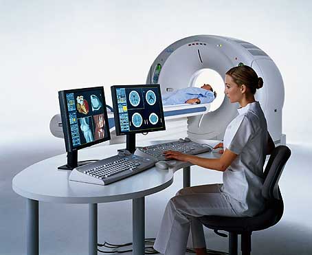
According to the method of obtaining information, angiography of cerebral vessels can be traditional X-ray, computer (based on X-ray images) and magnetic resonance imaging.
How is the examination going?
Important conditions
- Aseptic conditions for the procedure,
- The presence of a team of doctors: radiologist, anesthesiologist, cardioreanimatologist.
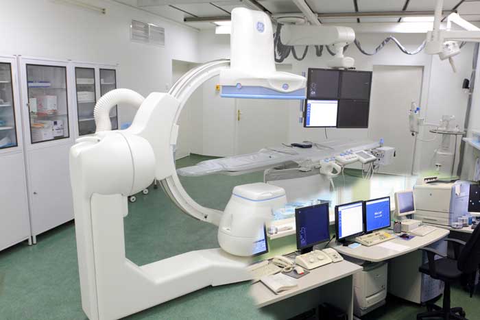
The very process of examining a patient takes about half an hour or an hour. The procedure is considered invasive, as a puncture is performed to access the artery, where a special catheter is inserted. Therefore, angiography of the brain is often combined with other interventions in the body, which occur through access through large blood vessels, for example, with the removal of an aneurysm.
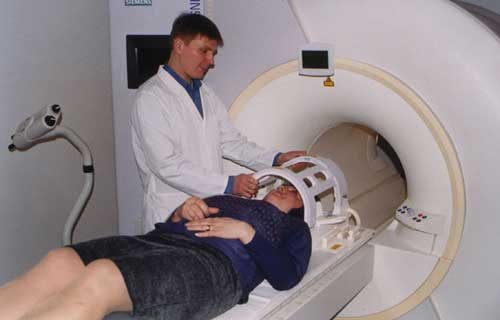
To avoid complications associated with the penetration of infection through the catheterization site, the skin is treated with an antiseptic solution. Next, local anesthesia is performed. Puncture (puncture) of the vessel is carried out with a special needle. A flexible catheter is inserted through this site to deliver contrast. As a rule, the puncture is done in places through which it is easy to "get" to the necessary vessels.
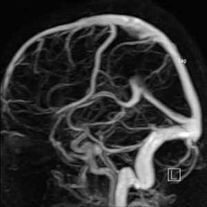
A contrast agent is injected into the blood through a special catheter. After contrasting, a series of X-ray images of the cerebral vessels is performed.
These images show different phases of blood circulation: capillary, arterial and venous. Modern medical equipment allows you to make layer-by-layer images for the formation of a three-dimensional image in the future using special computer programs.
When the exposure is completed, the patient's catheter is removed and the bleeding stops. Further, the information received is decrypted. The vascular surgeon and the radiologist are involved in decoding and setting or clarifying the diagnosis.
After the angiography procedure, the patient should remain under the supervision of medical professionals for some time.
Do I need special preparation for the procedure
Preparation for the procedure is very important for the quality of the procedure. Adult patients should be familiar with the process, the objectives of the study and its possible consequences. Only then can they make an informed decision, which is drawn up in writing. In the case when cerebral angiography is needed for a minor patient, all decisions are made by the parents.
Preparation stages
- Patient informing and written consent,
- Sedation on the eve of the procedure to relieve anxiety and tension,
- Ensuring that the study is carried out on an empty stomach (you need to refuse dinner the day before and breakfast on the day of the procedure).
In the presence of allergic reactions to contrast agents, but if it is necessary to carry out this type of examination, antiallergic agents can be used. If the patient is overly agitated, sedatives may be prescribed a second time, on the day of the procedure.
Advantages of the procedure
- The ability to create a three-dimensional image,
- Vascular imaging, which allows the doctor to detect blood clots, hematomas, aneurysms,
- The possibility of an individual approach to the patient, which means an accurate diagnosis.
The modes and programs of this type of examination can be very wide, so you can always choose the best option in each specific case. The absence of inaccuracies in the diagnosis allows us to timely and qualitatively identify problem areas in the vessels, to understand what is the cause of diseases or disorders in the work of the cerebral vessels. Adequate diagnosis is the main condition for correct treatment.
