Unfortunately, pathologies associated with disruption of the arteries of the circle of Willis are not uncommon in our time. These arteries are responsible for the blood supply to the brain. A change in the functional state of any of them can lead to serious disruptions in the functioning of brain structures, due to improper or inadequate blood supply. Brachycephalic arteries (BCA) include:
- general sleepy (2), which are further divided into external and internal;
- vertebrates (2);
- subclavian (2).
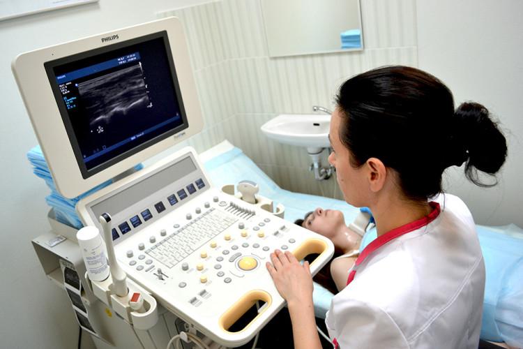
At the base of the brain, the brachiocephalic arteries form a circle of Willis so that blood can be evenly distributed throughout all brain structures. To date, the most informative, non-invasive, safe and therefore demanded method of ultrasound diagnostics of the brachiocephalic arteries of the brain is duplex ultrasound scanning (USDS). Duplex scanning makes it possible to assess not only the functional state, but also the architectonics of the artery of the circle of Willis. Research can be carried out at any required frequency. The applicability of the method does not depend on the patient's age.
Brachycephalic arteries are best viewed by duplex scanningDuplex scanning as a method for examining arteries
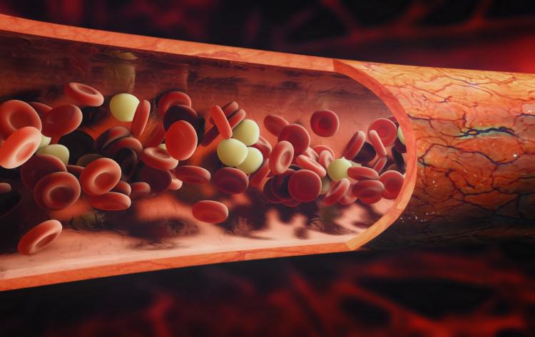
DS, as a method, combines the capabilities of the B-mode (visual interpretation of the physical state of the vessels and adjacent tissues) and doppleroscopy (the study of the qualitative and quantitative characteristics of blood flow). The result can be obtained in the Doppler spectrum, which can be complemented by color mapping.
Duplex allows you to determine any anomaly of the vascular bed (occlusion, stenosis, pathological tortuosity), as well as to establish the blood flow velocity, its change and the cause that affects the blood flow and its velocity (thrombus, embolus, atherosclerotic plaques). Modern equipment allows using DS to determine the state of blood flow in most of the existing vessels of the human body - from the main arteries to small subcutaneous vessels. The larger the vessel, the higher the reliability of the data on changes in its walls.
The results of the study of the great vessels are distinguished by a high degree of reliability. Duplex scanning allows you to establish both the oncopathology of the vascular wall and determine the type of violation of the intraluminal patency (thrombus, plaque).
Pros and cons of duplex scanning
The main advantages of DS as a method is the possibility of early diagnosis of vascular pathology at the stage of the absence of a clear clinical picture of the disease. The specificity of the data obtained makes it possible to assess the functional state of the vascular bed, with the determination of the degree of damage to the vascular wall. The doctor sees the results on the monitor in real time. If the patient's condition changes, for example, when he turns his head or tilts his neck, the doctor can find out the cause of this change immediately. It is also easy to track the effectiveness of the prescribed therapeutic procedures.
The analysis of duplex scanning data, as the analysis of projective techniques in psychology, depends on the qualifications, experience and intuitive intuition of the doctor-operator of the ultrasound machine. The subjective nature of the interpretation of the received ultrasound data is the main disadvantage of DS as a method. In addition to the doctor's experience, the accuracy of the ultrasound apparatus itself and the anatomical features of the structure of the organs and systems of the subject are of great importance.
 Duplex scanning is one of the most informative examination methods, but it requires a highly qualified doctor. Results are based on correct interpretation, intuitive flair and extensive specialist experience
 Indications for the appointment of duplex scanning
Due to its safety, ultrasound diagnostics of BCA can be prescribed both for the diagnosis of clinically expressed pathology, and as a preventive diagnosis. Duplex is assigned in the following cases:
- the presence of symptom complexes indicating the pathology of the arteries of the circle of Willis;
- pre- and postoperative studies;
- planned study of intra- and extracranial vessels.
In the event of the appearance of certain symptoms, a duplex scan may be prescribed to clarify the diagnosis. These symptoms include:
- vestibulopathy, unsteadiness when walking;
- pain and throbbing in the head and neck area;
- hearing impairment (noise and congestion in the ears) and vision (flashing flies, loss of visual field);
- sleep disorders (insomnia, hypersomnia);
- weakness, lethargy, emotional disorders;
- cognitive impairment;
- drops in blood pressure (hypo-, hypertension);
- fainting;
- pain when changing the position of the head, tilting the neck.
In addition to the above, a DS examination can be recommended if there are risk factors or known vascular pathologies. After undergoing operations that affected the cardiovascular system or during planned surgery.
These indications are:
- systemic vascular pathologies;
- the presence of a tumor process in the neck;
- hematological pathologies;
- surgical interventions on the vessels in the head and neck area;
- planned operations on the myocardium and brain structures;
- post-stroke conditions and TEA;
- compression of the arteries in the neck;
- dystonia;
- the presence of risk factors (diabetes, a sedentary lifestyle, emotionally and psychologically intense work, a genetic predisposition to vascular pathologies);
- addiction to alcohol and tobacco smoking;
- age range from 40 to 80 years old (and older);
- osteochondrosis / hernia / injury of the cervical spine;
- arrhythmias;
- insulin resistance and antiphospholipid syndrome;
- the presence of atherosclerotic signs.
 Vascular dystonia is a good reason to prescribe a duplex scan
 Preparing and conducting a scan
Duplex scanning does not require special training, diet, or any medications. If the patient is taking medications on an ongoing basis (hypertensive, antiarrhythmic drugs, etc.) or has had injuries in the neck, the doctor should be warned about this.
As a rule, the procedure is performed while lying down. A special pillow or roller can be placed under the patient's neck. For better contact of the sensor with the skin surface, a special hypoallergenic gel is used. The DS procedure takes approximately 40 minutes.
It is customary to begin the examination by examining the carotid artery. The transducer is positioned directly over the artery, then at an angle. It is visible up to the entrance to the cranium. The condition of the artery wall, blood flow parameters, the presence of foreign inclusions (blood clots, plaques) or formations (tumors), etc. are assessed.
Arteries are viewed not only in the longitudinal direction, but also in the transverse direction to improve visualization of stenoses, occlusions, blood clots, traumatic wall lesions, etc. Sometimes arteries can be abnormally located. This situation can affect the picture of the study and make the results insufficiently reliable and correct. This is an exceptional situation; in general, the method is very informative. The patient receives the result within a few minutes.
Arterial changes visualized by duplex scanning
With the duplex method of research, due to the possibility of visual display of the studied vessel, both the presence of thrombi, atherosclerotic formations, and their size, number and localization are revealed.
Also, echo signs of damage to the vascular walls, developmental anomalies (tortuosity, the presence of kinks, loops, elongated or shortened sections, underdevelopment) are clearly recognized, aneurysms, stenoses and vascular occlusions are diagnosed. The size of the vessel (its diameter), the state of the vascular wall (mobility, elasticity, thickness and uniformity of the surface), as well as changes in the lumen of the artery (narrowing, expansion) are determined with a sufficient degree of accuracy. In addition, the blood flow, its velocity characteristics and the direction of blood flow are recorded. Some indicators are presented in the table:
| Artery | General sleepy | Internal sleepy | External sleepy | Vertebrate |
| Diameter, mm | 4,2 - 6,9 | 3,0 - 6,3 | 3,0 - 6,0 | 2,0 - 4,4 |
| V systole, cm / sec | 50 - 104 | 32 - 100 | 37 - 105 | 20 - 61 |
| V diastole., Cm / sec | 9,0 - 36 | 9,0 - 35 | 6,0 - 27 | 6,0 - 27 |
| Resistance index | 0,6 - 0,8 | 0,5 - 0,8 | 0,6 - 0,9 | 0,6 - 0,8 |
Diseases Detected by Duplex Scanning
The DS technique makes it possible to determine the presence of both primary vascular lesions and secondary ones caused by changes associated with the pathology of human internal organs and systems. The main changes detected are:
- mechanical damage to the vascular wall (the presence of injuries, extravasal syndrome, deformities);
- atherosclerosis and thrombosis;
- discirculatory phenomena;
- aneurysms, arteriovenous shunts;
- vascular abnormalities;
- angiopathic disorders of any etiology.
 Diagnostics allows to identify primary and secondary vascular disorders, one of them is aortic aneurysm
 Survey results
The results of the examination are ready immediately after the end of the procedure, the analysis of the data and preparation of the study protocol is carried out by the doctor of the functional diagnostics office who performed the procedure. The effectiveness of the method directly depends on his qualifications. The decoding of the data is a description of the arteries (blood flow, wall conditions, presence / absence of pathological changes), indicating the norm. According to the data obtained, the attending physician can clarify the diagnosis, prescribe treatment or check the effectiveness of the already applied complex measures for the treatment of one or another vascular pathology.
The place of duplex scanning among other ultrasound methods
Ultrasound is a complex of methods (ultrasound, CDC, duplex and triplex scanning) that study the arteries and veins of the human body. Doppler ultrasound (Doppler) allows you to assess only the qualitative and quantitative characteristics of blood flow, to compare them with the norm, but does not allow you to indicate the reasons for the change, if any.
In this sense, duplex scanning is a much more advanced technique, i.e. the vessel is clearly visible on the monitor, and it is possible to determine the cause of the change in the blood flow rate and the patient's poor health with a sufficient degree of reliability. When using color mapping or triplex scanning, the quality of diagnostics is increased due to a clearer color display of the received data.
Duplex scanning of brachiocephalic arteries is one of the most modern, progressive and correct methods of examination to assess their condition. This method allows you to get a real-time visual and audio picture of the BCA state, assess the existing pathology or the danger of its development, and take timely steps towards the prevention and treatment of severe cerebrovascular accidents.
