Navigation
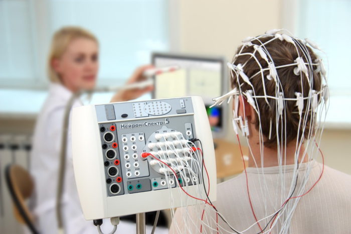
The encephalography of the brain is a method of diagnostic study of the organ, which allows you to estimate its electrical activity. A simple and painless procedure is needed to identify the enhanced convulsive readiness of the crust of the important department of the CNS. The encephalogram of the head, as they also call the EEG brain, is able to: confirm or refute the epilepsy, structural and exchange damage to the organ, to identify the cause of sleep disorders, assess the condition of tissues after a stroke. What is EEG in practice - a schematic recording of neurons of various brain departments with the help of electrodes that are put on the human head.
Indications and contraindications
EEG - The method of diagnosing violations is considered informative and safe, but it is carried out only for the appointment of a doctor in case of indications.
Even in suspected of the same disease, various patients have a decision on the feasibility of the approach is made by a specialist. For example, EEG in migraine in some cases helps to distinguish the disease from epilepsy, and in others - only provokes a new attack.
Indications for EEG
The need to pass the survey may be needed if the development of the pathological process is suspected, to assess the treatment carried out, to establish the optimal method of therapy. Representatives of a number of professions are obliged to attend sessions in order to verify the performance of the brain and their compliance with standards.
Indications for EEG in adults:
- confirmation of epilepsy, degenerative, vascular or inflammatory lesions of the organ;
- identifying the place of finding tumors, cyst, damage to tissue of traumatic type;
- the need to distinguish violations of vision or hearing from their simulation;
- evaluation of the work of the human brain, which is in a coma;
- identifying the causes of Lunatism, sleep disorders, confusion, dizziness, blood pressure jumps;
- setting the quality of vessels and fabric functionality.
In childhood, examination is carried out according to the same testimony. Another one is added to them - estimate the functional state of the CNS by studying the degree of ripening at a particular stage. In some cases, the technique allows you to identify the reasons for stuttering and speech disorders in the kid.
Contraindications

EEG is contraindicated in the period of colds.
From EEG will have to be closed to people in the period of ORZ, ORVI, with nasal labeling or cough. The session is not conducted with various impairment of psyche. If the study delivers too clear discomfort to a person and leads to hysterics due to inexplicable fears, it is better to try other diagnostic methods. In physiological plane, there are no contraindications to the approach. He does not cause unpleasant sensations and does not have any impact on the state of a person.
What shows the encephalogram of the brain
After removing the electrocardiogram of the head - another one of the options for the session name - the doctor receives a list of data. Each indicators have their own limits of norms and nuances of interpretation. They testify not only about the activity of the brain, but also capable of pointing to areas affected by the disease. Despite the fact that the main work on assessing the functioning of one of the sections of the central nervous system lies on the computer, the machine is not able to independently identify the problem and make a diagnosis.
Only an experienced specialist can understand that shows the EEG brain. Attempts to independently decipher the results of the survey can lead to neurosis or psychosis due to the occurrence of suspicion about the diagnosis. In some cases, the patients are additionally appointed by reoeczephalography (REG) of the body, which allows you to study in detail the peculiarities of its vessels.
BEA brain bioelectric activity
The indicator fixes the waves resulting from the transmission of pulses between the cells. According to the standard, they must be synchronous, sequential, without failures and amplification. Situations with the neurotic, unusually accelerated frequency of oscillations or amplitude data above adopted norms are characteristic of a number of physiological conditions and diseases.
Usually, pathological changes are observed at:
- injuries and concussions;
- inflammatory lesions of fabrics - encephalitis, meningitis and others;
- age-related changes in the brain - Parkinson's disease, Alzheimer;
- narrowing of the vacation lumen;
- irradiation and poisoning;
- the presence of epilepsy or migraine;
- structural changes in the hypothalamus and pituitary gland.
The change in the indicators of the bioelectric activity of the brain does not always turn out to be the result of the disease. The slower work of neural cells with a decrease in BEA is characteristic of depression. For this reason, electroencephalography should be carried out according to the testimony. When making a diagnosis, the results of additional research and data of the clinical picture are necessarily taken into account.
Rhythms electroencephalogram
Deciphering the electroencephalogram of the brain, special attention specialist draws to biorhythms. At the same time, it is taken into account in what situation and at what period of time the data was shot. The functionality of the CNS department, the mood of a person, the reception of drugs, the degree of activity of the body (period of sleep or wakefulness) is affected on rhythms.
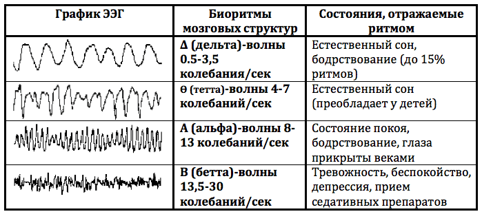
The majority of the alpha and beta rhythms, theta and delta indicators are important. Additional data is usually taken into account with more complex tests that are conducted against the background of mental or intellectual load.
Alpha Rhythm
In an adult, his indicators must be within 8-13 Hz with amplitude of up to 100 μV. Violation of rhythm, frequency instability, detection of paroxysms or arches, asymmetry between hemispheres indicate pathology. The cause of the phenomenon is often tumor, cyst, stroke, formation of a scar on fabric, brain injury acquired by dementia. Data failures in children can be registered with the delay in psychomotor development, neurosis, the availability of psychopathology.
Beta rhythm
In the frontal stakes are expressed more than in the rest of the departments. It is characterized by symmetric amplitude in hemispheres within 3-5 μV. The problems indicate the presence of paroxysms, excess of the amplitude indicator, asymmetry, the change in the rhythm schedule. The deviation from the norm may indicate such diseases of the brain as encephalitis, neurosis, concussion and the delay in the child's development.
Teta Rhythm and Delta Rhythm
Slow Thata and Delta Waves in people over 21 years old are not registered during the waking period. The first is fixed against the background of falling asleep, superficial sleep and during dreams. The second is characteristic of a deep disconnection. Up to the specified age, "malfunctions" in this area can be a variant of the norm, but for this certain factors must come together. Violated theta rhythm and delta rhythm - signs of the presence of tumor, neurosis, psychopathy, acquired dementia, asthenic syndrome, twilight state.
How to prepare for the encephalogram of the brain
Even adult people have concerns, but they are in vain. The process of influencing the contents of the cranial box by means of electrodes is absolutely safe and not accompanied by any sensations. Sessions can be carried out at least every day an unlimited number of times. Electrotomography does not imply complex and long-term preparations. In some cases, you can do without them.
Preparation for EEG for an adult man looks like this:
- 2 days before the session, it is necessary to refuse to take alcohol and stimulating drinks (coffee, cocoa, power engineering), chocolate consumption;
- it is necessary to take care of the purity of the hair, apply any means for laying is prohibited;
- 2 hours before the procedure, it is impossible to eat tightly and smoke;
- before the event, metal items (jewelry, piercing, studs) should be removed from the head.
Ideally, how to prepare for EEG should tell a doctor who has appointed the procedure. He must report, from receiving what drugs and for what period before the study should be refused. If this is not possible, the appropriate mark is made on the destination for a specialist who will decipher the results.
Our readers write
Subject: Get rid of headaches!
From whom: Irina N. (34 years) ( [Email Protected])
To: Administration Website

Hello! My name is
Irina, I want to express my gratitude to you and your site.
Finally, I was able to overcome the headache. We lead an active lifestyle, I live and rejoice every moment!
And here is my story
I do not know a single person who would not have bothered a periodic headache. I'm not an exception. All this has been written off on a sedentary lifestyle, non-normalized schedule, poor nutrition and smoking.
I usually have such a state on the change of weather, before the rain, and the wind turns me into a vegetable.
Fought with this with the help of painkillers. He appealed to the hospital, but I was told that most people and adults, and children and old people suffer like this. What is the most paradox, I have no pressure problems. It was worth reversed and everything: the head begins to hurt.
EEG results
After the completion of the procedure, the worker who removed the indicators receives data on an electronic or paper carrier. It compares all the results of the electroencephalogram to decipher the EEG. Then conclude and passes it into the patient's medical card. Today, many clinics provide customers with the opportunity to receive an EEG entry in electronic form to show her doctor or specialist in this area.
The final opinion of the EEG consists of three parts:
- characteristic of the activity of waves and their typical affiliation;
- conclusion according to the description and its decoding;
- detection of compliance of the results of the study of symptoms and an intended diagnosis.
When deciphering the EEG indicators of the brain, the age and features of the patient's condition, the clinical picture, the list of the therapeutic manipulations carried out are taken into account. The results of the study are extremely specific and almost not informative for a third-party person.
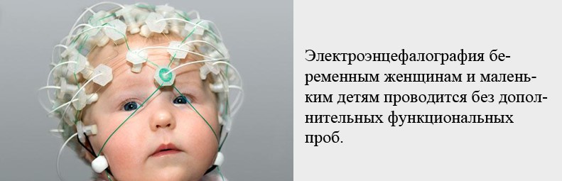
Features of the EEG in children
Removing these encephalograms of the brain in a child can deliver a lot of trouble to parents and specialists. The first must be remembered that the session is absolutely safe. The power of the current during the procedure is so insignificant that the little patient will not feel anything. The nervousness of the parents can adversely affect the toddlers and distort the results, so they must be controlled.
Preparation of the child to the procedure
EEG brain in children under the age of year is always held during sleep. At that moment, Kroch is on the hands of a father or mother. It is enough to wait for the desired moment and gently hold the session steps. Usually no more than 15-20 minutes goes to manipulation. Children under 3 years are also usually experienced the procedure during rest. The exception is calm and contact kids, but this is done only if it is impossible to wait for their sleeping.
Features of the preparation of children to the EEG brain depend on the age and condition of the patient:
- up to the year - it is necessary to take care of the purity of the head of the child and feed it immediately before visiting the specialist so that he fell asleep to the start of the session;
- up to three years - it is also necessary to wash the head of the baby and bring it into a diagnostic institution by the time when he has a dream in the plan;
- after three years - except for the head wash, you need to think about the distracting factor for crumbs. Removing the correct indicators of the work of the brain in a child is possible only if it is calm. The desired effect can be achieved by preparing the patient by creating an appropriate mood or favorite toy.
The procedure itself differs from an adult version of only the number of electrodes - they are used not more than 12 pieces. During the session, it is necessary to ensure that the head of the kid is in the same position, did not lean forward. In the case of older children, additional samples may be needed. The baby will ask to close and open their eyes, squeeze the fingers in the fist, listen to special sounds, inflate the balloon, look at the flashing light bulb.
Where to make the enencephalogram of the brain
Contrary to popular belief, for the passage of EEG, it is not necessary to apply to paid medical clinics. Private commercial institutions of neurological specialization provide such services to adults and children, but the cost of their services is often to exceed the average price of the price.
Where can I do EEG, depends on the age of the patient:
- adults can contact a neurological clinic, a city or district hospital by profile, a psychiatric dispensary;
- baby and teenagers up to 14 years old must be examined in specialized children's hospitals under the control of pediatricians.
At the appeal to budget organizations, only one drawback is usually a queue for such procedures. Sometimes you have to wait a few days and even weeks to get to the specialist. Sometimes, diagnostic cabinets are limited to the analysis, and the decoding and conclusion is sent to other specialists, which additionally takes time.
Cost of procedure
The price for holding the electroencephalogram of the brain depends on the type of medical institution, city, option and duration of the procedure. In the regions, the cost of the service during the wake period begins with 800-1000 rubles. In Moscow, prices for sessions start from 1,500 rubles. Monitoring during sleep will cost 8000-12,000 rubles in Moscow and 10-20% less in regional centers. These amounts include the costs of the work of medical personnel and the operation of equipment. Discounts on such services are dubious, do not trust such offers.
Even with the emergence of such methods for diagnosing diseases of the brain, like CT and MRI, the value of EEG has not decreased. A simple and safe survey sometimes helps to identify pathology where modern techniques are powerless. If the doctor recommends passing the procedure, do not refuse. Already in the process of session, an experienced specialist can detect degenerative changes in the tissues of the body. This will allow you to choose a suitable treatment and timely proceed to the implementation of the plan.
We draw conclusions
Strokes - the reason for almost 70% of all deaths in the world. Seven out of ten people die due to the blockage of the arteries of the brain. And the first and most important sign of the blockage of the vessels is a headache!
The blockage of the vessels is poured into the disease under the well-known name "Hypertension", here are only some of its symptoms:
- Headache
- Hardness participation
- Black dots before your eyes (flies)
- Apathy, irritability, drowsiness
- Fuzzy vision
- Sweating
- Chronic fatigue
- Entrance females
- Numbness and chills of fingers
- Pressure jumps
Attention! If at least 2 symptoms noticed - this is a serious reason to think about it!
The only means that has given a significant result ...
