Acoustic signals propagating in an external environment are perceived by the human brain as a result of a number of transformations produced at various levels of the auditory system.
The auditory analyzer is a single integrally functioning system consisting of three departments: a) peripheral, or receptor; b) medium, or conductive; c) central, or cortical.
It is characteristic that the input acoustic signal is first decomposed on some spectral-temporal components, which are then encoded in the form of multichannel pulse sequences. And such a registration obtained at the level of auditory nerve fibers is then used in the further decoding of the signals with the highest centers of the auditory system in the process of perception.
The peripheral department of the analyzer consists of special nerve cells that perceive a certain type of irritation. These cells are a receptor that is a special transformer (converter) of external irritation energy into the energy of nervous excitation. At the level of the peripheral audit system, the following functions are carried out:
1. These conditions for receiving the signal are created, under which maximum sensitivity is ensured with a valid signal-noise ratio.
2. The spectral-temporal multichannel decomposition of the signals to the components is carried out.
3. There is a transformation of a multichannel analog description of the signals into the pulse activity of the hearing nerve fibers.
The hearing body has a complex structure and performs the functions of the sound analyzer. Figure 2 shows schematically a human hearing body, which is divided into three parts - external, average, inner ear (snail). In Figure 3, there is a cross-section of human ear.
The conduction department consists of nerve fibers and cells of intermediate nerve centers in the spinal cord and the stem portion of the brain. The function of this department is to carry out nervous excitement from the receptor to the cortical end of the analyzer.
Fig. 2: A - outdoor ear; B - middle ear; B - Interior Ear (Snail)
Fig. 3. Cross cut of man's ear:
1 - ear sink; 2 - external hearing pass; 3 - Drumpand-ka; 4 - snail; 5 - hammer; 6 - anvil; 7 - striking; 8 - hearing tube; 9 - oval window; 10 - Round window; 11, 12, 13 - semicircular channels - respectively horizontal, vertical, rear; 14 - facial nerve; 15 - vestibular nerve; 16 - hearing nerve; 17 - temporal bone
Central, or cortical, department is the highest analyzer department. Here is the analysis and synthesis of irritation coming from the peripheral department of the auditory system.
The auditory system differs the sound and sound visible devices with certain functional purposes.
The sound conducting apparatus conducts sound oscillations to receptor cells and consists of an external and middle ear, the labyrinth windows of the inner ear and its liquid media.
A sounding machine transforms sound energy into nervous excitement and transmits it to the central analyzer department. It includes the hair cells of the ear, the auditory nerve, neural education and hearing centers in the temporal fraction of the brain.
Outdoor Ear
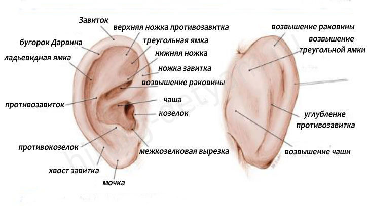
The outdoor ear (see Fig. 3 and 4) consists of a skin-cartilaginous shell and an external auditory passage ending with a drumpatch. The ear sink has a funnel form, which goes into the tube - the auditory pass; Equipped with six internal rudimentary muscles and three external. The front of the sink has a peculiar cartilage formation (goat) in the form of a protrusion limiting the outer hearing pass; It is adjacent to the cottage process, forming an exhaust fold. The upper part of the auricle forms curls; The lower part of it is an urchination - unlike other departments in its anatomical structure, it does not have cartilage, but has a fatty tissue.
Own sink plays the role of a collector of sound waves and participates in the localization of sounds. Acoustic dimensions showed that the pressure of the sound wave at the entrance to the outer hearing pass is almost twice the pressure in the free sound field.
Fig. 4.: Outdoor Ear:1 - curl; 2 - triangular fossa; 3 - anti-guns (antihelix); 4-sign of the opposite; 5 - ear sink; 6 - Antiragus; 7 - Ear Middle; 8 - kids; 9 - Foot Curl
The elevations and grooves of the surface of the ears are used in the rumor to fix the ear insert. In children, it is very soft, low-elastic, its deepening seems more embossed, and curls and the mole are expressed less clearly. The auditory passage in which the ear sink is becoming a winding canal in an adult is 22-27 mm long with a lumen of 5-8 mm. In children, it is significantly shorter, has a slick form of interfluent-cartilage education. As the child grows, the auditory pass becomes oval, and by 10-12 years, its shape and length approach the same dimensions as in an adult.
The outer part of this channel consists of cartilage, the inner is the bone department. The hearing pass is lined with skin with small hairs, sall and sulfur glands, which produce ear sulfur. Its cartilaginous part is movable, and when pulling off the shell, you can expand the lumen and change it to curvature, which must be taken into account in the manufacture of an auditory side.
The main functions of the outer ear: the localization of the sound source, the gain of high-frequency sounds, conducting sound waves to the eardrum, determination of the offset of the sound source in the vertical plane, the protection of the inner ear and maintain a stable temperature mode.
MIDDLE EAR
The middle ear is located in the thickness of the temporal bone and consists of a number of reporting cavities - the drum cavity, the cells of the mastoid process, the eardrum, the hearing bones, the hearing pipe (see Fig. 5). From the outer auditory pass, the average ear is separated by the eardrum, i.e. The drum cavity is between the eardrum and the ear labyrinth. The front wall is the most narrow, it leads to the hole of the Eustachius pipe, through which the drum cavity is reported to the cavity of the nasopharynx. The lower wall is a thin bone plate, which separates the drum cavity from the large blood vessel - the bulbs of the inner jugular vein. The rear wall of the drum cavity in its upper part has a hole leading to the system of air-capable cells of the presenter process. The upper wall is also a thin bone plate - separates the drum cavity from the medium cranial fox, where the temporal proportion of the brain is located. The inner wall of the drum cavity is simultaneously the outer wall of the ears labyrinth (inner ear) and separates the average ear from the internal one. On the labyrinth wall there is a protrusion (promontory) formed by the main curl of the snail.
Fig. 5. Secondary Ear: 1 - Muscle, pulling the eardrum; 2-shirts; 3 - anvil; 4 - aspiring muscle; 5 -liene nerve; 6 - a footing of the stirrup; 7 -braped membrane
Above the last, the oval window is located, the stupid closed by a plate, the front nerve canal passes from the front and front. Over the channel of the facial nerve is an extended part of the horizontal semicircular channel - ampoule. Rear and the book from the protrusion - a round window, which is closed with a thin elastic membrane, called the secondary eardrum.
In connection with the specified peculiarities of the anatomy of the drum cavity, it turns out to transition to the inflammatory process during the defeat of the middle ear (acute average otitis, the aggravation of chronic otitis):
. Through the upper wall of the cavity - on the brain shells and the brain (meningitis, meningoencephalitis, brain abscess) may occur);
. Through the bottom wall - on large blood vessels (inflammation and thrombosis of large blood vessels; thrombophlebitis, sinustrombosis may occur);
. through the inner wall - on the ear labyrinth (labyrinthitis);
. Through the back wall - on the mastoid process (inflammation of the mastoid process, mastoid).
The inflammatory process can move on the front nerve whose channel passes through the inner back wall of the drum cavity, as a result of which paresis or paralysis of the facial nerve occurs.
The outer wall of the drum cavity is the drummeal (Fig. 6), which is a dense fibrous membrane with a thickness of 0.1 mm, has a conical shape with elliptic circuits and an area of \u200b\u200babout 85 mm2 (of which only 55 mm2 are exposed to sound waves). With age, the shape and dimensions of the eardrum almost do not change. From the outside, it is covered with epidermis, with an inner - mucous membrane. Most of the eardrum consists of radial and circular collagen fibers, providing its tension. The central region resembles a cone with a deepening in the middle.
Fig. 6. Drumping membrane: 1, 2, 3, 4 - quadrants - respectively, revenge, front, rendering, leading; 5 - short process hammer; 6 - light cone; 7 - Handle hammer
The drum membrane is divided into two parts - stretched and relaxed. The first area is located in the center and below. Relaxed part, insignificant in size, is at the top. Due to the cone-shaped form and unequal tension in various sections, the drummeal has a minor own resonance and transmits sound waves of different frequency with almost the same force, without distortion.
The drum cavity is enclosed in the pyramid of the temporal bone and is a sloping space of incorrect shape. Its volume is 1-2 cm3, height 15-16 mm, width 4-6 mm. Mostly, the outer wall of the drum cavity is the drum membrane, the remaining parts are bone tissue, mainly the pyramids of the temporal bone. The inner wall of the drum cavity serves as the outer wall of the inner ear. It has two holes: the snail window (1 -2 mm diameter) and the runout window (3-4 mm diameter). The latter is closed by the foundation of the tears, the snail window is a fibrous membrane. On the inner wall of the drum cavity there is a bulge - a cape, or a promontory, which is formed by the main (basal) snail curl. The bone canal is located on top of it, in which there is a facial nerve, and over it and behind it - an ampoule of a horizontal semicircular channel. The upper wall of the drum cavity borders with the cavity of the skull; The rear is located a hole connecting the drum cavity with pneumatic cells of the mastoid process; In the front wall there is a mouth of an auditory pipe, which connects the drum cavity with the cavity of the nasopharynx.
The conditionally drum cavity is divided into three departments: the upper - abrasing space, or attic; Middle - Mesotimpanum; Nizhny - basement.
The upper part is located above the short process of the hammer, the average (mesotympanum) is located between the short process of the hammer and the lower wall of the outer auditory passage, the lower represents a slight recess, located below the level of attachment of the eardrum.
The drum cavity is lined with a mucous membrane, in which there is a small amount of mucous gland gland. The cavity contains three auditory bones and two miniature muscles - a muscle that pulls the drumpoint, and the muscles of austrian. The first starts from the front wall of the drum cavity, where it is attached to the bone half-chamber, then passing through the drum cavity, turns into a tendon and woven the hammer in the handle. The muscles of stupid originates from the rear wall and ends in the neck and the head of the stirrup.
Three bones of the sound system are located between the eardrum and the inner ear: hammer, anvil and stirrup (Fig. 7). Of these, the outdoor - hammer - woven by the handle in the fibrous layer of the eardrum and is connected to the medium bone - an anvil, which, in turn, is associated with the internal hearing bone - stirring. Hearing bones are interconnected and with a drummeal with small muscles and ligaments, which are covered with a mucous membrane, which is a continuation of the drum mucous membrane.
In the hammer (its length 9 mm), the head, neck, handle, short process diffuse. Anvil (mass 25-27 mg) consists of a body and two processes: short and long. In the stirrup, the head, neck, a foothold of the plate is distinguished. The latter is fixed with a bundle and inserted into the oval window of the aurorant labyrinth (the inner ear). The head of the hammer is connected to the body of anvil by means of a joint with a meniscus, and the long turnover of an anvil is connected to the head of the stupidity.
Along with the specified articulation of hearing bones, the hammer and anvil are attached to the wall of the drum cavity using a ligament. Due to the fact that the handle of the hammer is splitted with the drummeal, and the elder in the oval window is connected to the ears labyrinth, the specified sound system that responds to sound oscillations, transfers the oscillations of the eardrum on the liquid medium of the internal ear (perilimf and endolymph).
Fig. 7. Hearing bones: 1 - hammer; 2 - anvil; 3 - Stuffed
In the cavity of the middle ear there are two muscles involved in the mechanism of sounding. The first muscle, straining eardrum, begins in the cartilaginous department of the Eustachius pipe, passes from the inner wall of the drum cavity to the outer and attached to the top of the hammer handle. This muscle is innervated by a trigeminal nerve. The second muscle (aspiring) is located in the bone canal in the rear wall of the drum cavity and is attached to the cervix. This muscle is innervated by the facial nerve. By the time of the birth of a person, auditory bones reach their full development and do not have the ability to regenerate or restore, therefore their damage or destruction is irreversible.
In addition to the hearing bones and the internal muscles in the drum cavity there is still a sensitive nerve. He passes between the hammer and the anvil and provides taste sensations of the language.
The drum cavity is reported to the cavities of the mastoid process and with the Eustachius pipe, which are also constituted parts of the middle ear. The maternity process is a bone formation, which resembles an incorrect prism, limited by four walls and the base upwards, and the top down. The outer wall of the mining process has a triangular shape, the surface of the tip of the process is buggy, especially in the place where the sternum-clarifying muscle is attached to it. In the thicker of the deputyid proof, the system of interconnected aerial cells is connected, the value of which varies. The largest cell of the mastoid process, which is an air cavity, which is reported to the drum cavity, is called antrum (cave).
With an inflammatory process in the middle ear, the cellular structure of the maternity process is often disturbed or completely disappears. In contrast to the normal pneumatic structure, the mastoid proceedings in such cases acquires sclerotic.
Eustachieva, or auditory, pipe - a channel connecting the drum cavity with the cavity of the nasopharynx. His mouth is located in the front part of the front wall of the drum cavity, and in the nasopharynx, the opening of the Eustachius pipe is located on its side wall at the level of the rear end of the lower nose shell. The length of the Eustachius pipe in an adult is an average of 35-40 mm, and in children it is shorter, wider and is more horizontal, which facilitates the penetration of infection from the nasopharynx in the drum cavity and the possibility of inflammation of the middle ear (acute average otitis). The upper part of the pipe, which is connected to the drum cavity and takes the third part of its length, is formed by bone tissue, and the lower consists of cartilage and connective tissue. The surface of the Eustachius pipe is covered with a fiscal epithelium, through the ciliation of which it is cleaned from dust and various mechanical particles and bacteria, promoting them into the nasophalc. In a calm condition, the connective and cartilage departments of Eustachius pipe are in the falling form, and during swallowing the lumen of the pipe is revealed, and the air passes into the drum cavity, balancing pressure outside and inside it. The disclosure of the Eustachius pipe occurs due to the reduction of two muscles - pulling and lifting soft panels.
The mucous membrane of the drum cavity is innervated by the drum branch of the Language and Tripher Nerves. The drum nervous plexus, as well as the nerve fibers, coming from the plexus of the internal carotid artery, is of great importance in the sensitive innervation of the drum cavity. Motor innervation of the muscles of the drum cavity is carried out by trigeminal and face nerves. Arterial blood supply to the middle ear comes from the branches of the outer and internal carotid arteries.
In adult, the auditory pipe is directed a book that ensures the evacuation of liquids from the middle ear in the nasopharynk. In children, the auditory pipe is much shorter. Its growth occurs due to the development of the cartilage part, while the bone department remains unchanged. The hearing tube performs two main functions: the air pressure on both sides of the eardrum is aligned through it, which is a prerequisite for its optimal vibration, and it provides a drainage function.
Interior Ear
The inner ear, or an ear maze, is a bone-refigble formation in the form of a number of cavities and channels and consists of a bone maze (case) and the interputed labyrinth inside it.
Due to the complexity of the relationship of its structures, the inner ear is called the labyrinth. It is located in the thickness of the rocky part (pyramid) of the temporal bone and consists of a very compact bone tissue. The labyrinth is informed with the cavity of the skull (rear cranial pocket) through the inner hearing passage and the snail water supply, it borders with the drum cavity and separated from it the wall formed by the thread and the protrusion of the main curl of the snail, as well as the oval window, closed by the footpilling plate, and the round window, Tightened secondary membrane.
The ear labyrinth consists of three departments: front - snail, middle-running and rear - semicircular channels.
Fig. 8. A ear labyrinth (by L. V. Neuman): 1 - snail; 2 - anticipation; 3, 4, 5 -Pollar channels - respectively top, outdoor, rear
Figure 8 shows schematically the main components of the ears labyrinth, in Figure 9, a vertical cut of the snail is given. The transverse sections of the inner ear presented in Figures 10 and 11 illustrate the features of the complex structure of this dialing system.
Snail - bone formation having a form of a spiral channel located two and a half curls around the bone column (Fig. 9). Each subsequent curl is smaller than the previous one, so that this channel really resembles the sink sink sink. Channel length - about 22 mm. In ear snail, the lower (main) curls, the middle and the top, in which the bone canal passes (the total length of curls on average 3 cm) is distinguished. The bone column, around which snail curls are groaning, has a spiral comb, speaking into the cacia snail bone canal. From the large edge of the spiral ridge to the opposite wall of the bone snail, the main membrane is stretched, which together with the ridge divides the bone canal on the top (Staircase of the Travel) and the lower departments (drum staircase) (see Fig. 10). These departments are filled with intrarabium liquid (perilimph) and communicate with each other by the small hole located at the top of the snail. The drum staircase borders with a drum cavity, which is separated from the bone snail cavity with a circular window, closed by a secondary membrane. The staircase of the Thread Starts is communicated with the eve of the ears of the labyrinth and is separated from the drum cavity by the oval window, closed by a leading stupid plate.
From the free edge of the spiral ridge, along with the main membrane at an angle of 30 °, a thin elastic membrane, called a raisner membrane (see Figure 10, 11), is departed (see Fig. 10, 11), which divides the staircase in the runway into two parts: the proprietary staircase itself and the snail move.
Fig. 9. Snail (vertical cut)
Fig. 10. Interior ear. Cross cut Snail: 1 - Staircase of the Travel (filled with perilimph); 2 - the median staircase (filled with endolymph); 3 - Reisner Membrane; 4 - bone wall of the sniplog canal; 5 - internal hairs cells; 6 - external hairs cells; 7 - cover (tectorial) membrane; 8 - Basilar membrane; 9 - nerve fibers; 10 - drum staircase; 11 - spiral ganglia cells; 12 - Poles and Tunnel Cortiee Organ
Fig. 11. Transverse cut through a snail curl: 1 - the main membrane; 2 - fibers of the main nerve; 3 - the bone wall of the snail; 4 - auditory (hairs) cells; 5 - support cells; 6 - cover membrane; 7 - Risnerova Membrane; 8 - Pre-expert staircase; 9 - drum staircase; 10 - snail stroke and the Cortiev organ located in it
The latter is an eppection channel of a triangular shape, formed by a raisner membrane (from above), the main membrane (bottom) and the bone wall of the snail labyrinth, outside the epithelium covered. The snail move is filled with liquid - endolymph, which is different from Perilimphs to the chemical composition and physical properties. The labyrinth liquids - the Perelimf, located in the cavities of the Staircase of the Three or the drum staircase, and the endolymph, filling the snail stroke, are not communicated to each other.
The main membrane, being a continuation of a spiral curl, divides the bone snail channel on the runner of the runway and the drum staircase and consists of separate fibers that are in the radial transverse direction from the free edge of the bone spiral ridge to the outer wall of the aurous labyrinth. The number of these fibers reaches 15,000-25,000, and their length is non-etinak and increases in the direction of the snail to its top. The membrane itself has a kind of tape that is the most narrow at the bottom at the base and, gradually expanding, it turns out to be the best at the top, in the area of \u200b\u200bthe snail.
Inside the snipling, on the main membrane, there is a cortis (spiral) organ containing receptor hairs cells, which are the most important peripheral nerve elements of the auditory system. They transform mechanical oscillations into electrical potentials, as a result of which the fibers of the auditory nerve are excited.
Cortium organ from above is covered with a coating membrane, which, during oscillation of intrarabinary liquids, closely comes into contact with the hairs of sensitive cells, which causes the transformation of mechanical oscillations to auditory nerve impulses entering a rumor nerve and conductive nervous paths into the brain. Sensitive hairs of the Cortiene organ are associated with nerve fibers, which are coming from the two-pole cells of the spiral nerve node located in the bone canal at the base of the bone spiral plate. The nerve endings of the fibers, the number of which the average reaches 30,000, make up a snippene branch of the auditory nerve. The latter, together with the vestibular branch, forms a barrel of a hearing nerve, which with the front and intermediate nerves passes through the inner hearing passage in the brain, heading into the bridge corner.
In the central part of the ear maze (run-up) and the rear part of it (three semicircular channels) there is a peripheral receptor of the spatial (vestibular) analyzer, or an equilibrium organ, which is placed in the webbed part of the specified formations filled with endolymph. Theused semicircular channels (top, rear, outdoor), located inside the bone, lie in three mutually perpendicular planes and open on the eve of five holes. The presence of five holes is explained by the fact that three semicircular channels originate from the run-up (forming an empty expansion at the end) and they fall into it another, smooth end. But when the smooth ends of the upper and rear semicircles are connected together on the eve of the smooth ends of the upper and rear semicircles, constituting one general knee.
In ampoules of semicircular channels there are ampullular scallops, sensitive hairless nerve cells of which form the peripheral receptor apparatus of the spatial analyzer. The specified hairs have a large length, and when the endolymph is moved, arising from changing the position of the body in space, their displacement occurs inside the connecting labyrinth, which causes irritation of the vestibular nerve sprigs. On the eve of the nervous receptor formation of the vestibular nerve is the front and rear bags with sensitive nerve cells, covered with a olhed membrane containing calcium salts crystals. The displacement of the membrane due to the motion of the endolymph occurring as a result of the straightforward movement of the body in space, and the contact of it with hairs of sensitive nerve cells cause the stream of nerve pulses entering the vestibular nerve into the cerebral bark.
Rotational motions as a result of a similar mechanism determine the oscillations of the endolymph in the semicircular channel, the plane of which corresponds to the motion plane. As a result, irritation of sensitive hairless nerve cells is irritated in the appropriate semicircular channel, which also applies to the conductive paths of the vestibular system in the cerebral bark.
Nervous fibers, which come from ampullular neuro-sensitive formations and the vestibular receptor apparatus laid in the sheds of the thread, are connected to the vestibular branch of the auditory nerve, according to which the stream of nerve pulses is carried out in the central nervous system. The vestibular irritation of the peripheral receptor link is entered into the bark of the brain, resulting in sensations of body position in space and various motor reflex reactions that contribute to the preservation of equilibrium. In addition, in response to irritation of the vestibular apparatus, the rhythmic movements of the eyeballs in a certain direction (Nistagm) occur in a certain direction.
On the presence, the nature and degree of vestibular irritation and the function of the vestibular apparatus are judged by somatic and vegetative reactions arising from the rotation of the subject with the help of a special chair of the Barani (named Australian otolaryngologist Robert Bararan), creating a position corresponding to the deviation of the body, its fall, accompanied by the feeling Nausea and vomiting.
And the morphologists of this structure are called organelum and equilibrium (Organum Vestibulo-Cochleare). It allocate three departments:
- outdoor ear (external auditory passage, ears with muscles and ligaments);
- the middle ear (drum cavity, maternity appendages, hearing tube)
- (The membrane labyrinth located in the bone maze inside the bone pyramid).
1. The outer ear concentrates sound oscillations and directs them into the outer hearing aid.
2. In the auditory canal conducts sound oscillations for the eardrum
3. The eardrum is a membrane that vibrates under sound action.
4. The hammer is attached to the center of the eardrum with the help of ligaments, and its head is connected to an anvil (5), which, in turn, is attached to the tears (6).
Tiny muscles contribute to the transmission of sound, adjusting the movement of these bones.
7. Evstachiev (or auditory) pipe connects the middle ear with the nasopharynk. When the surrounding air pressure changes, the pressure on both sides of the eardrum aligns through the hearing tube.
The kortyov organ consists of a series of sensitive cells equipped with cells (12), which cover the basilar membrane (13). Sound waves are captured by hairsmaps and are converted into electrical impulses. Next, these electrical impulses are transmitted by a rumor nerve (11) in the head. The auditory nerve consists of thousands of finest nerve fibers. Each fiber begins from a certain section of the snail and transmits a certain sound frequency. Low-frequency sounds are transmitted by fibers emanating from the top of the snail (14), and high-frequency - on fibers associated with its base. Thus, the function of the inner ear is the transformation of mechanical oscillations to electrical, since the brain can only perceive electrical signals.
Outdoor Ear He is a sounding apparatus. The outer hearing pass holds sound oscillations to the eardrum. The eardrum separating the outdoor ear from the drum cavity, or the middle ear, is a thin (0.1 mm) partition, having a shape directed inside the funnel. The membrane fluctuates in the action of sound oscillations that came to it through the outer hearing pass.
Sound oscillations are travded with ear sinks (animals can turn to the sound source) and are transmitted according to the outer auditory passage to the eardrum, which separates the outdoor ear from the average. Calming the sound and the whole process of hearing with two ears - the so-called binaural hearing - it matters to determine the direction of the sound. Sound oscillations running on the side reach the nearest ear for several ten-thousand fractions of a second (0.0006 c) earlier than to another. This insignificant difference in the time of arrival of sound to both ears is enough to determine his direction.
Middle ear It is a sound hardware. It is an air cavity, which through an auditory (Eustachiev) pipe is connected to the cavity of the nasopharynx. Outbursions from the eardrum through the middle ear transmitted with each other 3 auditory bones - hammer, anvil and stirred, and the last vibration of a liquid in the inner ear transmits these fluctuations in the inner ear, - perilimph.
Due to the peculiarities of the geometry of auditory bones, the swords are transmitted by oscillations of the eardrum of reduced amplitude, but increased force. In addition, the surface ispidden is 22 times less than the eardrum, which increases its pressure on the membrane of the oval window in the same time. As a result, even weak sound waves acting on the eardrum are capable of overcome the resistance of the membrane of the oval window of the runway and lead to fluctuations in the liquid in the snail.
With strong sounds, special muscles reduce the mobility of the eardrum and auditory bones, adapting the hearing apparatus to such changes in the stimulus and protecting the inner ear of destruction.
Due to the combination of the nasopharynk air cavity hearing tube, the possibility of equalizing the pressure on both sides of the eardrum, which prevents it from a gap with significant changes in pressure in the external environment - when diving under water, rise to height, shots, etc. This ear borofunction .
In the middle ear there are two muscles: a straining eardrum and dying. The first of them, shrinking, enhances the tension of the eardrum and thereby limits its oscillations to the amplitude at strong sounds, and the second fixes the swollenly and thereby limits its movement. The reflex reduction of these muscles occurs after 10 ms after the start of a strong sound and depends on its amplitude. This inner ear is automatically protected from overloads. With instantaneous strong irritations (blows, explosions, etc.), this protective mechanism does not have time to work, which can lead to hearing impairments (for example, explosives and artilleryrs).
Interior Ear He is a sound permissive device. It is located in the pyramid of the temporal bone and contains a snail, which in a person forms 2.5 spiral turns. The ulital channel is divided into two partitions of the main membrane and the vestibular membrane for 3 narrow moves: the top (vestibular staircase), the middle (membered channel) and the lower (drum staircase). On the top of the snail there is a hole connecting the upper and lower channels into a single, from the oval window to the top of the snail and then to the round window. The cavity is filled with a liquid - peri-lymona, and the cavity of the mid-connecting channel is filled with a liquid of another composition - endolymph. In the middle of the channel, there is a sound-visible apparatus-cortiyev organ in which there are mechanoreceptors of sound oscillations - hairs cells.
The main way to deliver sounds to the ear is air. The sound of the silent drum blade, and then through the chain of hearing bones of oscillations are transmitted to the oval window. At the same time, the fluctuations in the air of the drum cavity, which are transmitted to the membrane of the circular window.
Another way to deliver sounds to snail is tissue or bone conductivity . In this case, the sound directly acts on the surface of the skull, causing its oscillations. Bone Sound Transmission Path It is gaining great importance if the vibrating item (for example, the pedalone's leg) is in contact with the skull, as well as for the diseases of the middle ear system, when the transmission of sounds violates through a chain of auditory bones. In addition to the airway, there is a tissue wave, or bone, path.
Under the influence of aerial sound oscillations, as well as in contact with the vibrators (for example, bone telephone or bone tune), with the cover of the bone of the skull come to oscillation (the bone maze begins to fluctuate). Based on the latest data (bekesy - bekesy, etc.), it can be assumed that the sounds spreading through the bones of the skull, only in the event excite the Cortis organ if they, similarly to air waves, cause the definite portion of the main membrane.
The ability of the bones of the skull to spend the sound explains why his person himself, recorded on the tape recorder, seems to be a record when playing a record, while others easily recognize. The fact is that the tape recorder reproduces your voice is not completely. Usually, talking, you hear not only those sounds that your interlocutors are heard (that is, those sounds that are perceived through air-fluid conductivity), but also those low-frequency sounds whose conductor is the bones of your skull. However, listening to the tape recorder of your own voice, you only hear what could be recorded - sounds, the conductor of which is air.
Binaural hearing . Man and animals have spatial hearing, i.e. the ability to determine the position of the sound source in space. This property is based on the presence of binaural hearing, or hearing with two ears. It is important for it and the presence of two symmetric half at all levels. The severity of binaural hearing in humans is very high: the position of the sound source is determined with an accuracy of 1 angular degree. The basis of this is the ability of the neurons of the auditory system to evaluate the interaural (intertpete) differences in the time of sound coming on the right and left ear and the intensity of the sound on each ear. If the sound source is located aside from the middle line of the head, the sound wave comes to one ear somewhat earlier and has a greater force than on the other ear. Evaluation of the remoteness of the sound source from the body is associated with the weakening of the sound and the change in its timbre.
With a separate stimulation of the right and left ear through the headphones, the delay between the sounds is already in 11 μs or the difference in the intensity of two sounds of 1 dB lead to the seeming shift of the localization of the sound source from the midline towards an earlier or stronger sound. In the auditory centers there is with acute setting to a certain range of interaural differences in time and intensity. Also found cells that react only to a certain direction of the sound of the sound source in space.
Rumor is one of the important senses. It is with the help of it we perceive the slightest changes in the surrounding world, we hear disturbing signals warning about the danger. Very important for all living organisms, although there are also those that cost without it.
In humans, the auditory analyzer includes the outer, average and from them in the auditory nerve information goes to the brain, where it is processed. The article will dwell on the structure, functions and diseases of the outer ear.
The structure of the outdoor ear
The human ear consists of several departments:
- Outdoor.
- Middle ear.
- Internal.
The outdoor ear includes:
Starting from the most primitive vertebral animals, whose hearing appeared, the structure of the ear gradually became more complicated. This is due to the general increase in the organization of animals. For the first time, the outer ear appears in mammals. In nature there are some types of birds with ear sink, for example, the eared owl.
Auricle
The outdoor human ear begins with ear shell. It almost completely consists of a cartilage tissue with a thickness of about 1 mm. It does not have a cartilage in its structure only it consists of adipose tissue and covered with skin.
The ear outdoor has concave with curls on the edge. It is a small deepening separated from the inner disgrace, from which the cavity of the ear shell is in the side of the auditory passage. At the entrance to the ears there is a kids.
Hearing pass
The next department, which has an outdoor ear, - Hearing passage. It is a 2.5 centimeter tube and a diameter of 0.9 cm. It is based on cartilage, resembling a chute, opening upwards. In the cartilage tissue there are sanitary gaps that border with salivary gland.

Crying is available only in the initial passage department, then it goes into bone tissue. The auditory pass itself is a little curved in the horizontal direction, so when examining the doctor in adults, the ear shell pulls back and up, and in children - back and down.
There are salted and sulfurges, which produce it to remove it contributes to the process of chewing, during which the passage walls are oscillation.
Ends the auditory passage of the eardrum, which blindly closes it.
Eardrum
Connects the outer and medium ear eradicularism. It is a translucent plate with a thickness of only 0.1 mm, its area is about 60 mm 2.

There is a drumpatch relative to the auditory passage slightly obliquely and drawn in the form of a funnel inside the cavity. She has the biggest tension in the center. It is already located
Features of the building outdoor ear in babies
When the baby appears on the light, its hearing body is not yet yet formed, and the structure of the outer ear has a number of distinctive features:
- Own sink soft.
- The ear of the ear and curl is practically not expressed, they are formed only by 4 years.
- In the auditory passage there is no bone part.
- Passage walls are located nearby.
- Located drumpipe horizontally.
- In size, the eardrum does not differ from those in adults, but it is much thicker and covered with mucous membrane.
The child grows, and with it there is a crevice of the organ of hearing. Gradually, it acquires all the features of an adult auditory analyzer.
Functions of outdoor ear
Each hearing analyzer department performs its function. The ear is intended primarily for the following purposes:

Thus, the functions of the outer ear are quite diverse, and the ear sink serves us not only for beauty.
Inflammatory process in the outer ear
Quite often, colds end in the inflammatory process within the ear. This problem in children is particularly relevant, since the hearing tube has short sizes, and the infection quickly from the nasal cavity or throat can penetrate the ear.
All inflammation in the ears can manifest itself in different ways, it all depends on the form of the disease. Distinguish several types:

You can cope at home only with the first two varieties, but the inner otitis requires inpatient treatment.
If you consider outdoor otitis, then it also happens two forms:
- Limited.
- Diffuse.
The first form arises, as a rule, as a result of inflammation of the hair follicle in the ears. In some way it is an ordinary furuncle, but only in the ear.
The diffuse form of the inflammatory process covers the entire passage.
Causes of Otita
The reasons that can provoke an inflammatory process in the outer ear, quite a lot, but among them are often the following:
- Bacterial infection.
- Fungal disease.
- Allergic problems.
- Incorrect gigien of the ears.
- An independent attempt to remove ear plugs.
- Hitting foreign bodies.
- Viral nature, although it happens very rarely.
The cause of pain in the outer ear in healthy people
It is not at all necessary if pain in the ear appears, the diagnosis of "Otitis" is made. Often such pains may occur for other reasons:
- Walking into windy weather without a headdress can provoke pain in the ear. Pressure is put on the ear shell and the bruise is formed, the skin acquires a blue color. This condition passes rather quickly after entering a warm room, treatment is not required.
- Fans of swimming also have a frequent satellite. Because during classes, water falls into the ears and irritates the skin, it can lead to edema or outdoor otitis.
- Excessive sulfur accumulation in the ears can cause not only a feeling of lagging, but also pain.
- The lack of sulfur sulfur with sulfur glands, on the contrary, is accompanied by a feeling of dryness, which can also cause pain.
As a rule, if the otitis does not develop, all unpleasant feelings in the ear take place independently and additional treatment do not require.
Manifestations of outdoor otita
If the doctor diagnoses the defeat of the auditory passage and the ear shell, the diagnosis is the outdoor otitis. Its manifestations may be as follows:
- The pain is of different intensity, from very little to preventing sleeping at night.
- Such a state can last a few days, and then slightly.
- In the ears there is a sense of lagging, itching, noise.
- During the inflammatory process of hearing acuity, it may decrease.
- Since otitis is an inflammatory disease, the body temperature may rise.
- Skin covers near ear can acquire a reddish tint.
- Pressing the ear pain is enhanced.
The inflammation of the outer ear must treat the ENT doctor. After examining the patient and determining the stage and the severity of the disease, drugs are prescribed.
Therapy limited Otita
Treatment of this form of the disease is usually carried out surgically. After the introduction of an anesthetic drug, the furuncle and the removal of the pus is opened. Already after this procedure, the patient's condition is significantly improved.
For a while you will have to take antibacterial drugs in the form of droplets or ointments, for example:
- "Regus".
- Candibiotic.
- "Levomecole."
- "Calestoderm-B".
Usually after the course of antibiotics, everything comes to normal, and the patient fully recovers.
Diffuse Otita therapy
Treatment of such a form of the disease is carried out only conservatively. All drugs appoint a doctor. Usually the course includes a set of measures:
- Reception of antibacterial droplets, for example, "offloxacin", "neomycin".
- Anti-inflammatory drops "Otipaks" or "Otreleks".
- Antihistamine preparations ("citrines", "claritin") help to remove swelling.
- For the removal of pain syndrome, NPB is prescribed, for example, "diclofenak", "Nurofen".
- To increase immunity, the reception of vitamin and mineral complexes is shown.
During treatment it is necessary to remember that any warming procedures are contraindicated, they can be appointed only by a doctor at the stage of recovery. If all the recommendations of the doctor are observed and passed a full course of therapy, then you can be sure that the outdoor ear will be great.
Otitis treatment in children
In kids, physiology is such that the inflammatory process is very quickly reckoned from the nasal cavity in the ear. If it is noticed in time that the child is worried about the eye, then the treatment will be short and simple.

The doctor usually does not appoint antibiotics. All therapy is to receive antipyretic drugs and painkillers. Parents can be recommended not to engage in self-medication, but adhere to the recommendations of the doctor.
Drops that bought on the recommendation of the girlfriends can only harm your child. When the kid is ill, appetite is usually reduced. It is impossible to force it through force, it is better to drink more to drink so that the toxins are removed from the body.
If the child is too often more otitis, there is a reason to talk to the pediatrician about vaccination. In many countries, you already make such a vaccination, it will protect the outdoor ear against inflammatory processes that are caused by bacteria.
Prevention of inflammatory diseases of the outdoor ear
Any inflammation of the outer ear can be prevented. For this, only some simple recommendations must be followed:

If the pain in the ear do not cause great anxiety, it does not mean that we should not go to the doctor. Launched inflammation can turn around much more serious problems. Timely treatment will allow you to quickly cope with otitis of the outdoor ear and will save from suffering.
Ear is a complex organ that performs two functions: a hearing by which we perceive sounds and interpret them, thus communicating with the environment; and maintaining equilibrium of the body.
Auricle - captures and directs the sound waves into the internal auditory canal;
Rear LabyrinthOr semicircular canals - directs traffic to the head and brain to regulate the balance of the body;
Front Labyrinth, or snail - contains sensory cells, which, catching the vibration of sound waves, transform mechanical pulses into the nervous;
Auditory nerve - sends common nerve impulses to the brain;
Bones of middle ear: hammer, anvil, rapidly - get vibrations from auditory waves, strengthen them and transferred to the inner ear;
External auditory passage - catches the sound waves coming from the outside, and directs them to the middle ear;
Eardrum - the membrane, vibrating sound waves from entering it and transmitting vibrations along the fire chain in the middle ear;
Eustachian tube - Canal connecting the drumppe with a throat and allows you to maintain
equilibrium pressure created in the middle ear pressure with the surrounding environment.
The ear is divided into three departments whose functions are different.
; The outer ear consists of the pinna and external auditory canal, its purpose is to capture the sounds;
; Middle ear is located in the temporal bone, inner ear is separated from the movable membrane - the eardrum - and has three joint bones: the hammer, anvil and stirrup involved in the transfer of the cochlea sound;
The inner ear, also called the labyrinth, is formed from two departments that perform various functions: anterior labyrinth, or a snail, where the Cortis is a body, responsibility for rumor, and rear maze, or semicircular channels in which pulses are produced involved in maintaining equilibrium Body (article "Equilibrium and hearing")

The internally ear, or the labyrinth, consists of a very durable bone skeleton, an ear capsule, or a bone maze, inside of which a membrane mechanism with a structure similar to a bone, but consisting of membrane tissue. The inner ear is hollow, but filled with liquid: there is a perilimph between the bone maze and the membrane, while the maze itself is filled with endolymph. The front labyrinth, the bone shape of which is called snail, contains structures that generate auditory impulses. The rear labyrinth involved in the regulation of body equilibrium has a bone skeleton consisting of a cubic part, the run-up and three channels in the form of an arc - semicircular, each of which includes space with a flat plane.
Snail, called so because of its spiral shape, contains a membrane consisting of channels filled with liquid: the central channel of the triangular cross section and a curl containing endolymph, which is located between the exterior staircase and the drum staircase. These two stairs are partially separated, they go to large snail channels covered with thin membranes separating the inner ear from the medium: the drum staircase begins with an oval window, while the runner of the rundder reaches a rounded window. A snail having a triangular shape consists of three faces: the top, which is separated from the ladder of the eve of the membrane of the raisner, the bottom, separated from the drum staircase, the main membrane, and the side, which is attached to the sink and is a vascular groove that produces endolymph. Inside the snail there is a special hearing agency - Cortiev (the sound perception mechanism is described in detail in the article "
There is nothing surprising that the person is considered the most perfect sensual organ of the auditory apparatus. Inside it contains the highest concentration of nerve cells (over 30,000 sensors).
Human apparatus of man
The structure of this apparatus is very complex. People are understood by the mechanism for which the perception of sounds is carried out, but scientists still do not quite aware of the feeling of hearing, the essence of the conversion of signals.
In the structure of the ear allocate such basic parts:
- outdoor;
- average;
- internal.
Each of the above areas is responsible for performing specific work. The outer part is considered a receiver that perceives the sounds from the external environment, the average - an amplifier, an internal transmitter.
The structure of the ear of man
The main components of this part:
- auditory pass;
- own sink.
The ear sink consists of cartilage (he is characterized by elasticity, elasticity). From above it covers skin cover. Downstairs is a glass. This site has no cartilage. It includes fatty tissue, skin. Ear feel quite sensitive organ.
Anatomy
Small elements of the auricle are presented:
- curl;
- kids;
- disabled;
- curl legs;
- antiques.
Kosch is a specific coating that lins the auditory passage. Inside it contains glands, which are considered vital. They allocate the secret protecting against many agents (mechanical, thermal infectious).
The end of the pass is presented in a kind of impasse. This specific barrier (eardrum) is required to separate the outer, middle ear. It begins to hesitate when hitting sound waves about him. After hitting the wave of sound about the wall, the signal is transmitted further, towards the middle part of the ear.
Blood to this site goes on two branches of the arteries. Blood outflow is performed by veins (v. Auricularis Posterior, V. RetromandiBularis). are located in the front, in the back of the ear. They take place of lymphs.
In the photo the structure of the outdoor ear
Functions
We indicate significant functions that are fixed behind the outer part of the ear. It is capable of:
- take sounds;
- transmit sound to the middle of the ear;
- direct the wave of sound to the inner part of the ear.
Possible pathology of the disease, injury
Note the most commonly encountered diseases:
Average
The middle ear plays a huge role when the signal is gained. Strengthening is possible due to the auditory bones.
Structure
We indicate the main components of the middle ear:
- drum cavity;
- hearing (Eustachiev) pipe.
The first component (eardrum) contains within the chain, which includes a small bone. The smallest bones play an important role in the transmission of sound oscillations. The drum membrane consists of 6 walls. Its cavity contains 3 auditory bones:
- hammer. Such a bone is endowed with a rounded head. This is the connection with the handle;
- anvil. It includes a body, processes (2 pcs.) Different lengths. With stirring its compound is performed by means of a minor oval thickening, which is located at the end of a long process;
- stirrup. In its structure, a small head is isolated, carrying the articular surface, anvil, legs (2 pcs.).
Artery go to the drum cavity from a. Carotis Externa, being her branches. The lymphatic vessels are directed to the nodes located on the side wall of the pharynx, as well as to those nodes that are localized behind the ear shell.
The structure of the middle ear

Functions
Bones from the chain are needed for:
- Sound.
- Transmission of oscillations.
The muscles placed in the middle ear region specialize in performing various functions:
- protective. Muscular fibers protect the inner ear from sound irritation;
- toning. Muscular fibers are needed to maintain a chain of hearing bones, tone of the eardrum;
- accommodation. The sound conducting machine adapts to the sounds endowed with various characteristics (power, height).
Pathology and diseases, injuries
Among popular middle ear diseases, we note:
- (perforated, non-perforated,);
- Qatar middle ear.
Acute inflammation can appear in injuries:
- otitis, mastoiditis;
- otitis, mastoiditis;
- , Mastoidite, manifested with wounds of temporal bone.
It happens complicated, uncomplicated. Among specific inflammation, we indicate:
- syphilis;
- tuberculosis;
- exotic diseases.
Anatomy of outdoor, middle, inner ear in our video:
We indicate the significant importance of the vestibular analyzer. It is necessary for regulating the position of the body in space, as well as for the regulation of our movements.
Anatomy
The periphery of the vestibular analyzer is considered a plot of the inner ear. In its composition, we highlight:
- semicircular channels (these parts are placed in 3 planes);
- stavtocytic organs (they are represented by bags: oval, round).
Planes are called: horizontal, frontal, sagittal. Two pouches are the threshold. Round pouch is near curl. The oval bag is placed closer to semicircular channels.
Functions
Initially, the analyzer is excited. Due to the vestibular-spinal nervous bonds, somatic reactions occur. Such reactions are needed to redistribute muscle tone, support for body equilibrium in space.
The relationship between vestibular nuclei, cerebellum determines movable reactions, as well as all reactions to coordinate movements that appear when performing sports, labor exercises. To maintain equilibrium, vision, muscular and articular innervation are very important.

