The cornea - the most convex part of the visual apparatus - is responsible for the refractive function and is an integral part of the perception of surrounding information.
Corneal edema is a common condition that occurs for a variety of reasons. With edema, the patient experiences a lot of unpleasant sensations. The surrounding objects seem blurry to him, the focus is blurred. In this article, you will learn about the causes and symptoms and treatments for corneal edema.
Definition of disease
The cornea of the eye is the main component of the refractive system. This convex-concave lens, which is no more than one millimeter thick, has 6 transparent layers.
The cornea not only refracts light, but also protects the eyes from negative external influences, for example, from dust particles floating in the air. With its high sensitivity, the cornea saves the eye from clogging by closing the eyelashes, as well as washing away particles with tear fluid. With the development of the lesion, its properties change, light transmission decreases, photophobia develops, vision decreases significantly, especially in the morning and evening hours.
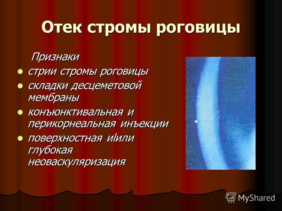
As a result of the pathological process, edema in the cornea can contribute to the destruction of the substance of the stratum corneum, and then to its necrosis.
Causes of occurrence
The causes of corneal edema can be as follows:
Symptoms
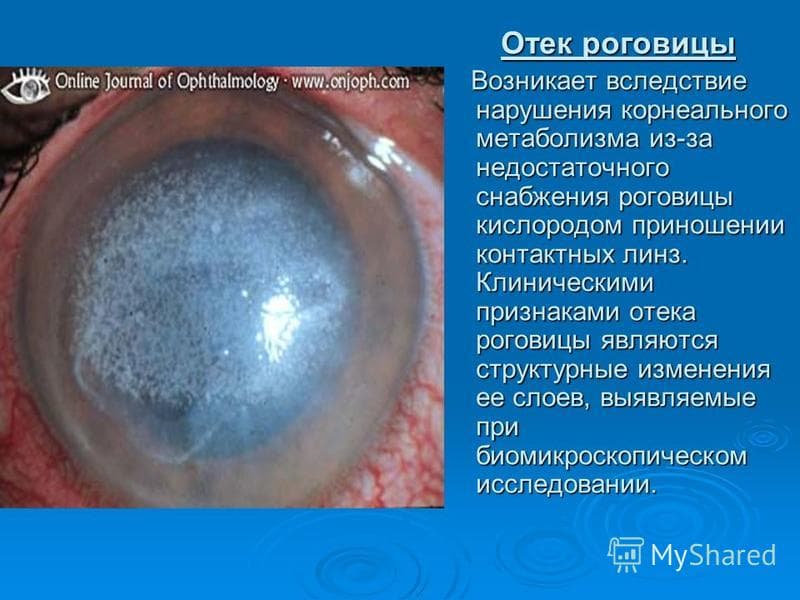
Corneal edema manifests itself in the formation of folds and vertical lines in its layers. Violation of its transparency and thickening leads to the appearance of a veil before the eyes and a decrease in visual acuity, and while wearing contact lenses, a person begins to experience discomfort.
With constant and prolonged edema, the body begins to compensate for the violation by the appearance of a network of blood vessels in the cornea. This changes the structure of the main part of the cornea - the stroma; , penetration of lipids and violation of the transparency of the cornea occurs.
Corneal edema may be accompanied by symptoms such as:
Often, corneal edema is asymptomatic, and this pathology can be detected only when examined by an ophthalmologist.
Possible complications
If the edema is advanced and chronic, vascularization occurs, that is, new blood vessels are formed inside the cornea. This sign can only be noticed during a biomicroscopic examination.
Corneal edema also leads to a significant decrease in vision. If corneal edema becomes chronic, then surgical intervention is often required.
Treatment
Therapy completely depends on the cause that provoked the pathology. Diagnosis is carried out by an ophthalmologist. To exclude infections, laboratory tests are prescribed. Assessment of the degree of corneal edema is carried out using a technique called in medicine (thickness measurement using ultrasound or optics). The optometrist, if necessary, can prescribe a Schirmer test, which will determine the level of tear fluid produced by the eye. 
In a medical way
The tactics of treatment with medications is chosen depending on the cause that provoked the corneal edema.
Reason - contact lenses
If contact lenses are the source of the problem, the first thing to do is to stop using them until the symptoms disappear completely.
A bacterial infection is often the result of improper lens wear. Bacteria such as Staphylococcus aureus, Pseudomonas aeruginosa, amoebic infection provoke corneal edema.
Treatment in this case consists in the local application of antibacterial agents, such as,. The antibiotics contained in these preparations will quickly and effectively help the patient.

Levofloxacin is used for corneal edema
Cause - complication after cataract surgery
Corneal edema after cataract surgery sometimes occurs the next day after the procedure. The cause of edema in this case is a large amount of fluid that passes through the eye during crushing and flushing of the lens to be replaced. The denser the cataract and the lower the vision, the more likely the development of postoperative corneal edema.
As a rule, corneal edema after surgery does not require additional treatment. Disappears on its own within 1-2 weeks.
In rare cases, swelling is relieved with injections and procedures, which, if necessary, are prescribed by the attending physician. 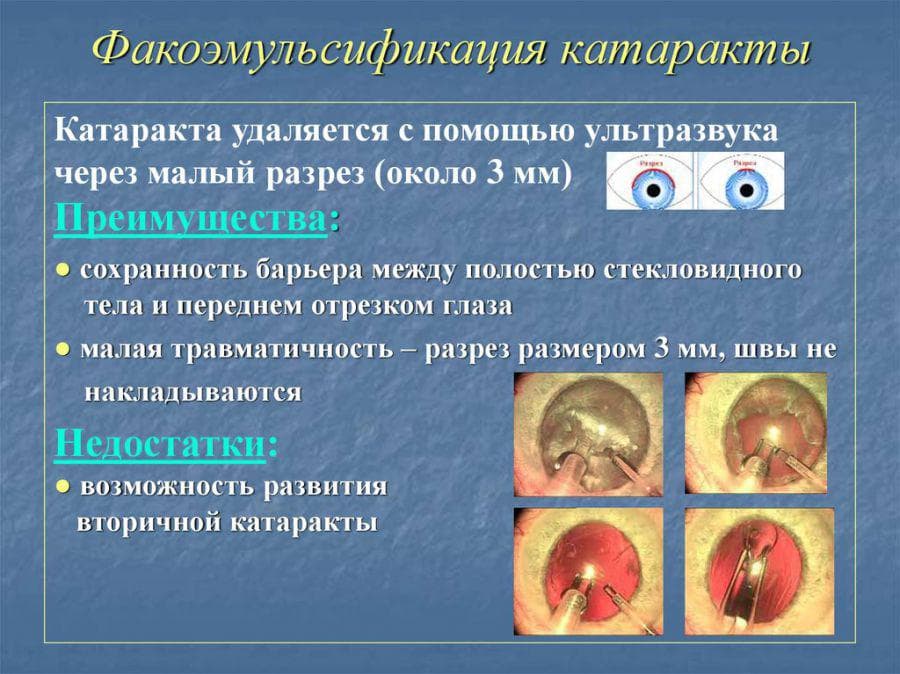
Infections
Treatment of infectious diseases that cause corneal edema requires antifungal or. Local remedies (eye drops) are usually used, but in more severe conditions, pills or intravenous injections are prescribed.
For viral diseases, drugs containing interferon (for example) are used, as well as artificial tears.

Ophthalmoferon is used for viral diseases of the cornea of the eye
For bacterial infections, antibacterial agents are indicated (Moxifloxacin, Levofloxacin).

Moxifloxacin is used for bacterial infections
Allergic reaction
To remove an allergic corneal edema, the first step is to identify and eliminate contact with the allergen (cosmetics, dust, animal hair, pollen, perfumery). To relieve symptoms, you should take an antihistamine (Diazolin, Suprastin, Diphenhydramine).

Diazolin is an antihistamine
Corneal edema after injury
Corneal trauma is quite common. Minor trauma does not require treatment. If the damage is significant, then the doctor must be called immediately. Before help arrives, you need to blink frequently (if a foreign body does not interfere with this) and rinse the eye with clean water. 
In case of injury, do not rub your eyelids with your fingers, do not yourself pull out a foreign body that has pierced the eye.
Surgically
If the methods of conservative treatment do not help, then the doctor may recommend surgery. In case of violations in the cornea, its transplant is performed, and in some modern clinics, the cornea is compacted with ultraviolet light.
Folk remedies
With inflammation and swelling in the eye, you can use traditional medicine recipes as an additional treatment. Below are the most popular recipes:

Prophylaxis
Preventive measures against corneal edema:
- Compliance with the rules of hygiene when caring for the face;
- Use of hypoallergenic quality cosmetics;
- Regular measurement of the level of intraocular pressure in patients over 45 years old;
- Eye protection with special goggles to avoid injury to the organ of vision and the appearance of symptoms of puffiness during hazardous work.
An important role in the prevention of pathological conditions of the stratum corneum is played by the correct selection of contact optics. Lenses must be of high quality, allowing oxygen to pass to the eyes. Use your lenses correctly.

Choose cosmetics for eyelids and eyelashes from the point of view of health safety, it should not contain allergens that cause edema.
After removing cataracts, glaucoma and other surgical interventions in different parts of the eye, do not burden the organs of vision with computer work or reading, so as not to cause a relapse.
The job must be selected that does not require strong physical activity, inclinations. During sleep, it is necessary to lie down so that the head is above the legs, which will ensure the necessary outflow of blood.
It is forbidden to go in for swimming or go to the sauna after the treatment of edema.
If these rules are followed, repeated puffiness of the cornea of the eye can be avoided.
Video
conclusions
Most often, corneal edema is, which has a different origin. It is very important to establish the cause of the swelling state with the help of medical diagnostics, after which it is possible to carry out treatment aimed at effectively eliminating the cause of the disease.
In group B, we took into account the complications associated with the presence of the CM in the CT cavity: corneal degeneration, secondary hypertension (acute hypertension requiring additional basal iridectomy, the so-called "silicone block"), rhematogenous retinal detachment after removal of the CM, which was removed to all patients within 2 to 6 months. Intraoperative laser coagulation in panretinal volume (PLC) was performed in the presence of total (extensive) tractional retinal detachment for the purpose of retinopexy. Considering that laser coagulation can affect the frequency of retinal detachment after removal of the CM, the number of patients who underwent PLC was approximately the same in the subgroups.
Inflammation of the eye and cataracts. Cataract bg
Cancer Headache ARI, otitis media and tonsillitis Toothache Inflammation of the eye and cataracts
Elena Denisova
ophthalmologist
Freshly brewed tea - honey - onions
“If a patient has signs of eye inflammation (redness, itching), then in conditions when there is no doctor or pharmacy nearby, it is possible to recommend rinsing the eyes. tea... This is a public ambulance device. I do not recommend rinsing with sleeping tea - it is better to use freshly brewed, strained, cooled, black or green tea without aromatic additives. Freshly brewed tea contains tannin, which relieves signs of inflammation such as redness and swelling by constricting blood vessels. It is important not to overdo it, because too frequent use of tea can dry out the edges of the eyelids, which in turn will contribute to inflammation. Everything is good in moderation. You can rinse in different ways, I would recommend using a dropper if you have one at hand, but you should not touch the eyedropper with the eyedropper, and also touch the pipette tip with your hands. You can rinse with cotton swabs (discs), from the outer corner of the eye to the inner one, with separate swabs for each eye, or drip from a cotton ball, moistening it abundantly.
There is a clever trick that stimulates the healing of the newly operated cornea.
Abundant irrigation of the anterior surface of the eye with tear fluid stimulates healing better than any drops. The tear has a bactericidal effect: it contains the substance lysozyme. You can, of course, remember something sad and cry, but a much more effective way is after the operation. cut onions... It contains phytoncides that will irritate the eyes, causing profuse tearing. The cornea heals great. "
Inflammation of the cornea
My husband is 47 years old, he has the 2nd group of visual disability - corneal dystrophy of both eyes from the age of 15. In 2001, 5 operations were performed to straighten the cornea, but to no avail - within 1-2 months after the operation, everything returned to its original state. In addition, in 2011, an operation was performed on the left eye to replace the lens (cataract - both eyes) (Excimer, Novosibirsk). The operation went without complications, the vision on this eye improved, however, on the other it began to fall. A year later, a general deterioration began - there was a strong photophobia, lacrimation, "sand in the eyes", "scratching" of the eyelids, and the eyes hurt so much that it was impossible to open. Symptoms sometimes subside, but then only get worse. In the MNTK "Eye Microsurgery" them. Fedorova (Novosibirsk) recommended only a corneal transplant, but with a very ambiguous prognosis for the result. They also prescribed Balarpan drops, but we could not remove the inflammation with drops. The symptoms of inflammation persist with a constant exacerbation as early as 2 months.
Details
Chief physician of the Excimer ophthalmological clinic in Novosibirsk, doctor of medical sciences, academician of the Russian Academy of Natural Sciences and the Russian Academy of Medical Sciences, doctor of the highest category, Honored Doctor of the Russian Federation.
Permanent participant of Russian and international congresses of refractive and cataract surgeons. He specializes in microsurgical operations of varying degrees of complexity, and is a leading ophthalmic surgeon at the Excimer clinic in Novosibirsk.
Corneal diseases
Corneal inflammation or keratitis is a serious disease leading to a violation of the transparency of the cornea, a sharp decrease in vision and the spread of the inflammatory process deep into the eye.
The cause of keratitis is infection. The most dangerous are herpes virus, fungi, protozoal infection, blue-purulent bacillus, pseudomonas. It will help to determine the quality diagnosis of vision.
Corneal infection provokes trauma, which can be caused by both simple mechanical trauma to the eye and wearing lenses.
Viral keratitis often develops against the background of a decrease in the general immunity of the body.
As a rule, keratitis is accompanied by redness of the eye, lacrimation, photophobia, and sharp pains.
Decreased vision and pain in the eye cause severe discomfort and completely make it impossible to lead a normal life.
Keratitis is treated with antibiotics, antiviral and anti-inflammatory drugs specific to each specific infection. The treatment is usually long and does not always lead to a complete cure. After severe keratitis, corneal opacity remains forever - a thorn, significantly reducing vision and impairing the cosmetic appearance of a person.
Many keratitis are recurrent (repetitive) in nature, and each subsequent attack of inflammation causes more and more changes in the cornea.
The most difficult to treat are herpetic, protozoal and fungal keratitis. Sometimes drug therapy is powerless.
Today, a new method of treating severe inflammatory diseases of the cornea has appeared. This technique is called corneal cross-linking.
The essence of the method lies in the fact that with the help of a special device - the Sailer lamp, complete sterilization of the cornea is performed (i.e., the entire infection in the thickness of the cornea is destroyed) with the help of specially focused homogenized monochrome ultraviolet radiation.
At the same time, corneal edema caused by the inflammatory process is eliminated.
One procedure is enough to stop the inflammatory process and eliminate the infection.
After the procedure, the pain disappears, the eye calms down, the cornea becomes transparent.
There is hope today for those suffering from severe keratitis!
Cross-linking is also used for epithelial-endothelial corneal dystrophies. which are one of the rare but serious complications of cataract surgery. Decompensation of the cornea after surgery leads to its edema, a violation of the transparency and integrity of the surface layer. As a result, vision is significantly reduced, pain, photophobia and lacrimation appear. Patients are forced to constantly carry out anti-inflammatory treatment or resort to corneal transplant.
With the help of cross-linking, corneal edema is eliminated by thickening the layers of collagen fibers, vision is improved, discomfort and pain disappear or significantly decrease.
Historical reference
In 2008, Professor Sailer received the highest award in ophthalmology, the Kellman Prize, for his innovative invention.
The cross-linking method has been used since 1996 for the treatment of severe corneal diseases.
Medical Center "AS" has certified the Sailer lamp in the Ministry of Health of Ukraine, which helps restore vision in Ukraine.
Medical Center "AS" is officially certified for the application of the method, the clinic's specialists have been trained and have the appropriate diplomas.
Medical Center "AS" is certified as an official center for training in cross-linking techniques for Ukrainian doctors.
In the recovery period, some patients are faced with such a manifestation as edema after cataract surgery. The more advanced the stage of the disease, the higher the likelihood of its occurrence. It can last from 1 to 15 days and requires supervision by a specialist. Only a doctor will be able to determine whether the condition needs correction or is a variant of the normal rehabilitation of the body.
Edema after cataract surgery: causes
In the process of phacoemulsification, the clouded lens is crushed by ultrasound. Decomposition products are washed out with a large amount of liquid. At the stage of "mature" and "overripe" cataracts, the edema of the eye is more pronounced - the nucleus and cortical layers are denser than at the previous stages, and for their destruction, ultrasound exposure of greater power is required, the tissues are subjected to more intensive processing.
The condition of the cornea also affects. If it is weakened, a violation of the outflow of fluid in the patient can be observed before surgery and is a consequence of destructive changes in the eye.
Other causes may be inflammatory processes, postoperative infections, concomitant pathologies of the eye tissues.
Symptoms
The cataract was removed, the edema of the eye is manifested by the following symptoms:
- blurry vision;
- feeling of "fog";
- inability to focus;
- increased sensitivity to light.
Effects
Being under the supervision of a doctor, the patient is protected from possible negative consequences. If the swelling is caused by mechanical stress, it will go away on its own at the end of the rehabilitation period.
An experienced ophthalmologist will distinguish between a postoperative violation of the pumping function of the epithelium (in other words, the ability to pump fluid from tissues) from bullous keratopathy. This is a rare complication (occurs in 0.1% of cases) when small bubbles form in the cornea, which are treated with hypertonic ointments and corrective lenses.
Differentiation with cystic macular edema is also required. Complication manifests itself in 1% of cases after phacoemulsification (ultrasound crushing) and in 20% of cases after the extracapsular technique (when the nucleus of the lens is removed while preserving the capsule).
People with diabetes mellitus, mature and overripe stages of the disease, who have a capsule rupture or loss of the vitreous during surgery, are predisposed to it (treatment of an advanced disease is fraught with these consequences).
Both conditions, unlike the previous one, require treatment. Modern medicine is able to successfully correct them with timely medical supervision.
Various reasons lead to corneal edema. One of them is surgery to remove cataracts - a disease associated with clouding of the lens. During the operation, the transparent body of the pupil is replaced with an artificial implant. An artificial lens allows a person to preserve their eyesight for many years. In most cases, the operations are successful. Patients recover quickly. But sometimes corneal edema occurs.
The cornea is the anterior convex part of the eyeball. It is a natural lens with high refractive power. The cornea consists of a transparent stroma and specific bodies. It has five layers.
The cornea has a number of functions:
- refracts light
- protects the visual organs from the negative effects of the environment (dust, dirt, etc.).
In a healthy state, the cornea is transparent. The edema that occurs after the operation leads to pathological changes. The cornea becomes cloudy. Its refractive function decreases. A person sees objects in a blurred form. The focus shifts. The swollen tissues put pressure on other organs. If the process is not stopped in a timely manner, the edema will lead to the destruction of the stratum corneum. This will be followed by tissue necrosis. It will become impossible to stop the process.
Corneal edema symptoms
Corneal edema does not go unnoticed. The first sign of edema is a change in visual acuity. The patient complains that he does not see well. Captivity appears before the eyes. The use of contact lenses is uncomfortable. Even with the naked eye, folds and stripes are visible on the cornea. With prolonged edema, a mesh of blood vessels appears on the cornea.

Symptoms of corneal edema also include:
- distortion of the image,
- photophobia,
- pain in the eyes (burning and stinging),
- feeling of a foreign body (more often sand),
- redness of the eyeball.
Various eye diseases lead to edema of the cornea of the eyeball. Symptoms are similar regardless of the cause. In rare cases, the disease is asymptomatic.
Causes of corneal edema
There are various causes of corneal edema. The most common cause of puffiness is increased intraocular pressure. Increased turgor leads to disruption of metabolic processes in the organ of vision. The outflow of fluid is hindered. Swelling occurs.

There are other causes of corneal edema:
- Congenital pathology. With endothelial dystrophy, cells of the posterior epithelium die off. The main symptom of this disease of the cornea of the eye is a decrease in visual acuity in the morning.
- Mechanical damage. They occur when foreign objects enter the eyes and lead to swelling.
- Corneal injury. The most common cause of injury is chemical eye burns. To avoid them, great care should be taken when working with acids and alkalis.
- Inflammation of the lining of the eyes. Inflammation is the result of fungal diseases, reduced immunity, infection with Staphylococcus aureus. To avoid this, touch your eyes only with clean hands.
- Infectious and viral diseases: conjunctivitis, blepharitis, keratitis and others, often stimulate corneal edema.
- Allergic edema. It is caused by long-term use of certain types of drugs. Allergy occurs due to the use of low-quality cosmetics. At the first signs of allergic edema, you should stop taking the drug and the use of cosmetics.
- The use of contact lenses sometimes leads to swelling. If puffiness of the cornea of the eye appears, it is necessary to remove contact lenses and consult an ophthalmologist.
- Glaucoma is a group of diseases of the organs of vision that occurs against the background of an increase in intraocular pressure and leads to a decrease in visual acuity and atrophy of the optic nerve. Its consequence is corneal edema.
- Astigmatism - a pathology that leads to a distorted shape of the lens, is the cause of edema. A person with astigmatism sees objects vaguely. The clarity of the image depends on the degree of the disease.
- Strabismus. The visual axes deviate from the natural direction. The eyes see an object from different angles. There is no single image.
- Eye surgery. In particular, with regard to cataracts, sometimes it leads to edema.
Diagnostic measures
At the first signs of corneal edema, you should contact your local ophthalmologist. The doctor will prescribe a diagnosis, and then treatment. Research begins with a study of the patient's complaints and a visual examination of the eye. This is followed by a tissue biopsy. Laboratory tests can exclude or confirm the presence of bacterial and viral lesions.

Schirmer's test gives an idea of the amount of tear fluid. Further treatment depends on the cause of the swelling. If necessary, the ophthalmologist can refer the patient to a neurologist, endocrinologist or nephrologist.
Treatment
Corneal edema requires timely treatment. Treatment of edema is prescribed by a doctor. It depends on the cause of the disease. Treat corneal edema with medical and surgical methods. Traditional medicine has its own recipes for getting rid of. But they should be treated with caution and used only after consulting an ophthalmologist. It should be remembered that traditional medicine refers to auxiliary methods.
In a medical way
As mentioned above, the treatment is closely related to the cause that led to the edema and impairment of the phantom of the cornea:
- Wearing contact lenses. If the swelling is caused by them, then the use of uncomfortable optics should be discontinued. Eye drops are instilled into the eyes with a moisturizing effect.
- Allergy. In this case, an allergen blocker is prescribed. First you need to find out what the allergic reaction went to.
- Viruses and infections. The exact cause of the infection is established. Which led to swelling. The patient is prescribed eye drops and ointments containing antibiotics and antiviral components. For the treatment of eye infections, ointments are prescribed: Actovegin, Hydrocortisone, Demazol, Oxolinic ointment. The medicine is selected by an ophthalmologist.
- Mechanical damage. Eliminate the cause of edema. Drops are used to repair damaged tissue.
- Surgery to remove cataracts and replace the lens. After replacing the lens, planned treatment is carried out, which is aimed at overcoming postoperative complications. The patient is prescribed special eye drops. They are dripped three times a day for 10 days. Further, it is recommended to protect your eyes from foreign bodies and to bury albucid for prevention.
No matter how hard a person tries, everything cannot be predicted and avoided. Complications sometimes occur after cataract surgery. One of them is corneal edema. The cause of the swelling is the large amount of fluid that passes through the eye. The greater the opacity of the lens, the higher the risk of postoperative edema.

The edema that appears resolves on its own after 14 days. In rare cases, the patient is prescribed injections.
Surgically
After cataract surgery, patients are at risk of endothelial dystrophy. This is a rare complication. But if it happened, then the patient needs surgical intervention. Keratoplasty is performed - a corneal transplant surgery. It helps to restore the transparency of the cornea. The operation is performed in one step. The patient is covered with a protective bandage and sent home.

Recovery after transplantation takes 12 months. The stitches are removed after six months.
Folk remedies
Traditional medicine has tools that can help with edema of the cornea of the eye. And, although these funds are auxiliary in nature, sometimes they are very effective. They help to get rid of puffiness in the postoperative period:
- Honey has long been used in folk medicine. Eye drops are prepared from it. A teaspoon of honey is diluted with boiled water and stirred well. The cooled mixture is instilled into the eyes, 1-2 drops. This is a simple and good remedy after cataract surgery.

- Finely chopped onions and horseradish are poured with boiling water. Allow to cool and brew. Wet cotton pads are moistened and applied to the eyes.
Attention! Only lotions do this. Do not drip into your eyes!
