articulationes synoviales are the most advanced types of bone connections. They are distinguished by great mobility and a variety of movements.
Joint structure
Each joint includes articular surfaces of bones covered with cartilage, an articular capsule, and an articular cavity with a small amount of synovial fluid. Some joints also have auxiliary formations in the form of articular discs, menisci and articular labrum.
The articular surfaces, fades articulares, in most cases of articulating bones correspond to each other - they are congruent (from the Latin congruens - corresponding, coinciding).
Articular cartilage, cartilago articularis, is usually hyaline, in individual joints (temporomandibular) it is fibrous, and has a thickness of 0.2-6.0 mm.
The articular capsule, capsula articularis, is attached to the articulating bones near the edges of the articular surfaces or at some distance from them; it firmly fuses with the periosteum, forming a closed articular cavity.
The articular cavity, cavum articulare, is a slit-like space between the articular surfaces covered with cartilage.
Articular discs and menisci, disci et menisci articulares, are cartilaginous plates of various shapes that are located between articular surfaces that do not completely correspond to each other (incongruent). The disc is usually a solid plate, fused along the outer edge with the articular capsule, and, as a rule, divides the articular cavity into two chambers (two floors).
Menisci
These are semi-lunar-shaped, continuous cartilaginous or connective tissue plates that are wedged between the articular surfaces.
The articular lip, labrum articulare, is located along the edge of the concave articular surface, complements and deepens it (for example, in the shoulder joint). It is attached with its base to the edge of the articular surface, and with its inner concave surface facing the joint cavity.
Shapes of articular surfaces
resemble segments of the surfaces of various geometric bodies: a cylinder, an ellipse, a ball. Accordingly, joints are distinguished according to the shape of the articular surfaces: cylindrical, ellipsoidal and spherical. There are also variants of the indicated forms of joints. For example, a type of cylindrical joint would be a trochlear joint, a spherical joint would be a cup-shaped and flat joint.
The shape of the articular surfaces determines the number of axes, around which movement occurs at a given joint. Thus, the cylindrical shape of the articular surfaces allows movement only around one axis, and the ellipsoidal shape allows movement around two axes. In joints with spherical articular surfaces, movements are possible around three or more mutually perpendicular axes.
Thus, there is a certain interdependence between the shape of the articulating surfaces and the number of axes of motion.
Biomechanical classification of joints:
1) joints with one axis of movement (uniaxial);
2) joints with two axes of movement (biaxial);
3) joints with many axes of movement, of which three are main (multiaxial, or triaxial).
JOINT
In anatomy, a joint is an articulation (connection) of two or more bones. In mammals, joints are usually divided into three groups: synarthrosis - immobile (fixed); amphiarthrosis (half-joints) - partially mobile; and diarthrosis (true joints) - mobile. Most joints are movable joints.
Fixed joints. Synarthrosis is a direct connection of two bones without a gap between them. The connection may involve a thin layer of fibrous connective tissue or cartilage. There are four types of synarthrosis in the skull. Sutures are connections between the flat bones of the skull; a typical example is the suture between the parietal and frontal bones. Schindylosis is a form of synarthrosis in which the plate of one bone enters a gap or notch in another bone. The vomer (median bone of the facial skull) and the palatine bone are connected in this way. Gomphosis is a type of synarthrosis in which the conical process of one bone enters the depression of another bone. There is no such articulation of two bones in the human body, but this is how the teeth are connected to the jaw. Synchondrosis is a continuous connection of bones through cartilage; it is typical for young and is found, for example, between the ends and the middle part of long tubular bones; in adults, these cartilages ossify. A similar articulation between the sphenoid bone, located in the middle of the base of the skull, and occipital bone persists in the child for several years after birth.
Partially movable joints usually have a fibrocartilaginous disc or plate (this includes intervertebral discs) between two bone elements, or the bones are connected to each other by dense inelastic ligaments. The first type is called symphysis, the second - syndesmosis. The articulations between the vertebral bodies in the form of intervertebral discs are typical symphyses, and the articulation between the upper ends of the fibula and tibia of the leg is an example of syndesmosis.

Movable joints are the most common in animals. In joints of this type (true joints), the bony surfaces are covered with articular cartilage, and the joint itself is enclosed in a capsule of fibrous connective tissue, lined from the inside with a synovial membrane. The cells of this membrane secrete a lubricating fluid that facilitates movement in the joint. Diarthrosis includes block-shaped and cylindrical (rod, rotational) joints, as well as spherical, flat (movements are sliding), saddle-shaped and condylar (ellipsoidal).
Block joints. A typical example is the joints between the phalanges of the fingers. Movements are limited to one plane: forward - backward. The bones lie in a straight line, and strong lateral ligaments prevent them from moving laterally. The temporomandibular joint also belongs to the block-shaped joint, although sliding movements are also possible in it. The knee and ankle joints allow slight rotation, so they are not typical locking joints, although the main movement in them is forward and backward.

There are two types of cylindrical joints. Examples are the joint between the first and second cervical vertebrae (atlas and axis) and the articulation between the head radius And ulna. In the atlantoaxial joint, the odontoid process of the second cervical vertebra enters the ring-shaped opening of the first cervical vertebra and is held in place by ligaments so that movement is limited to rotation around the process. At the articulation between the head of the radius and the ulna, the ring consists of the radial notch of the ulna and the round ligament that holds the head of the radius so that it can rotate. In other words, in the atlantoaxial joint the rod (odontoid) is fixed and the ring rotates around it, but in the radioulnar joint the ring is fixed and the rod rotates inside it.

Ball and socket joints provide the greatest range of motion: both rotation and flexion are possible, so that the limb can describe a cone; movement is limited only by the size of the articulating surfaces. Examples are the shoulder and hip joints. Both consist of a cup-shaped depression in which a ball-shaped head is located.

Flat joints. This is the simplest form of joint; as a rule, it is formed by two flat sections of bone. The range of motion is limited by the ligaments and bony processes at the edges of the articulating surfaces. Some flat joints consist of a slightly concave and slightly convex surface. These include the wrist and ankle joints, the sacroiliac joint, and the articular processes of the vertebrae.

The saddle joints resemble a rider in a saddle, who can move forward and backward and sway from side to side. But without rising in the stirrups, the rider will not be able to make a rotational movement, and even then his legs will get in the way; rotation is also impossible in the saddle joint. This type of joint is found in humans only at the base of the thumb: this is the carpometacarpal joint, where the first metacarpal bone serves as the saddle, and the trapezium bone of the wrist serves as the rider.
Condylar joints. They are similar in action to saddle-shaped ones, i.e. flexion - extension, adduction - abduction, as well as arc movement are possible in them. Rotation is not possible. This type includes, for example, the wrist joint between the radius, scaphoid and lunate bones of the wrist.
Articulations in invertebrates. Invertebrates have many types of joints, but they have their own characteristics. Thus, at the junction of mollusk shells there are often small processes in the form of denticles that prevent the shell valves from rotating relative to each other or from separating them. If the joints of mammals are controlled by two groups of opposing muscles, then the shell valves can be controlled by only one muscle, balanced on the opposite side by elastic connective tissue. In insects, crabs, crayfish and other arthropods, the body is covered with chitin, a dense leathery substance. In certain areas of their cover there are joints that allow the mutual movement of body parts. In these places, the epidermis is folded inward, forming folds, and is not covered with chitin. In some echinoderms, namely sea urchins, many articulations are located between the calcareous plates that cover the body and form the chewing apparatus (the so-called Aristotelian lantern), and these plates are connected in the same way as the parietal bones of the human skull. The spines, especially pronounced in sea urchins of the genus Arbacia, are attached to the exoskeleton by ball-and-socket joints controlled by two groups of muscles, one arranged circularly and the other radially. In the Aristotelian lantern there is a peculiar swinging joint between two elements: the arch of the jaw and the bracket; contraction of the muscles on the outer side of the lantern lowers the outer end of the bracket, respectively, its inner side rises and lifts the roof of the lantern, thereby creating a pump effect.
Joint diseases.
Any inflammatory process in the joints is called arthritis. There are many types of arthritis, the causes of which are infection, degenerative processes, tumors, trauma or metabolic disorders. At rheumatoid arthritis joints are swollen, painful and stiff. The most commonly affected joints are the hand joints, knee and hip joints, and the spine. The cause of the disease remains unclear. Synovitis - inflammation of the synovial membrane - is a very painful condition that occurs as a result of injury or infection in the joint capsule. Dislocations are often a complication of joint disease. Common injuries include sprains and joint dislocations with partial ligament tears. Injuries to intra-articular cartilage are very painful, especially in the knee joint. Adhesions that arise in the joint lead to ankylosis - immobility and fusion of the joint.
see also ARTHRITIS.

Collier's Encyclopedia. - Open Society. 2000 .
Synonyms:See what "JOINT" is in other dictionaries:
JOINT, in anatomy, the place where BONES join. In movable joints such as the knees, elbows, spine, fingers and toes, the bones are separated from each other by pads of CARTILAGE. In immobile joints, cartilage may be present in... ... Scientific and technical encyclopedic dictionary
Diarthrosis, joint, knee Dictionary of Russian synonyms. joint noun, number of synonyms: 10 ankles (2) ... Synonym dictionary
- (articulatio), diarthrosis (diarthrosis), a structure that provides movable articulation of vertebrate bones. Simple S. are formed by two bones, complex S. by several. Basic elements of a typical C: surfaces of articulating bones, covered with cartilaginous... ... Biological encyclopedic dictionary
A movable connection between bones that allows them to move relative to each other. Auxiliary formations of ligaments, menisci and other structures... Big Encyclopedic Dictionary
JOINT, joint, man. A movable joint (see joint in 3 meanings), the place where the ends of bones are connected by cartilaginous plates and ligaments. Ushakov's explanatory dictionary. D.N. Ushakov. 1935 1940 ... Ushakov's Explanatory Dictionary
JOINT, huh, husband. Movable connection of the ends of bones in humans and animals. Joint pain. | adj. articular, oh, oh. C. rheumatism. Ozhegov's explanatory dictionary. S.I. Ozhegov, N.Yu. Shvedova. 1949 1992 … Ozhegov's Explanatory Dictionary
Etc. see compose. Dahl's Explanatory Dictionary. IN AND. Dahl. 1863 1866 … Dahl's Explanatory Dictionary
See COMPOSITION V. V. Vinogradov. History of words, 2010 ... History of words
- ... Wikipedia
A movable connection between bones that allows them to move relative to each other. The main elements of articulation: the surfaces of articulating bones, covered with cartilaginous tissue; cavity with articular fluid; bag insulating the cavity. Some S. have... Great Soviet Encyclopedia
Books
- Ultrasound diagnostics. Knee joint, A. N. Sencha, D. V. Belyaev, P. A. Chizhov, The book is based on many years of experience in researching the knee joint in a multidisciplinary clinic with developed rheumatological and orthopedic services. The authors provide an unbiased… Category: Ultrasound. ECG. Tomography. X-ray Publisher:
1125 0
Perfect glide for mindless movements
When you see another “snake woman” in “Minute of Fame”, twisting her body almost into pigtails, you understand that the structure of joints and bones that is standard for other people is not about her. About which dense tissues there may be a question - they simply are not here!
However, even she has hard tissues - many joints, bones, as well as structures for their connections, according to the classification, divided into several categories.
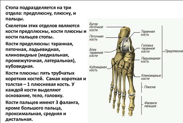
Classification of bones
There are several types of bones depending on their shape.
Tubular bones have a medullary cavity inside and are formed from compact and spongy substances, performing supporting, protective and motor roles. Divided into:
- long(bones of the shoulders, forearms, thighs, legs), having a biepiphyseal ossification;
- short(bones of both wrists, metatarsals, digital phalanges) with a monoepiphyseal type of ossification.

Bones have a spongy structure, with a predominance of spongy substance in the mass with a small thickness of the covering layer of compact substance. Also divided into:
- long(including costal and sternum);
- short(vertebral bones, carpals, tarsals).
They belong to the same category bone formations sesamoids, located near the joints, participating in their strengthening and promoting their activity, not having a close connection with the skeleton.
Flat shaped bones including categories:
- flat skull(frontal and parietal), acting as protection and formed from two outer plates of a compact substance with a layer of spongy substance located between them, having connective tissue origin;
- flat bones of both limb girdles(scapular and pelvic) with a predominance of spongy substance in the structure, acting as support and protection, with origin from cartilaginous tissue.
Bones of mixed (endesmal and endochondral) origin with different structures and tasks:
- forming the base of the skull;
- clavicular
Only the bones do not live on their own - they are connected to each other by joints in the most ingenious ways: two, three, at different angles, with varying degrees of sliding against each other. Thanks to this, our body is provided with incredible freedom of static and dynamic poses.

Synarthrosis VS diarthrosis
But not all bone joints should be considered diarthrosis.
According to the classification of bone joints, these are not included the following types articulations:
- continuous (also called adhesions, or synarthrosis);
- semi-mobile.
The first gradation is:
- synostoses- fusion of the boundaries of the bones with each other until complete immobility, zigzag “zippers” of sutures in the cranial vault;
- synchondrosis- fusion through a cartilaginous layer, for example, an intervertebral disc;
- syndesmoses- strong “stitching” with a connective tissue structure, the interosseous sacroiliac ligament, for example;
- synsarcoses- when connecting bones using a muscle layer.
The tendon membranes stretched between the paired formations of the forearms and shins, holding them dead next to each other, are also not joints.

As well as semi-movable joints (hemiarthrosis) in the form of the pubic symphysis with a small (incomplete) cavity-gap in the thickness of the fibrocartilaginous suture, or in the form of sacroiliac amphiarthrosis with real articular surfaces, but with an extremely limited range of movements in the semi-joints.
Structure and functions
Joint (continuous or synovial junction) can only be considered a movable joint of bones that has all the necessary attributes.
In order for all dysarthrosis to move, there are special education and auxiliary elements.

Diagram of the structure of the knee joint
If on one bone it is a head, which has a pronounced roundness in the form of a thickening - the epiphysis of the terminal section, then on the other bone it is associated with it, it is a depression exactly corresponding to it in size and shape, sometimes significant (this in the pelvic bone is called “vinegar” due to its vastness). But there may also be an articulation of one bone head with a structure on the body-diaphysis of another, as is the case in the radioulnar joint.
In addition to the perfect fit of the shapes that form the joint, their surfaces are covered with a thick layer of hyaline cartilage with a literally mirror-smooth surface to glide over each other flawlessly.
But smoothness alone is not enough - the joint should not fall apart into its component parts. Therefore, it is surrounded by a dense elastic connective tissue cuff - a capsule bag, similar to a lady's muff for warming the hands in winter. In addition, it is held together by ligamentous apparatus of varying strength and muscle tone, ensuring biodynamic balance in the system.
A sign of true dysarthrosis is the presence of a full-fledged articular cavity filled with synovial fluid produced by cartilage cells.
The classic and simplest in structure is the shoulder. This is the gap of the joint between its bursa and two bone ends that have surfaces: the round head humerus and the articular cavity on the scapula, which matches it in configuration, filled with synovial fluid, plus ligaments that hold the entire structure together.
Other dysarthroses have a more complex structure - in the wrist, each bone is in contact with several neighboring bones at once.
Spine as a special case
But the relationships between the vertebrae - short-columnar bones with a complex surface topography and many structures for varying degrees of movable adhesion with neighboring formations - are particularly complex.
 The spine has a structure reminiscent of a rosary, only its “beads” are the bodies of each of the adjacent bones, which are connected to each other through hemiarthrosis (synchondrosis) based on a cartilaginous disc. Their spinous processes, overlapping each other like tiles, and the arches, forming a container for the spinal cord, are fastened with rigid ligaments.
The joints between the transverse processes of the vertebrae with flat surfaces (as well as the costovertebral ones, formed through the costal heads and articular cavities on the vertebral bodies located laterally) are quite real, having all the necessary attributes: working surfaces, cracks, capsules and ligaments.
In addition to connections with each other and with the ribs, the vertebrae form a fusion in the sacrum area, turning this group into a monolith, to which the “tail”-coccyx is attached through real joints - the formation is quite mobile, especially during childbirth.
Dysarthroses are the beginning of the pelvic girdle, formed by the bones of the same name, which are closed in a ring by the pubic symphysis in the front and center.
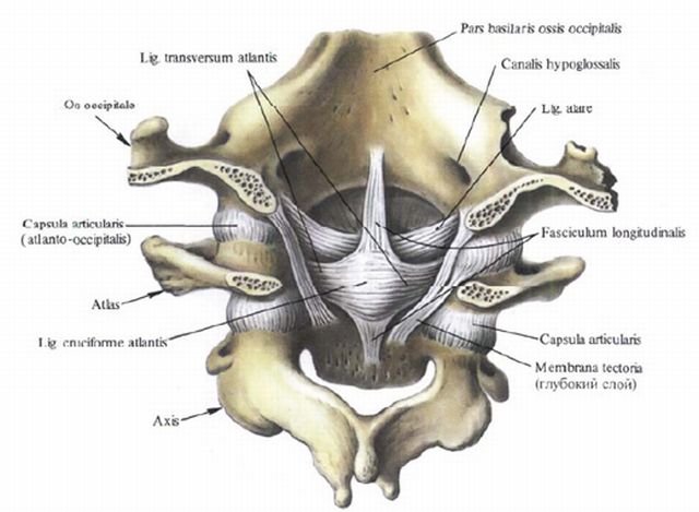
In addition to the intervertebral joints, there are other joints in the supporting column system: a combination that forms one unpaired and two paired components of the atlanto-axial connection (between the 1st and 2nd vertebrae) and paired atlanto-occipital joints (between the 1st vertebra and the occipital bone).
Due to this very structure, the spine is an incredibly flexible formation, having a large degree of freedom of movement and at the same time extremely strong, bearing the entire weight of the body. In addition to its supporting function, it also plays a protective role, serving as a canal through which the spinal cord passes and is involved in hematopoiesis.
The spectrum of damage to vertebral joints is diverse: from injuries (with different categories and displacements) to metabolic-dystrophic processes leading to varying degrees of spinal stiffness (and similar conditions), as well as infectious lesions (in the form of them, lues, brucellosis).
Detailed classification
The above classification of bone joints does not include the taxonomy of joints, which has several options.
According to the number of articular surfaces, the following categories are distinguished:
- simple, with two surfaces, as in the joint between the phalanges of the first finger;
- complex when there are more than two surfaces, for example, in the elbow;
- complex with the presence of internal cartilaginous structures dividing the cavity into non-insulated chambers, as in the knee;
- combined in the form of a combination of joints isolated from each other: in the temporomandibular joint, the intraarticular disc divides the working cavity into two separate chambers.
According to the functions performed, joints with one, two and multiple axes of rotation (one-, two- and multi-axial) are distinguished, depending on the shape they look like:

Examples of uniaxial joints are:
- cylindrical – atlantoaxial median;
- trochlear – interphalangeal;
- helical – shoulder-ulnar.
Structures of complex shape:
- ellipsoid, like the radiocarpal lateral;
- condylar, like the knee;
- saddle-shaped, like the metacarpal joint of the first finger.
Multi-axis are represented by varieties:
- spherical, like the shoulder;
- cup-shaped - a deeper variation of spherical (like hip);
- flat (like intervertebral ones).

Radioulnar cylindrical joint
There is also separate category tight joints (amphiarthrosis), differing in the shape of their surfaces, but similar in other respects - they are extremely stiff due to the strong tension of the capsules and a very powerful ligamentous apparatus, therefore their sliding displacement relative to each other is almost imperceptible.
Characteristics, design and functions of the main joints
With all the abundance of joints in the human skeleton, it is most logical to consider them as separate groups - categories of joints:
- skulls;
- spine;
- limb girdles (upper and lower).
Cranial joints
In accordance with this position, the skeleton of the skull includes two diarthrosis:
- temporomandibular;
- atlanto-occipital.
The first of these paired connections is created with the participation of the heads of the bones of the lower jaw and the working cavities on the temporal bones.
The joint consists of two synchronously functioning formations, although spaced on opposite sides of the skull. According to its configuration, it is condylar and belongs to the category of combined due to the presence of a cartilaginous disc dividing its volume into two chambers isolated from each other.
Thanks to the existence of this diarthrosis, freedom of movement of the lower jaw in three planes is possible and its participation both in the process of primary food processing and in swallowing, breathing and the formation of speech sounds. The jaw also serves as a means of protecting the oral organs from damage and is involved in creating the relief of the face. It can be subject to both injury and infection during the development of acute (mumps) and exacerbation of chronic (tuberculosis) diseases.
The configuration of the paired atlanto-occipital region is also condylar. It serves to connect the skull (its occipital bone with convex working surfaces) with the spine through the first two cervical vertebrae, acting as one whole, on the first of which - the atlas - there are working fossae. Each half of this synchronously operating formation has its own capsule.
Being biaxial, the atlas allows you to make head movements both according to the frontal and sagittal axes - both nodding and tilting left and right, providing freedom of orientation and the fulfillment of a social role by a person.
The main pathology of atlanto-occipital diarthrosis is injury as a result of a sharp tilting of the head and the development of osteochondrosis and other metabolic-dystrophic conditions due to the long-term preservation of a forced posture.
Shoulder girdle
Considering the description of the spine proposed above, moving on to diarthrosis of the shoulder girdle, it should be understood that the connections  the clavicle with the sternum and the scapula with the clavicle are synarthrosis. The real joints are:
- brachial;
- elbow;
- radiocarpal;
- carpometacarpal;
- metacarpophalangeal;
- interphalangeal.
The spherical shape of the head of the humerus is the key to almost complete circular freedom of rotation of the upper limb, therefore the humerus is a multi-axial joint. The second component of the mechanism is the scapular cavity. All other attributes of diarthrosis are also present here. Shoulder connection most susceptible to damage (due to the large degree of freedom), and to a much lesser extent to infections.

The shoulder joint is the most mobile in the entire musculoskeletal system
The complex structure of the elbow is due to the articulation of three bones at once: the humerus, radius and ulna, which have a common capsule.
The shoulder-elbow joint is trochlear: the shoulder block enters the notch on the ulna, the humerus-radius is the result of the head of the humeral condyle entering the fossa of the head of the radius bone with the formation of a spherical working area.
Movements in the system are carried out according to two axes: flexion-extension, and also due to the participation of the proximal radioulnar joint, rotation (pronation and supination) is possible, because the head of the radius rolls along the groove on the ulna.
Problems of the elbow joint include damage, as well as inflammatory conditions (in acute and exacerbation chronic infections), dystrophy due to professional sports.
The distal radioulnar joint is a cylindrical joint that provides vertical rotation of the forearm. In the working cavity there is a disk that separates the said joint from the cavity of the carpal joint.
Diseases of the elbow area:
- instability;
- stiffness.
 By means of a capsule covering the lower epiphysis of the radius and the first row of carpal bones, an ellipsoidal configuration of the wrist joint is formed. This is a complex articulation with sagittal and frontal axes of rotation, allowing both adduction-abduction of the hand with its circular rotation, and extension-flexion.
The most common diseases:
- damage (in the form of bruises, fractures, sprains, dislocations);
- synovitis;
- varying degrees of severity of tunnel syndrome;
- arthritis and hip;
- knee;
- ankle;
- tarsometatarsal;
- metatarsophalangeal;
- interphalangeal.
The shape of the hip multiaxial joint is cup-shaped, with the participation of the head femur and the sciatic cavity, which provides adduction and abduction of the hip back and forth and medially to laterally, as well as its rotation.
TZB is susceptible to damage (due to the high degree of freedom) and damage from microbial flora, most often brought here hematogenously (tuberculosis, brucellosis, gonorrhea).
The most common diseases of the hip area: 
- bursitis;
- tendinitis;
- femoroacetabular impingement syndrome; .
- extension-flexion;
- slight vertical abduction-adduction (in flexion position).
- subtalar;
- talocaleonavicular;
- calcaneocuboid;
- wedge-scaphoid.
The structure of diarthrosis allows for:
Most frequent disorder functions - (external or internal), as well as disruption of metabolic processes in the body and blood circulation in the lower extremities.
The tarsal area is formed by a “mosaic” of joints:
These are connections of a combined or flat configuration (the first two are cylindrical and spherical).
Metatarsal diarthrosis is represented by various ( for the most part, flat) joints that form a support for the arches of the foot, made by metatarsophalangeal (trochle-shaped) joints.
Also, the block-shaped interphalangeal joints of the feet provide the toes with a sufficient level of mobility and flexibility (patients who have lost both arms draw and even sew with their feet) without sacrificing strength.
Small joints of the feet are characterized by damage due to metabolic-dystrophic processes in the body, with disorders of local and general blood supply and as a result of chronic injuries in the form of wearing high-heeled or simply tight shoes.
The existence of different ways of connecting bones, as well as the diversity of the articular surfaces themselves, understanding their structure and function allows a person not only to live and act, but also to treat the musculoskeletal system (and, if necessary, even replace structures that have become unusable with artificial ones).
The whole truth about: human joints anatomy and other interesting information about treatment.
Human joints are the basis of every body movement. They are found in all bones of the body (the only exception is the hyoid bone).
Their structure resembles a hinge, due to which the bones slide smoothly, preventing their friction and destruction.
A joint is a movable connection of several bones, and in the body there are more than 180 of them in all parts of the body.
They are immobile, partially movable, and the main part is represented by movable joints.
The degree of mobility depends on the following conditions:
- volume of connecting material;
- type of material inside the bag;
- shapes of bones at the point of contact;
- the level of muscle tension, as well as ligaments inside the joint;
- their location in the bag.
How is the joint structured? It looks like a bag of two layers that surrounds the junction of several bones. The bursa seals the cavity and promotes the production of synovial fluid.
It, in turn, acts as a shock absorber for bone movements.
Together they perform three main functions of the joints: they help stabilize the body position, are part of the process of movement in space, and ensure the movement of parts of the body in relation to each other.
Basic elements of a joint
The structure of human joints is complex and is divided into the following basic elements: cavity, capsule, surface, synovial fluid, cartilage, ligaments and muscles. We'll talk briefly about each below.
- The joint cavity is a slit-like space, which is hermetically sealed and filled with synovial fluid.
- Joint capsule - consists of connective tissue that envelops the connecting ends of the bones. The capsule is formed on the outside from a fibrous membrane, but inside it has a thin synovial membrane (a source of synovial fluid).
- Articular surfaces have a special shape, one of them is convex (also called the head), and the second is pit-shaped.
- Synovial fluid. its function is to lubricate and moisturize surfaces, and also plays an important role in fluid exchange. It is a buffer zone during various movements (pushing, jerking, squeezing). Provides both sliding and divergence of bones in the cavity. A reduction in the amount of synovium leads to a number of diseases, bone deformations, loss of a person’s ability to perform normal physical activities and, as a result, even disability.
- Cartilage tissue (thickness 0.2 - 0.5 mm). The surfaces of the bones are covered with cartilage tissue, the main function of which is shock absorption during walking and sports. The anatomy of cartilage is composed of connective tissue fibers that are filled with fluid. This, in turn, nourishes the cartilage when it is at rest, and during movement it releases fluid to lubricate the bones.
- Ligaments and muscles are auxiliary parts of the structure, but without them the normal functionality of the entire body is impossible. With the help of ligaments, bones are fixed without interfering with movements of any amplitude due to their elasticity.
The inert protrusions around the joints also play an important role. Their main function is to limit the range of motion. As an example, consider the shoulder. There is a bony tubercle in the humerus. Due to its location next to the process of the scapula, it reduces the range of motion of the arm.
Classification and types
In development human body, way of life, mechanisms of human interaction and external environment, the need to perform various physical actions and various types of joints were obtained. The classification of joints and its basic principles are divided into three groups: the number of surfaces, the shape of the end of the bones, and functionality. We'll talk about them a little later.
The main type in the human body is the synovial joint. Its main feature is the connection of bones in the bag. This type includes shoulder, knee, hip and others.
There is also a so-called facet joint. Its main characteristic is the limitation of rotation to 5 degrees and tilt to 12 degrees.
The function also consists of limiting the mobility of the spine, which helps maintain the balance of the human body.
By structure
In this group, the classification of joints occurs depending on the number of bones that connect:
- A simple joint is a connection between two bones (interphalangeal bones).
- Complex – a connection of more than two bones (elbow). The characteristics of such a connection imply the presence of several simple bones, while the functions can be implemented separately from each other.
- Complex joint - or two-chamber, which contains cartilage that connects several simple joints (lower jaw, radioulnar). Cartilage can separate the joints either completely (disc shape) or partially (meniscus in the knee).
- Combined - combines isolated joints that are placed independently of each other.
According to the shape of the surfaces
The shapes of the joints and the ends of the bones have different shapes geometric shapes(cylinder, ellipse, ball).
Depending on this, movements are carried out around one, two, or three axes. There is also a direct relationship between the type of rotation and the shape of the surfaces.
- Cylindrical joint - the surface has the shape of a cylinder, rotates around one vertical axis (parallel to the axis of the connected bones and the vertical axis of the body). This species may have a rotational name.
- Block joint - a cylinder-shaped joint (transverse), one axis of rotation, but in the frontal plane, perpendicular to the connected bones. Characteristic movements are flexion and extension.
- Helical is a variation of the previous type, but the axes of rotation of this form are located at an angle other than 90 degrees, forming helical rotations.
- Ellipsoidal - the ends of the bones have the shape of an ellipse, one of them is oval, convex, the second is concave. Movements occur in the direction of two axes: bend-unbend, abduct-addite. The ligaments are perpendicular to the axes of rotation.
- Condylar is a type of ellipsoidal. The main characteristic is the condyle (a rounded process on one of the bones), the second bone is in the shape of a depression, and can differ significantly in size from each other. the axis of rotation is represented by the frontal one. The main difference from the block-shaped one is the strong difference in the size of the surfaces, from the ellipsoidal one - the number of heads of connecting bones. This type has two condyles, which can be located either in the same capsule (similar to a cylinder, similar in function to the trochlear one) or in different capsules (similar to the ellipsoidal one).
- Saddle-shaped - formed by connecting two surfaces as if “sitting” on each other. One bone moves lengthwise, while the second moves across. Anatomy involves rotation around perpendicular axes: flexion-extension and abduction-adduction.
- Ball-and-socket joint - the surfaces are shaped like balls (one convex, the other concave), due to which people can make circular movements. Basically, rotation occurs along three perpendicular axes, the intersection point being the center of the head. The peculiarity is a very small number of ligaments, which does not interfere with circular rotations.
- Cup-shaped - the anatomical appearance involves a deep depression of one bone that covers most of the area of the head of the second surface. As a result, there is less free mobility compared to the spherical one. Necessary for greater joint stability.
- Flat joint - flat ends of bones of approximately the same size, interaction along three axes, the main characteristic is a small range of movements and surrounded by ligaments.
- Tight (amphiarthrosis) - consists of bones of different sizes and shapes that are closely connected to each other. Anatomy - inactive, the surfaces are represented by tight capsules, non-elastic short ligaments.
By nature of movement
Due to their physiological characteristics, joints perform many movements along their axes.
In total, there are three types in this group:
- Uniaxial - which rotate around one axis.
- Biaxial - rotation around two axes.
- Multi-axis - mainly around three axes.
In addition, there are also different types of movements in the joints:
- Flexion and extension.
- Rotation in and out.
- Abduction and adduction.
- Circular movements (surfaces move between axes, the end of the bone draws a circle, and the entire surface draws the shape of a cone).
- Sliding movements.
- Removal from one another (for example, peripheral joints, distance of fingers).
The degree of mobility depends on the difference in the size of the surfaces: the larger the area of one bone over another, the greater the range of movement.
Ligaments and muscles can also inhibit range of motion.
Their presence in each type is determined by the need to increase or decrease the range of motion of a certain part of the body.
"An Illustrative Review of Anatomy"
In the next video you can visually study the anatomy and see how the joints on the skeleton work.
Source: https://prospinu.com/anatomija/stroenie-sustava.html
Structure and functions of joints
Joint- is a movable articulation of two or more skeletal bones.
Joints unite the bones of the skeleton into a single whole. More than 180 help a person move various joints. Together with bones and ligaments, they are classified as the passive part of the musculoskeletal system.
Joints can be compared to hinges, the task of which is to ensure smooth sliding of bones relative to each other.
In their absence, the bones will simply rub against each other, gradually collapsing, which is a very painful and dangerous process.
In the human body, joints play a triple role: they help maintain body position, participate in the movement of body parts relative to each other, and are organs of locomotion (movement) of the body in space.
The main elements that are present in all so-called true joints are:
- articular surfaces (ends) of connecting bones;
- joint capsule;
- joint cavity.
The joint cavity is filled with synovial fluid, which is a kind of lubricant and promotes free movement of the articular ends.
Based on the number of articular surfaces, they are distinguished:
- a simple joint having only 2 articular surfaces, for example interphalangeal joints;
- a complex joint having more than two articulating surfaces, such as the elbow joint. A complex joint consists of several simple joints in which movements can be performed separately;
- a complex joint containing intra-articular cartilage that divides the joint into 2 chambers (bicameral joint).
Classification of joints is carried out according to the following principles:
- by the number of articular surfaces;
- according to the shape of the articular surfaces;
- by function.
The articular surface of the bone is formed by hyaline (less often fibrous) articular cartilage. Articular cartilage is tissue filled with fluid.
The surface of the cartilage is smooth, strong and elastic, capable of absorbing and releasing liquid well.
The thickness of articular cartilage is on average 0.2-0.5 millimeters.
The joint capsule is formed by connective tissue. It surrounds the articulating ends of the bones and on the articular surfaces passes into the periosteum.
The capsule has a thick outer fibrous fibrinous membrane and an inner thin synovial membrane, which secretes synovial fluid into the joint cavity.
The ligaments and tendons of the muscles strengthen the capsule and promote movement of the joint in certain directions.
The auxiliary formations of the joint include intra-articular cartilage, discs, menisci, lips and intracapsular ligaments.
The blood supply to the joint comes from a widely anastomosing (branched) articular arterial network formed by 3-8 arteries.
The innervation (supply of nerves) of the joint is carried out by a nervous network formed by sympathetic and spinal nerves. All articular elements, except hyaline cartilage, have innervation.
They contain significant amounts of nerve endings that carry out pain perception, as a result of which they can become a source of pain.
Joints are usually divided into 3 groups:
- synarthrosis - motionless (fixed);
- amphiarthrosis (half-joints) - partially mobile;
- diarthrosis (true joints) - mobile. Most joints are movable joints.
According to the World Health Organization, every 7th person on the planet suffers from joint pain. Between the ages of 40 and 70 years, joint diseases are observed in 50% of people and in 90% of people over 70 years of age.
A synovial joint is a joint in which the ends of the bones meet in the articular capsule. These include most human joints, including weight-bearing joints - the knee and hip joints.
Joints are divided into simple and complex. Simple bones are formed by 2 bones, while complex bones are formed by more than 2 bones. If several independent joints are involved in the movement, as in the lower jaw when chewing, such joints are called combined.
A combined joint is a combination of several joints isolated from each other, located separately, but functioning together.
These are, for example, both temporomandibular joints, proximal and distal radioulnar joints, and others.
In shape, the articular surfaces resemble segments of the surfaces of geometric bodies: a cylinder, an ellipse, a ball. Depending on this, cylindrical, ellipsoidal and spherical joints are distinguished.
The shape of the articular surfaces determines the volume and direction of movements around 3 axes: sagittal (runs from front to back), frontal (runs parallel to the plane of support) and vertical (perpendicular to the plane of support).
Circular motion is a sequential movement around all axes. In this case, one end of the bone describes a circle, and the entire bone - a cone shape.
Sliding movements of the articular surfaces are also possible, as well as moving them away from each other, as is, for example, observed when stretching the fingers.
The function of a joint is determined by the number of axes around which movements occur.
The following main types of joint movements are distinguished:
- movement around the frontal axis - flexion and extension;
- movements around the sagittal axis - adduction and abduction movements around the vertical axis, that is, rotation: inward (pronation) and outward (supination).
The human hand contains: 27 bones, 29 joints, 123 ligaments, 48 nerves and 30 named arteries. We move our fingers millions of times throughout our lives. The movement of the hand and fingers is provided by 34 muscles; only when moving the thumb, 9 different muscles are involved.
Shoulder joint
It is the most mobile in humans and is formed by the head of the humerus and the articular cavity of the scapula.
The articular surface of the scapula is surrounded by a ring of fibrocartilage - the so-called articular lip. The tendon of the long head of the biceps brachii muscle passes through the joint cavity.
The shoulder joint is strengthened by the powerful coracohumeral ligament and surrounding muscles - deltoid, subscapularis, supra- and infraspinatus, teres major and minor.
The pectoralis major and latissimus dorsi muscles also take part in shoulder movements.
The synovial membrane of the thin articular capsule forms 2 extra-articular inversions - the tendons of the biceps brachii and subscapularis.
The anterior and posterior arteries that envelop the humerus and the thoracoacromial artery take part in the blood supply to this joint; the venous outflow is carried out into the axillary vein.
The outflow of lymph occurs in the lymph nodes of the axillary region. The shoulder joint is innervated by branches of the axillary nerve.
- brachial bone;
- shoulder blade;
- collarbone;
- joint capsule;
- folds of the joint capsule;
- acromioclavicular joint.
The shoulder joint is capable of movement around 3 axes. Flexion is limited by the acromion and coracoid processes of the scapula, as well as the coracobrachial ligament, extension by the acromion, coracobrachial ligament and joint capsule.
Abduction in the joint is possible up to 90°, and with the participation of the upper limb belt (when the sternoclavicular joint is included) - up to 180°. Abduction stops when the greater tuberosity of the humerus rests on the coracoacromial ligament.
The spherical shape of the articular surface allows a person to raise his arm, move it back, and rotate the shoulder along with the forearm and hand in and out. This variety of hand movements was a decisive step in the process of human evolution.
The shoulder girdle and shoulder joint in most cases function as a single functional formation.
Hip joint
It is the most powerful and heavily loaded joint in the human body and is formed by the acetabulum of the pelvic bone and the head of the femur.
The hip joint is strengthened by the intraarticular ligament of the femoral head, as well as the transverse ligament acetabulum, covering the neck of the femur.
From the outside, the powerful iliofemoral, pubofemoral and ischiofemoral ligaments are woven into the capsule.
The blood supply to this joint is through the circumflex femoral arteries, branches of the obturator and (variably) branches of the superior perforating, gluteal and internal pudendal arteries.
The outflow of blood occurs through the veins surrounding the femur into the femoral vein and through the obturator veins into the iliac vein. Lymphatic drainage occurs in the lymph nodes located around the external and internal iliac vessels.
The hip joint is innervated by the femoral, obturator, sciatic, superior and inferior gluteal and pudendal nerves.
The hip joint is a type of ball-and-socket joint.
It allows movements around the frontal axis (flexion and extension), around the sagittal axis (abduction and adduction) and around the vertical axis (external and internal rotation).
This joint is experiencing heavy load, therefore it is not surprising that its lesions occupy first place in the general pathology of the articular apparatus.
Knee-joint
One of the largest and most complex human joints. It is formed by 3 bones: the femur, tibia and fibula. Stability of the knee joint is provided by intra- and extra-articular ligaments.
The extra-articular ligaments of the joint are the fibular and tibial collateral ligaments, the oblique and arcuate popliteal ligaments, the patellar ligament, and the medial and lateral suspensory ligaments of the patella.
The intra-articular ligaments include the anterior and posterior cruciate ligaments.
The joint has many auxiliary elements, such as menisci, intra-articular ligaments, synovial folds, and bursae. Each knee joint has 2 menisci - external and internal.
The menisci look like crescents and play a shock-absorbing role. The auxiliary elements of this joint include synovial folds, which are formed by the synovial membrane of the capsule.
The knee joint also has several synovial bursae, some of which communicate with the joint cavity.
Everyone had to admire the performances of artistic gymnasts and circus performers. People who are able to climb into small boxes and bend unnaturally are said to have gutta-percha joints.
- femur
- tibia
- synovial fluid
- internal and external menisci
- medial ligament
- lateral ligament
- cruciate ligament
- patella
The shape of the joint is a condylar joint. It allows movements around 2 axes: frontal and vertical (with a bent position in the joint). Flexion and extension occur around the frontal axis, and rotation occurs around the vertical axis.
The knee joint is very important for human movement. With each step, by bending, it allows the foot to step forward without hitting the ground. Otherwise, the leg would be carried forward by raising the hip.
Source: http://meddoc.com.ua/stroenie-i-funkcii-sustavov/
Human joints
The basis of the structure of a living organism is the skeleton, which includes movable joints, as well as bone and cartilage tissue.
Human joints are important and necessary in order to walk and perform complex and coordinated movements in everyday work and professional activities.
Arthrology is a complex science that studies all types of anastomoses with bones, a brief general explanation of which is mandatory for everyone.
Types, their anatomy and structure
A good example of studying the structure of bone anastomoses in the human body is the synovial joint. Clinical human anatomy divides all structural components into 2 types:
- Essential elements:
- articular surfaces - areas on the bones with which they come into contact (head and socket);
- articular cartilage - protects against destruction due to friction;
- capsule - is a protection, responsible for the production of synovium;
- cavity - a gap between surfaces filled with liquid;
- synovium - softens bone friction, nourishes cartilage, supporting metabolism.
- Supporting education:
- cartilaginous disc - a plate that divides the cavity into two halves.
- menisci - play the role of a shock absorber, located in the knee;
- labrum - a border of cartilage around the glenoid cavity;
- ligamentous connective apparatus - controls movements;
- large and minor muscles.
Functions and tasks
The joints create shock absorption during human physical activity.
Different types of human joints and their varied anatomical design are of fundamental importance for a number of functional duties performed by bone joints. All actions are divided into performing functions such as:
- The combination of bones, teeth and cartilage with each other makes them a strong shock absorber of movement.
- Preventing bone destruction.
- Performing axial movements, including:
- frontal - flexion, extension;
- sagittal - adduction, abduction;
- vertical - supination (outward movement), pronation (inward);
- circular movements - moving the stroke from axis to axis.
- Physical activity of a person, which ensures the correct structure of the joint.
- Maintaining the position of the skeleton.
- Influence on the growth and development of the body.
Classification, its principles
There are many compounds in the body, each has its own characteristics and performs specific functions.
Most convenient in clinical practice The classification of joints into types and types is considered, which is successfully depicted in the table.
It did not include the continuous intercartilaginous connections of the ribs, starting from the 6th to the 9th.
| View | Characteristic | Type | Location Features |
| Fibrous | Connective tissue with collagen | Suture | Skull sutures |
| Syndesmoses | Connects the radius and ulna of the forearm | ||
| Nail-shaped | Teeth | ||
| Cartilaginous | The structure contains hyaline cartilage or disc | Synchondrosis | Joint of rib and manubrium of sternum |
| Symphyseal or semi-joints | Pubic symphysis, intervertebral joints | ||
| Synovial | The joint connects the cavity, capsule, accessory ligaments, synovial fluid, bursa, tendon sheaths | Flat (sliding) | Sacroiliac |
| Block-shaped | Elbow, knee, humeroulnar (helical joint) | ||
| Ball | Sternocostal (cup-shaped) | ||
| Hinged (cylindrical joint) | Connects the tooth epistotheus and atlas | ||
| Condylar | Metacarpophalangeal fingers | ||
| Saddle | Metacarpal thumb | ||
| Elliptical | Radiocarpal |
Connection types
Joints are also divided according to the following criteria:
Joints can be classified according to the degree of mobility.
- Mobility:
- synarthrosis - immovable;
- amphiarthrosis - inactive;
- diarthrosis - mobile.
- Axes of motion:
- uniaxial joints;
- biaxial;
- triaxial.
- Biomechanical properties:
- simple;
- difficult;
- complex.
Major joints in the human body
Hip
The articulation connects the femur to the pelvic bone.
Connects parts of the pelvic bone with the head of the femur, which are covered with cartilage and synovial membrane. Ball-and-socket, paired, multi-axial joint of the lower extremities.
Axes of movement - frontal, sagittal, vertical, circular rotation. The articular capsule is attached in such a way that the acetabular lip and femoral neck are located in the articular cavity.
The connecting component element is represented by the ligament of the femoral head, pubofemoral, iliofemoral, ischiofemoral and circular zone.
Knee design diagram
The complex, condylar, largest joint on the limbs of the lower girdle is made with the participation of the patella, the proximal edge of the tibia and the distal edge of the femur. The anatomical ligaments of the knee joint are represented by three groups:
- Lateral - collateral tibial and tibial.
- Extracapsular (posterior) - patellar ligament, arcuate, supporting lateral-medial, popliteal.
- Intracapsular - transverse knee ligament and cruciate.
Provides rotation and movement in the frontal axis. It has a number of synovial bursae, the number and size of which are individual.
The folds of the synovial membrane accumulate adipose tissue. The surfaces of the joint are covered with a cartilaginous layer.
A distinctive feature is the presence of outer and inner crescent-shaped parts of the cartilage, which are called menisci.
Ankle
The joint is more often injured in people actively involved in sports.
A movable joint in which the distal epiphyses (bottom) of the fibula and tibia are connected to the human foot, namely the talus.
Block-shaped, involved in movements of the frontal and sagittal axes. The ligaments are represented by two groups: the lateral, which includes the talofibular and calcaneofibular ligaments, and the medial, or deltoid ligament.
Ankle joint - main area injury in athletes who move continuously.
Saddle
A type of synovial anastomosis, reminiscent of a rider on a horse - consistent with the name. Another bone is mounted on a bone similar in shape to a saddle. They are more flexible than others.
A striking example of a joint that the human musculoskeletal system has is the metacarpal joint of the thumb. Here the trapezium bone acts as a saddle, and the 1st metacarpal bone is located on it.
Opposite thumb on the upper extremities - distinguishing feature a person, which sets him apart from the animal world, and thanks to which he has the opportunity to do work, including mastering new professions.
Paired elbow
A complex mobile articulation of the humerus with the radius and ulna, which consists of 3 joints surrounded by one capsule. Among them:
- brachioradial - a spherical joint, responsible for movements in two axes along with the elbow;
- humeroulnar - block-shaped, screw-shaped;
- proximal radioulnar - type 1 rotator joint.
The joint has a complex structure and has the most big size in the upper limbs.
The largest joint of the upper half of the body, which provides movement of the upper limbs and corresponds to their number.
Anatomically, it is considered block-shaped with helical slides; lateral movements are impossible in it.
Auxiliary elements are represented by two collateral ligaments - radial and ulnar.
Globular
This includes the hip and shoulder joints of the bones (multi-axial structures), which have the greatest mobility. The name of this group was determined by an obligatory bone element resembling a ball: in the 1st example it is the head of the humerus, in the 2nd example it is the head of the femur.
The general structural elements are represented by a spherical head at the end of one bone and a cup-shaped depression on the second. The shoulder joint has the greatest range of free movement in the skeleton; it is simple in structure, while the hip joint is less mobile, but stronger and more resilient.
Block-shaped
Types of joints that are classified as synovial. This includes the knee, elbow, ankle and less complex parts that have good mobility - the interphalangeal joints of the arms and legs.
These joints, to the extent of their characteristics, are endowed with less force and hold a small mass, which is standard for their structure - small ligaments, hyaline cartilage, a capsule with a synovial membrane.
Elliptical
The wrist joint is of the ellipsoidal type.
The type of joint, also known as planar, is formed by bones with an almost smooth surface.
In the joint space, the synovium, which is produced by the membrane, constantly functions. These moving joints contribute to limited range of motion in all directions.
Representatives of the group are the intervertebral, carpal, and carpometacarpal joints in the human body.
Condylar
A separate subspecies of the ellipsoid class. It is considered a transitional type from block-shaped.
A distinctive feature from the 1st is the discrepancy in the shape and size of the connecting surfaces, from the ellipsoidal one - the number of heads of the structure.
There are two examples of such joints in the body - the temporomandibular and the knee, the latter moves around 2 axes.
Diagnosis of joint diseases
Based on the following methods and techniques:
Goniometry allows you to determine how much a person can move a joint.
- Complaints.
- History of the disease.
- General examination, palpation.
- Goniometry is a characteristic of the free range of motion.
- Mandatory laboratory tests:
- general blood analysis;
- blood biochemistry, C-reactive protein, erythrocyte sedimentation reaction, antinuclear antibodies, uric acid are especially important;
- General urine test.
- Radiation research methods:
- X-ray;
- arthrography;
- Radionuclide.
Treatment of ailments
Therapy is effective only if the diagnosis is correct and if the diagnosis is not late. The table of main diseases highlights the cause that should be treated. When there are foci of infection, antibiotics are prescribed.
In the autoimmune process, immunosuppressants are used - monoclonal antibodies, corticosteroids, cytostatics. Degenerative conditions are corrected with chondroprotectors.
Take nonsteroidal anti-inflammatory drugs that affect calcium levels and bone strength. Rehabilitation is provided by physical therapy and physiotherapy.
Surgical treatment is used after conservative methods have been exhausted, but it does not guarantee complete blocking of any pathological process.
Source: https://OsteoKeen.ru/fiziologia/sustavy-cheloveka.html
Structure and functions of joints
The joints of our body are a true masterpiece of engineering. They combine sufficient simplicity and compactness of design with high strength. However, many aspects of their function are not fully understood.
There are more than 230 joints in the human body. They are represented in the skeleton everywhere where clearly defined movements of body parts occur: flexion and extension, abduction and adduction, rotation...
The joints of the bones must a priori be mobile so that a person can realize motor function, and at the same time reliably fastened together. The role of such “fastenings” is performed by joints.
And despite the fact that the size and shape of the joints are extremely diverse, the design of any of them has mandatory elements.
These are, first of all, two - at least - bones, because a joint is nothing more than a way of connecting bones, which experts call intermittent. (There is also a continuous connection.
So, for example, the bones of the skull and vertebral bodies are connected).
The intermittent joint allows the articulating bones to move relative to each other, with the help of muscles, of course. The articular surfaces of the bones are not the same.
In their shape they can resemble a ball, ellipse, cylinder and other geometric shapes.
Both articulating surfaces are “applied” with a high-strength material - cartilage, the thickness of which is different joints ranges from 0.2 to 6 millimeters.
By appearance Uniform, smooth and shiny cartilage under an electron microscope resembles a sponge with very fine pores.
Cartilage tissue is formed by chondrocyte cells and intercellular substance, through which the chondrocytes are supplied nutrients, water, oxygen.
Observations have shown that the fibers of the intercellular substance can change their direction, adapting to long-term loads. This dynamic fibers increase the wear resistance of cartilage tissue.
The joint of the bones is surrounded by an articular capsule. Outer layer The capsule is durable, fibrous: its inner surface is covered with a layer of endothelial cells that produce a viscous, transparent, yellowish color fluid - synovium.
Synovia in the joint, as they say, the cat cried: from one to three milliliters. But its significance is difficult to overestimate. Firstly, it is an excellent lubricant: by moisturizing the articular surfaces, it reduces friction between them and thereby prevents their premature wear.
At the same time, the synovium strengthens the joint, creating adhesive force between the articular surfaces. It, like a buffer, softens the shocks that the bones experience when walking, jumping, and various movements.
Synovial fluid also plays a significant role in providing nutrition to cartilage tissue.
It has been established that each joint maintains its characteristic level of synovium. But its composition is not always the same. For example, with an increase in the speed of movement in a joint, the viscosity of the synovium decreases, thereby further reducing the friction between the articular surfaces of the bones.
By studying the function of the synovial membrane, scientists came to the conclusion that it works as a biological pump. Experimenters discovered narrowly differentiated type A and B cells in this membrane.
Type B cells specialize in the production of hyapuronic acid, which gives the synovium its wonderful ability to promote “friction-free movement.”
Type A cells are a kind of cleaners: they suck out waste products of cell activity from the synovial fluid.
However, experts only know general scheme the devices and actions of this living pump. Its main “knots” and features of its work have yet to be studied.
The function of the biological pump is closely related to the maintenance of constant negative pressure inside the articular cavity.
This pressure is always lower than atmospheric pressure (which increases the adhesion force between the articular surfaces, they fit closer to each other), but the person does not feel it.
However, we all know people whose joints become sensitive to changes in atmospheric pressure with age. But what explains this sensitivity is not entirely clear to researchers.
The design of most joints is not limited to mandatory elements and includes various discs, menisci, ligaments and other “technical improvements” that nature has created in the process of evolution. In the knee joint, for example, there are two menisci: external and internal.
Thanks to these crescent-shaped cartilages, rotational and flexion-extension movements are performed in the joint; they also serve as buffers that protect the articular surfaces from sudden shocks.
Their role in the physiology and mechanics of the knee joint is so great that the menisci are sometimes called a joint within a joint.
The function assigned to the joint dictates the design. The most convincing evidence of this is the joints of the hand.
In the process of human labor activity, the articular and ligamentous apparatus of the hand has reached constructive perfection.
Various combinations of joints - and there are more than twenty of them in the hand, including trochlear joints. ellipsoidal, spherical, saddle-shaped - allow differentiated movements.
Or, for example, joints such as the shoulder and hip. Both are spherical, both are simple, since each is composed of two bones.
Try raising your arm up to the side. Easily! Now lift your leg.
But this is much more complicated, right? Why? Yes, because in the shoulder joint the relatively large head of the humerus corresponds to a small articular cavity of the scapula: the head is approximately three times larger than the cavity.
Its capacity is increased by a fibrocartilaginous ring, the so-called articular labrum, which is attached to the edge of the cavity. This structure allows movement in the shoulder joint in almost all directions.
IN hip joint such a range of movements is not provided. The main thing here is the strength of the structure: after all, the joint constantly has to experience significant dynamic and static loads.
In this joint, the socket of the pelvic bone almost completely covers the head of the femur, which naturally limits the range of movement.
But this is not the only reason why the hip joint is less mobile than the shoulder joint.
If in the shoulder joint the capsule is very spacious and weakly stretched, then in the hip joint it is less voluminous and very strong, in some places even reinforced by additional ligaments.
Why doesn’t it cost gymnasts, acrobats, ballet and circus performers not only to raise their legs vertically up, but also to perform more complex movements? This is yet another proof of the plasticity of the musculoskeletal system and its enormous potential.
What are the secrets of this plasticity and high performance of joints? Experts are conducting research that will help answer this and other questions.
The results of scientific research are not only of theoretical interest. Practical medicine is interested in them: surgery, orthopedics, transplantology.
Source: https://krasgmu.net/publ/anatomija/stroenie_i_funkcii_sustavov/95-1-0-1066
Structure and functions of joints and bones: detailed classification with photos and videos
Perfect glide for mindless movements
When you see another “snake woman” in “Minute of Fame”, twisting her body almost into pigtails, you understand that the structure of joints and bones that is standard for other people is not about her. What kind of dense fabrics can we talk about - they simply aren’t here!
However, even she has hard tissues - many joints, bones, as well as structures for their connections, according to the classification, divided into several categories.
Classification of bones
There are several types of bones depending on their shape.
Tubular bones have a medullary cavity inside and are formed from compact and spongy substances, performing supporting, protective and motor roles. Divided into:
- long (bones of the shoulders, forearms, thighs, legs), having a biepiphyseal ossification;
- short (bones of both wrists, metatarsals, digital phalanges) with a monoepiphyseal type of ossification.
Bones have a spongy structure, with a predominance of spongy substance in the mass with a small thickness of the covering layer of compact substance. Also divided into:
- long (including costal and sternum);
- short (vertebral bones, carpals, tarsals).
This category also includes sesamoid bone formations, located near the joints, participating in their strengthening and facilitating their activity, but not having a close connection with the skeleton.
Flat shaped bones including categories:
- flat cranial (frontal and parietal), acting as protection and formed from two outer plates of a compact substance with a layer of spongy substance located between them, having connective tissue origin;
- flat bones of both girdles of the limbs (scapular and pelvic) with a predominance of spongy substance in the structure, acting as support and protection, with origin from cartilaginous tissue.
Bones of mixed (endesmal and endochondral) origin with different structures and tasks:
- forming the base of the skull;
- clavicular
Only the bones do not live on their own - they are connected to each other by joints in the most ingenious ways: two, three, at different angles, with varying degrees of sliding against each other. Thanks to this, our body is provided with incredible freedom of static and dynamic poses.
Synarthrosis VS diarthrosis
But not all bone joints should be considered diarthrosis.
According to the classification of bone joints, the following types of joints do not include these:
- continuous (also called adhesions, or synarthrosis);
- semi-mobile.
The first gradation is:
- synostosis - fusion of the boundaries of bones with each other until complete immobility, zigzag “zippers” of seams in the cranial vault;
- synchondrosis - fusion through a cartilaginous layer, for example, an intervertebral disc;
- syndesmoses - strong “stitching” of a connective tissue structure, the interosseous sacroiliac ligament, for example;
- synsarcoses - when connecting bones using a muscle layer.
The tendon membranes stretched between the paired formations of the forearms and shins, holding them dead next to each other, are also not joints.
As well as semi-movable joints (hemiarthrosis) in the form of the pubic symphysis with a small (incomplete) cavity-gap in the thickness of the fibrocartilaginous suture, or in the form of sacroiliac amphiarthrosis with real articular surfaces, but with an extremely limited range of movements in the semi-joints.
Structure and functions
A joint (discontinuous or synovial joint) can only be considered a movable joint of bones that has all the necessary attributes.
In order for all dysarthrosis to move, there are special formations and auxiliary elements in them in strictly defined places.
If on one bone it is a head, which has a pronounced roundness in the form of a thickening - the epiphysis of the terminal section, then on the other bone it is associated with it, it is a depression exactly corresponding to it in size and shape, sometimes significant (this in the pelvic bone is called “vinegar” due to its vastness). But there may also be an articulation of one bone head with a structure on the body-diaphysis of another, as is the case in the radioulnar joint.
In addition to the perfect fit of the shapes that form the joint, their surfaces are covered with a thick layer of hyaline cartilage with a literally mirror-smooth surface to glide over each other flawlessly.
But smoothness alone is not enough - the joint should not fall apart into its component parts. Therefore, it is surrounded by a dense elastic connective tissue cuff - a capsule bag, similar to a lady's muff for warming the hands in winter. In addition, it is held together by ligamentous apparatus of varying strength and muscle tone, ensuring biodynamic balance in the system.
A sign of true dysarthrosis is the presence of a full-fledged articular cavity filled with synovial fluid produced by cartilage cells.
The classic and simplest in structure is the shoulder. This is the gap of the joint between its bag and two bone ends that have surfaces: the round head of the humerus and the articular cavity on the scapula that matches it in configuration, filled with synovial fluid, plus ligaments that hold the entire structure together.
Other dysarthroses have a more complex structure - in the wrist, each bone is in contact with several neighboring bones at once.
Spine as a special case
But the relationships between the vertebrae - short-columnar bones with a complex surface topography and many structures for varying degrees of movable adhesion with neighboring formations - are particularly complex.
The spine has a structure reminiscent of a rosary, only its “beads” are the bodies of each of the adjacent bones, which are connected to each other through hemiarthrosis (synchondrosis) based on a cartilaginous disc. Their spinous processes, overlapping each other like tiles, and the arches, forming a container for the spinal cord, are fastened with rigid ligaments.
The joints between the transverse processes of the vertebrae with flat surfaces (as well as the costovertebral ones, formed through the costal heads and articular cavities on the vertebral bodies located laterally) are quite real, having all the necessary attributes: working surfaces, cracks, capsules and ligaments.
In addition to connections with each other and with the ribs, the vertebrae form a fusion in the sacrum area, turning this group into a monolith, to which the “tail”-coccyx is attached through real joints - the formation is quite mobile, especially during childbirth.
Sacroiliac dysarthrosis is the beginning of the pelvic girdle, formed by the bones of the same name, which are closed in a ring by the pubic symphysis in front and in the center.
In addition to the intervertebral joints, there are other joints in the supporting column system: a combination that forms one unpaired and two paired components of the atlanto-axial connection (between the 1st and 2nd vertebrae) and paired atlanto-occipital joints (between the 1st vertebra and the occipital bone).
Due to this very structure, the spine is an incredibly flexible formation, having a large degree of freedom of movement and at the same time extremely strong, bearing the entire weight of the body. In addition to its supporting function, it also plays a protective role, serving as a canal through which the spinal cord passes and is involved in hematopoiesis.
The spectrum of damage to vertebral joints is diverse: from injuries (with various categories of fractures and displacements) to metabolic-dystrophic processes leading to varying degrees of spinal stiffness (osteochondrosis and similar conditions), as well as infectious lesions (in the form of tuberculosis, lues, brucellosis).
Detailed classification
The above classification of bone joints does not include the taxonomy of joints, which has several options.
According to the number of articular surfaces, the following categories are distinguished:
- simple, with two surfaces, as in the joint between the phalanges of the first finger;
- complex when there are more than two surfaces, for example, in the elbow;
- complex with the presence of internal cartilaginous structures dividing the cavity into non-insulated chambers, as in the knee;
- combined in the form of a combination of joints isolated from each other: in the temporomandibular joint, the intraarticular disc divides the working cavity into two separate chambers.
According to the functions performed, joints with one, two and multiple axes of rotation (one-, two- and multi-axial) are distinguished, depending on the shape they look like:
- cylindrical;
- block-shaped;
- helical;
- ellipsoidal;
- condylar;
- saddle-shaped;
- globular;
- cup-shaped;
- flat.
Examples of uniaxial joints are:
- cylindrical – atlantoaxial median;
- trochlear – interphalangeal;
- helical – shoulder-ulnar.
Structures of complex shape:
- ellipsoid, like the radiocarpal lateral;
- condylar, like the knee;
- saddle-shaped, like the metacarpal joint of the first finger.
Multi-axis are represented by varieties:
- spherical, like the shoulder;
- cup-shaped - a deeper variation of spherical (like hip);
- flat (like intervertebral ones).
There is also a separate category of tight joints (amphiarthrosis), differing in the shape of their surfaces, but similar in other respects - they are extremely stiff due to the strong tension of the capsules and a very powerful ligamentous apparatus, therefore their sliding displacement relative to each other is almost imperceptible.
Characteristics, design and functions of the main joints
With all the abundance of joints in the human skeleton, it is most logical to consider them as separate groups - categories of joints:
- skulls;
- spine;
- limb girdles (upper and lower).
Cranial joints
In accordance with this position, the skeleton of the skull includes two diarthrosis:
- temporomandibular;
- atlanto-occipital.
The first of these paired connections is created with the participation of the heads of the bones of the lower jaw and the working cavities on the temporal bones.
The joint consists of two synchronously functioning formations, although spaced on opposite sides of the skull. According to its configuration, it is condylar and belongs to the category of combined due to the presence of a cartilaginous disc dividing its volume into two chambers isolated from each other.
Thanks to the existence of this diarthrosis, freedom of movement of the lower jaw in three planes is possible and its participation both in the process of primary food processing and in swallowing, breathing and the formation of speech sounds. The jaw also serves as a means of protecting the oral organs from damage and is involved in creating the relief of the face. It can be subject to both injury and infection during the development of acute (mumps) and exacerbation of chronic (tuberculosis, gout) diseases.
The configuration of the paired atlanto-occipital region is also condylar. It serves to connect the skull (its occipital bone with convex working surfaces) with the spine through the first two cervical vertebrae, acting as one, on the first of which - the atlas - there are working fossae. Each half of this synchronously operating formation has its own capsule.
Being biaxial, the atlas allows you to make head movements both according to the frontal and sagittal axes - both nodding and tilting left and right, providing freedom of orientation and the fulfillment of a social role by a person.
The main pathology of atlanto-occipital diarthrosis is injury as a result of a sharp tilting of the head and the development of osteochondrosis and other metabolic-dystrophic conditions due to the long-term preservation of a forced posture.
Shoulder girdle
Taking into account the description of the spine proposed above, moving on to diarthrosis of the shoulder girdle, it should be understood that the connections of the clavicle with the sternum and the scapula with the clavicle are synarthrosis. The real joints are:
- brachial;
- elbow;
- radiocarpal;
- carpometacarpal;
- metacarpophalangeal;
- interphalangeal.
The spherical shape of the head of the humerus is the key to almost complete circular freedom of rotation of the upper limb, therefore the humerus is a multi-axial joint. The second component of the mechanism is the scapular cavity. All other attributes of diarthrosis are also present here. The shoulder joint is most susceptible to damage (due to the large degree of freedom), and to a much lesser extent to infections.
The complex structure of the elbow is due to the articulation of three bones at once: the humerus, radius and ulna, which have a common capsule.
The shoulder-elbow joint is trochlear: the shoulder block enters the notch on the ulna, the humerus-radius is the result of the head of the humeral condyle entering the fossa of the head of the radius bone with the formation of a spherical working area.
Movements in the system are carried out according to two axes: flexion-extension, and also due to the participation of the proximal radioulnar joint, rotation (pronation and supination) is possible, because the head of the radius rolls along the groove on the ulna.
Problems of the elbow joint include damage, as well as inflammatory conditions (with acute and exacerbation of chronic infections), dystrophy due to professional sports.
The distal radioulnar joint is a cylindrical joint that provides vertical rotation of the forearm. In the working cavity there is a disk that separates the said joint from the cavity of the carpal joint.
Diseases of the elbow area:
- arthrosis:
- instability;
- stiffness.
By means of a capsule covering the lower epiphysis of the radius and the first row of carpal bones, an ellipsoidal configuration of the wrist joint is formed. This is a complex articulation with sagittal and frontal axes of rotation, allowing both adduction-abduction of the hand with its circular rotation, and extension-flexion.
The most common diseases:
- damage (in the form of bruises, fractures, sprains, dislocations);
- tenosynovitis;
- synovitis;
- styloiditis;
- varying degrees of severity of tunnel syndrome;
- arthritis and arthrosis;
- osteoarthritis.
The joints of the small bones of the upper limb are combinations of flat and saddle joints (carpometacarpal) with spherical joints (metacarpophalangeal) and block-shaped joints (interphalangeal joints). This design provides strength to the base of the hand, and mobility and flexibility to the fingers.
Pelvic girdle
Diarthrosis of the pelvic girdle includes:
- hip;
- knee;
- ankle;
- tarsometatarsal;
- metatarsophalangeal;
- interphalangeal.
The shape of the multiaxial hip joint is cup-shaped, with the participation of the head of the femur and the ischial cavity, providing adduction and abduction of the hip forward-backward and medially-laterally, as well as its rotation.
TZB is susceptible to damage (due to the high degree of freedom) and damage from microbial flora, most often brought here hematogenously (tuberculosis, brucellosis, gonorrhea).
The most common diseases of the hip area:
- coxarthrosis;
- bursitis;
- tendinitis;
- femoroacetabular impingement syndrome;
- Perthes disease.
The knee joint (trochlear joint) is formed by the participation of the femoral condyles and the concave surface of the tibia. In addition to the powerful ligamentous apparatus, support in the front is created by a sesamoid formation - the patella.
The inner surface is supplemented until it fully matches the articular surfaces with menisci and ligaments. The available movements are flexion-extension and partly rotation.
Pathologies that affect the knee:
- injuries (especially patellar dislocation);
- arthritis;
- arthrosis;
- bursitis;
- knee "mouse".
The creation of the ankle (classical trochlear) joint involves the head-trochlear of the talus and the notch formed by the “fork” of both tibia bones.
The structure of diarthrosis allows for:
- extension-flexion;
- slight vertical abduction-adduction (in flexion position).
The most common dysfunction is ankle fractures (external or internal), as well as metabolic disorders in the body and blood circulation in the lower extremities.
The tarsal area is formed by a “mosaic” of joints:
- subtalar;
- talocaleonavicular;
- calcaneocuboid;
- wedge-scaphoid.
These are connections of a combined or flat configuration (the first two are cylindrical and spherical).
Metatarsal diarthrosis is represented by various (mostly flat) joints that form a support for the arches of the foot, made by metatarsophalangeal (trochle-shaped) joints.
Also, the block-shaped interphalangeal joints of the feet provide the toes with a sufficient level of mobility and flexibility (patients who have lost both arms draw and even sew with their feet) without sacrificing strength.
Small joints of the feet are characterized by damage due to metabolic-dystrophic processes in the body, with disorders of local and general blood supply and as a result of chronic injuries in the form of wearing high-heeled or simply tight shoes.
The existence of different ways of connecting bones, as well as the diversity of the articular surfaces themselves, understanding their structure and function allows a person not only to live and act, but also to treat the musculoskeletal system (and, if necessary, even replace structures that have become unusable with artificial ones).
Anatomy of the human knee joint and care for it
The knee joint is the largest and most complex in its structure in the human body; its anatomy is extremely complex, because it must not only support the weight of the entire owner’s body, but also allow him to perform a wide variety of movements: from dance steps to the lotus position in yoga.
- Functions
- Connecting components
- Knee muscles
Such a complex structure, an abundance of ligaments, muscles, nerve endings and blood vessels makes the knee very vulnerable to various diseases and damage. One of the most common causes of disability is injuries to this joint.
It consists of the following formations:
- bones - femur, tibia and patella,
- muscles,
- nerve endings and blood vessels,
- menisci,
- cruciate ligaments.
Functions
The knee joint in its structure is close to hinge joints. This allows not only to bend and straighten the lower leg, but also to perform pronation (inward rotation) and supination (outward movement), turning the bones of the lower leg.
Also, when flexing, the ligaments relax, and this makes it possible not only to rotate the lower leg, but also to make rotational and circular movements.
Bone components
The knee joint consists of the femur and tibia, these tubular bones are connected to each other by a system of ligaments and muscles, in addition, in the upper part of the knee there is a rounded bone - the patella or kneecap.
The femur ends in two spherical formations - the femoral condyles and, together with the flat surface of the tibia, form a connection - the tibial plateau.
The patella is attached to the main bones by ligaments and is located in front of the kneecap. Its movements are ensured by sliding along special grooves on the femoral condyles - the pallofemoral recess. All 3 surfaces are covered with a thick layer of cartilage tissue, its thickness reaches 5-6 mm, which provides shock absorption and reduces thorns during movement.
Connecting components
The main ligaments, together with the bones that make up the knee joint, are the cruciate ligaments. In addition to them, on the sides there are lateral collateral ligaments - medial and lateral. Inside there are the most powerful connective tissue formations - the cruciate ligaments. The anterior cruciate ligament connects the femur and the anterior surface of the tibia. It prevents the tibia from moving forward during movement.
The posterior cruciate ligament does the same thing, preventing the tibia from moving posterior to the femur. Ligaments provide connection between bones during movement and help to maintain it; rupture of the ligaments leads to the inability to make voluntary movements and lean on the injured leg.
In addition to the ligaments, in the knee joint there are two more connective tissue formations that separate the cartilaginous surfaces of the femur and tibia - menisci, which have very great importance for its normal functioning.
Menisci are often called cartilage, but in their structure they are closer to ligaments. Menisci are rounded plates of connective tissue found between the femur and the tibial plateau. They help to correctly distribute the weight of a person’s body, transferring it to a large surface and, in addition, stabilize the entire knee joint.
Their importance for the normal functioning of the joint is easy to understand by looking at the structure of the human knee - the photo makes it possible to see the menisci located between the spherical epiphysis of the femur (lower part) and the flat surface of the tibia.
Knee muscles
The muscles located around the joint and ensuring its functioning can be divided into three main groups:
- anterior muscle group - hip flexors - quadriceps and sartorius muscles,
- posterior group – extensors – biceps, semimembranosus and semitendinosus muscles,
- medial (inner) group - hip adductors - thin and adductor magnus muscles.
- One of the most powerful muscles in the human body is the quadriceps. It is divided into 4 independent muscles, located on the front surface of the femur and attached to the kneecap. There, the muscle tendon turns into a ligament and connects to the tibial tuberosity. The intermedius muscle, one of the branches of the quadriceps muscle, also attaches to the knee capsule and forms the knee muscle. Contraction of this muscle promotes leg extension and hip flexion.
- The sartorius muscle is also part of the muscles of the knee joint. It starts from the anterior iliac axis, crosses the surface of the femur and goes along the inner surface to the knee. There she goes around him with inside and is attached to the tibial tuberosity. This muscle is two-part and therefore participates in flexion of both the thigh and lower leg, as well as in the inward and outward movement of the lower leg.
- Thin muscle - starts from the pubic joint, goes down and attaches to the knee joint. It helps with hip adduction and ankle flexion.
In addition to these muscles, the tendons of the biceps femoris, tendinous, semimembranosus and popliteus muscles pass through the knee joint. They provide adducting and abducting movements of the lower leg. The popliteus muscle is located directly behind the knee and helps with flexion and internal rotation.
Innervation and blood supply of the knee
The knee joint is innervated by branches sciatic nerve, which is divided into several parts and innervates the lower leg, foot and knee. The knee joint itself is innervated by the popliteal nerve, it is located behind it, and is divided into the tibial and peroneal branches.
The tibial nerve is located on the back of the leg, and the peroneal nerve is located in front. They provide sensory and motor innervation to the lower leg.
The blood supply to the knee joint is carried out using the popliteal arteries and veins, whose course follows the course of the nerve endings.
What are the risks of injury?
Depending on which component of the knee is damaged, injuries, diseases and pathologies are classified. It can be:
- dislocations,
- fractures of the bones surrounding the joint,
- inflammatory and dystrophic diseases,
- damage to the tissues inside and around the joint, that is, cartilage, capsules, ligaments, and adipose tissue.
Comfortable and painless movement in the knee area is possible thanks to the meniscus of the knee joint. It is a cartilage tissue-lining, mainly consisting of collagen fibers (about 70% of the composition). Its main role is to cushion and reduce friction between bone surfaces. For example, when the knee bends, about 80% of the load is taken by the meniscus. Despite its strength, under overload (similar to what professional athletes experience), the meniscus in the knee can be injured, which complicates and limits a person’s mobility. Let's take a closer look at its structure, as well as the diagnosis and prevention of pathologies associated with it.
Structure and functions of the meniscus
The anatomy of the knee joint is quite complex and includes cartilage, menisci (also called crescent cartilages) and cruciate ligaments. The knee joint is not the only one where the meniscus is located: it is also present in the sternoclavicular, acromioclavicular and temporomandibular joints. However, it is knee meniscus more susceptible to injury than others. It is a triangular cartilaginous formation and is located between the tibia and femur. The structure of cartilage is fibrous, and it itself thickens in the outer part.
How many menisci are there in the knee? In each knee joint there are 2 types:
- External (lateral). It has a ring-shaped surface. It is more mobile than the medial meniscus, which is why it is less likely to be injured.
- Internal (medial) meniscus. It has C-shape and resembles an open ring. In some people it forms a disk shape (see photo for better understanding). Larger in size than the lateral one. The presence of a centrally attached tibial collateral ligament leads to a decrease in its mobility and, as a result, to a greater number of injuries.
The meniscus is attached to the capsule of the knee joint, the arteries of which supply it with nutrition (the so-called “red zone”). It is divided into the body, the anterior horn and the posterior horn.
The location and structure of the meniscus is tailored to a number of functions. This is a kind of protective cushion that prevents joints from wearing out and allows them to support body weight, evenly distributing pressure over the joint surface. It performs the following tasks:
- shock absorption when moving;
- joint stabilization;
- load distribution and reduction of pressure on the joint surface;
- informing the brain about the position of the joint in the form of signals;
- reducing friction between the tibia and femur;
- limitation of the range of motion of the cartilage;
- providing joint lubrication with synovial fluid.
Crescent cartilages have elasticity due to the presence of elastin and special protein compounds in their composition (in total they account for about 30%, the rest is collagen fibers). Strength is due to the ligaments that firmly connect them to the bones. Of the 12 ligaments of the knee joint, the transverse, anterior and posterior meniscofemoral ligaments interact with the meniscus.
Meniscal damage
Damage reduces the mobility of the knee joint, causing discomfort and pain. They can be of the following nature:
- Degenerative-dystrophic changes. Common to people over 45 years of age and part of the aging process. The fibers begin to gradually deteriorate, tissue nutrition with blood and synovial fluid is reduced, and the structure of the cartilage is weakened. The cause may also be certain diseases (gout, arthritis, rheumatism), metabolic failure, or hypothermia.
- Traumatic changes. They can occur at any age due to overload. Athletes and manual workers, predominantly male, are primarily at risk. The cause is careless movements such as jumping, spinning or deep squats. This can lead to tears of the outer or inner meniscus, pinching of the outer part of the cartilage pad, and tear of the medial meniscus. In rare cases, the injury is caused directly by a contusion, such as a blow to the knee.
The damage can be isolated, but more often it affects other elements in the knee joint, such as ligaments and joint capsules. You can recognize an injury by the following symptoms:
- increasing pain;
- inability to lean on your leg;
- decreased mobility;
- swelling;
- hematoma (for some types of damage);
- weakness in the upper thigh;
- accumulation of joint fluid;
- clicking in the joint when moving, etc.
Depending on the nature of the lesion, different types of ruptures are distinguished: complete, incomplete, horizontal, combined, radial, with or without displacement. Most often, tears occur in the posterior horn of the internal meniscus.
Interestingly, children under the age of 14 practically do not encounter such injuries: at this age, the cartilage lining is very elastic, which helps to avoid damage.
Diagnosis and treatment
A doctor can diagnose meniscus damage in several ways. Today the following methods are used:
- Arthroscopy (an invasive method in which a special device is inserted into the joint to see the condition of the meniscus on a monitor).
- Computed tomography (CT, used primarily to detect damage to bone structures).
- X-ray.
- Magnetic resonance imaging (MRI).
- Palpation.
The methods differ in the accuracy of the data obtained. MRI gives one of the best results: accuracy is more than 85%. The traumatologist selects the type of diagnosis based on specific situation, sometimes a combination of them is required.
To solve the problem of the meniscus, in some cases they resort to surgical intervention. Previously, it was practiced to remove it (complete meniscatomy), but now it has been replaced by partial intervention (partial meniscatomy).
A conservative type of treatment is also used, which includes physiotherapy (massage, recreational exercises, some procedures) and the use of chondroprotective drugs.
Knowing what the meniscus is and what important functions it performs allows you to take measures to prevent diseases associated with it.
First of all, these are thoughtful and standardized physical activity, a balanced diet, avoiding hypothermia and sudden careless movements. During active sports, properly selected shoes, bandages and knee pads will help if necessary.
Human bone is so hard that it can support about 10 thousand kilograms, but if the skeleton consisted of only one hard bone, our movement would be impossible. Nature solved this problem by simply dividing the skeleton into many bones and creating joints—the places where the bones intersect.
Human joints do enough important function. Thanks to them, the bones, teeth and cartilage of the body are attached to each other.
Types of human joints
They can be classified by functionality:
A joint that does not allow movement is known as synarthrosis. Skull sutures and gomphos (the connection of the teeth to the skull) are examples of synarthrosis. The connections between bones are called syndesmoses, between cartilages - synchordroses, and bone tissue - syntostoses. Synarthrosis is formed using connective tissue.
Amphyarthrosis allows slight movement of the connected bones. Examples of amphiarthrosis are intervertebral discs and the pubic symphysis.
The third functional class is free-moving diarthrosis. They have the highest range of motion. Examples: elbows, knees, shoulders and wrists. Almost always these are synovial joints.
The joints of the human skeleton can also be classified according to their structure (according to the material from which they are composed):
Fibrous joints are made of tough collagen fibers. These include the sutures of the skull and the joint that connects the ulna and radius bones of the forearm together.
Human cartilaginous joints consist of a group of cartilages that connect the bones together. Examples of such joints would be the joints between the ribs and costal cartilage, and between the intervertebral discs.
The most common type, a synovial joint, is a fluid-filled space between the ends of the connected bones. It is surrounded by a capsule of tough, dense connective tissue covered with a synovial membrane. The synovial membrane that makes up the capsule produces an oily synovial fluid whose function is to lubricate the joint, reducing friction and wear.
There are several classes synovial joints, for example, ellipsoidal, block-shaped, saddle-shaped and spherical.
Ellipsoidal joints connect smooth bones together and allow them to slide past each other in any direction.
Locking joints, such as the human elbow and knee, limit movement in only one direction so that the angle between the bones can be increased or decreased. Restricted movement in the trochlear joints provides more strength and strength to the bones, muscles and ligaments.
Saddle joints, such as between the first metacarpal bone and a trapezium bone, allow the bones to rotate 360 degrees.
The human shoulder and hip joint are the only ball-and-socket joints in the body. They have the freest range of motion and are the only ones that can turn on their own axis. However, the disadvantage of ball and socket joints is that their free range of motion makes them more susceptible to dislocation than less mobile human joints. Fractures are more common in these places.
Some synovial types of human joints need to be considered separately.
Trochlear joint
Trochlear joints are a class of synovial joints. These are the human ankles, knee and elbow joints. Typically, a trochlear joint is a ligament of two or more bones where they can only move in one axis to bend or straighten.
The simplest trochlear joints in the body are the interphalangeal joints, located between the phalanges of the fingers and toes.
Because they bear little body weight and mechanical force, they are composed of simple synovial material with tiny additional ligaments for reinforcement. Each bone is covered with a thin layer of smooth hyaline cartilage, designed to reduce friction at the joints. The bones are also surrounded by a capsule of tough fibrous connective tissue covered by a synovial membrane.
The structure of a person's joint is always different. For example, the elbow joint is more complex, formed between the humerus, radius and ulna bones of the forearm. The elbow is subjected to more heavy loads than the joints of the fingers and toes, so it contains several strong accessory ligaments and unique bone structures that strengthen its structure.
The ulnar and radial accessory ligaments help support the ulna and radius bones and strengthen the joints. The human legs also consist of several large block-like joints.
Elbow-like ankle joint located between the tibia and fibula in the tibia and the talus in the leg. The branches of the tibia fibula form a bony socket around the talus to limit the movement of the leg along one axis. Four additional ligaments, including the deltoid, hold the bones together and strengthen the joint to support the body's weight.
Located between the thigh of the leg and the tibia and fibula of the leg, the knee joint is the largest and most complex trochlear joint in the human body.
The elbow joint and ankle joint, which have similar anatomy, are most often susceptible to osteoarthritis.
Ellipsoidal joint
The ellipsoid joint, also known as the planus joint, is the most common form of synovial joint. They are formed near bones that have a smooth or almost smooth surface. These joints allow the bones to slide in any direction - up and down, left and right, diagonally.
Due to their structure, ellipsoidal joints are flexible, while their movement is limited (to prevent injury). Elliptical joints are covered by a synoval membrane, which produces fluid that lubricates the joint.
Most ellipsoidal joints are located in the appendicular skeleton between the carpal bones of the wrist, between the carpal joints and metacarpal bones of the hand, and between the bones of the ankle.
Another group of ellipsoidal joints is located between the faces of twenty-six vertebrae in the intervertebral joints. These joints allow us to flex, extend, and rotate our torso while maintaining the strength of the spine, which supports the body's weight and protects the spinal cord.
Condylar joints
There is a separate type of ellipsoidal joint - the condylar joint. It can be considered a transitional form from a block-shaped joint to an ellipsoidal one. The condylar joint differs from the trochlear joint by a large difference in the shape and size of the articulating surfaces, as a result of which movement around two axes is possible. The condylar joint differs from the ellipsoidal joint only in the number of articular heads.
Saddle joint
The saddle joint is a type of synovial joint where one of the bones is formed like a saddle and the other bone rests on it, like a rider on a horse.
Saddle joints are more flexible than ball and saddle joints.
The best example of a saddle joint in the body is the carpometacarpal joint of the thumb, which is formed between the trapezius bone and the first metacarpal bone. In this example, the trapezium forms a rounded saddle on which the first metacarpal bone sits. The carpometacarpal joint allows a person's thumb to easily cooperate with the other four fingers of the hand. The thumb is, of course, extremely important to us, as it is what allows our hand to firmly grasp objects and use many tools.
Ball and socket joint
Ball and socket joints are a special class of synovial joints that have the highest freedom of movement in the body due to their unique structure. The human hip joint and shoulder joint are the only ball-and-socket joints in the human body.
The two main components of a ball and socket joint are the ball-and-socket bone and the cup-shaped bone. Consider the shoulder joint. Human anatomy is designed in such a way that the spherical head of the humerus ( upper bone hands) fits into the glenoid cavity of the scapula. The glenoid cavity is a small, shallow notch that gives the shoulder joint the greatest range of motion in the human body. It is surrounded by a ring of hyaline cartilage, which acts as a flexible reinforcement to the bone, while muscles called the rotator cuff hold the humerus inside the socket.
The hip joint is slightly less mobile than the shoulder, but is a stronger and more stable joint. Additional stability of the hip joint is needed to support a person's body weight on the legs while performing activities such as walking, running, etc.
At the hip joint, the rounded, almost spherical head of the femur (femur) fits snugly into the acetabulum, a deep depression in the pelvic bone. A fairly large number of tough ligaments and strong muscles hold the head of the femur in place and resist the most severe stresses in the body. The acetabulum also prevents hip dislocation by limiting the movement of the bone within it.
Based on all of the above, you can create a small table. We will not include the structure of the human joint. So, the first column of the table indicates the type of joint, the second and third - examples and their location, respectively.
Human joints: table
|
Joint type |
Examples of joints |
Where are they located? |
|
Block-shaped |
Knee, elbow, ankle joint. The anatomy of some of them is shown below. |
Knee - between the femur, tibia and patella; ulna – between the humerus, ulna and radius; ankle - between the lower leg and foot. |
|
Ellipsoidal |
Intervertebral joints; joints between the phalanges of the fingers. |
Between the edges of the vertebrae; between the phalanges of the toes and hands. |
|
Globular |
Hip and shoulder joint. Human anatomy pays special attention to this type of joint. |
Between the femur and pelvic bone; between the humerus and scapula. |
|
Saddle |
Carpometacarpal. |
Between the trapezium bone and the first metacarpal bone. |
To make it clearer what human joints are, we will describe some of them in more detail.
Elbow joint
Human elbow joints, the anatomy of which has already been mentioned, require special attention.
The elbow joint is one of the most complex joints in the human body. It is formed between the distal end of the humerus (more precisely, its articular surfaces - the trochlea and condyle), the radial and trochlear notches of the ulna, as well as the head of the radius and its articular circumference. It consists of three joints at once: the humeroradial, humeroulnar and proximal radioulnar.
The glenohumeral joint is located between the trochlear notch of the ulna and the trochlea (articular surface) of the humerus. This joint is a trochlear joint and is uniaxial.
The humeroradial joint is formed between the condyle of the humerus and the head of the humerus. Movements in the joint occur around two axes.
The promaximal radioulnar connects the radial notch of the ulna and the articular circumference of the head of the radius. It is also single-axis.
There is no lateral movement in the elbow joint. In general, it is considered a trochlear joint with a helical sliding pattern.
The largest joints in the upper body are the elbow joints. Human legs also consist of joints that simply cannot be ignored.
Hip joint
This joint is located between the acetabulum on the pelvic bone and the femur (its head).
This head is covered with hyaline cartilage almost throughout its entire length, except for the fossa. The acetabulum is also covered with cartilage, but only near the semilunar surface; the rest of it is covered with a synoval membrane.
The hip joint includes the following ligaments: the ischiofemoral, iliofemoral, pubofemoral, orbicularis, and the ligament of the femoral head.
The iliofemoral ligament originates at the inferior anterior ilium and ends at the intertrochanteric line. This ligament is involved in maintaining the body in an upright position.
The next ligament, the ischiofemoral ligament, begins at the ischium and is woven into the capsule of the hip joint itself.
A little higher, at the top of the pubic bone, the pubofemoral ligament begins, which goes down to the capsule of the hip joint.
Inside the joint itself is a ligament of the head of the femur. It begins at the transverse ligament of the acetabulum and ends at the fossa of the femoral head.
The circular zone is made in the form of a loop: it is attached to the lower anterior ilium and surrounds the neck of the femur.
The hip and shoulder joints are the only ball-and-socket joints in the human body.
Knee-joint
This joint is formed by three bones: the patella, the distal end of the femur and the proximal end of the tibia.
The knee joint capsule is attached to the edges of the tibia, femur and patella. It is attached to the femur under the epicondyles. On the tibia it is fixed along the edge of the articular surface, and the capsule is attached to the patella in such a way that its entire anterior surface is outside the joint.
The ligaments of this joint can be divided into two groups: extracapsular and intracapsular. There are also two lateral ligaments in the joint - the tibial and fibular collateral ligaments.
Ankle joint
It is formed by the articular surface of the talus and the articular surfaces of the distal ends of the fibula and tibia.
The articular capsule is attached to the edge of the articular cartilage almost along its entire length and departs from it only on the anterior surface of the talus. On the lateral surfaces of the joint there are its ligaments.
The deltoid, or medial ligament, consists of several parts:
– posterior tibiotalar, located between the posterior edge of the medial malleolus and the posterior medial parts of the talus;
– anterior tibiotalus, located between the anterior edge of the medial malleolus and the posteromedial surface of the talus;
– tibiocalcaneal part, extends from the medial malleolus to the support of the talus;
– the tibial navicular part, originates from the medial malleolus and ends at the dorsum of the navicular bone.
The next ligament, the calcaneofibular ligament, extends from the outer surface of the lateral malleolus to the lateral surface of the neck of the talus.
Not far from the previous one is the anterior talofibular ligament - between the anterior edge of the lateral malleolus and the lateral surface of the neck of the talus.
And the last, posterior talofibular ligament originates at the posterior edge of the lateral malleolus and ends at the lateral tubercle of the process of the talus.
In general, the ankle joint is an example of a trochlear joint with a helical motion.
So, now we have an exact idea of what human joints are. Joint anatomy is more complex than it seems, as you can see for yourself.
Growth and development of bones occurs before the age of 20-25 in men and at 18-21 in women. Human joints, as an integral organ, make it mobile, facilitate the movement of body parts relative to each other, and protect internal organs. There are more than 180 of them in the human body, each of which performs its own function.
Human joint anatomy
The connection of bones is the interaction of the articular surface, the synovial cavity, and the auxiliary apparatus. Sliding in them is ensured by fibrous and hyaline cartilages. The joint capsule has two parts: an internal synovial membrane and an external fibrous membrane. Its main function is to release synovium onto the articular surfaces and protect them. The conformity of the surfaces is ensured by auxiliary elements, which include ligaments, muscle tendons, and cartilage. Anatomical classification joints and characteristics - consists of many levels.
The structure of the joint and its function is determined by the types of tissues that form them
The skeleton is a passive part of the movement apparatus and is a system of levers of movement and support. Consequently, its individual elements must be naturally connected to each other in a movable manner, which would allow the body to move in space. Movable bone joints are primarily characteristic of the limb bones - the thoracic and pelvic bones.
At the same time, part of the skeleton serves as a support and protection for the soft parts of the body and internal organs, so the individual elements of the skeleton must be connected motionlessly. Examples include the bones of the skull and chest cavity. Based on this, we can note a wide variety of types of connections of skeletal bones, depending on the function performed and in connection with the historical development of a particular organism. Thus, all types of bone connections can be divided into two large groups: continuous or synarthrosis (synarthrosis) and intermittent, or diarthrosis (diarthrosis). Science studies the connection of skeletal bones syndesmology(syndesmologia).
Types of continuous bone connectionsThere are five types of continuous bone connections.
1. synsarcosis (synsarcosis) - connection of bones with the help of muscles. For example, the scapula is connected to the torso by the trapezius, rhomboid, serratus ventral, and atlantoacromial muscles. The humerus is connected to the body by the latissimus dorsi, internal and superficial pectoral and brachiocephalic muscles. This connection ensures maximum mobility of the connecting parts.
2. syndesmosis (syndesmosis) - connection of bones using fibrous connective tissue. There are several types of syndesmoses:
· ligaments (ligamentum) - formed by bundles of collagen fibers. In this way, the radius and ulna bones of the forearm, the fibula and tibia of the lower leg are connected. Ligaments are a very strong connection, ranking second after bones in strength. With age, the strength of ligaments increases. However, prolonged absence of physical activity leads to a decrease in the tensile strength of the ligaments;
· membranes (membrana) - formed by flat plates of collagen fibers. For example, the broad pelvic ligament, which connects the sacrum to the pelvic bone, or the membranes of the occipitoatlas joint;
· seams (sutura) - formed by connective tissue and located between the lamellar bones of the skull. There are several types of seams: 1) smooth or flat(sutura plana) - are a fragile connection. They are located between the paired nasal bones, nasal and incisive, nasal and maxillary, 2) gear(sutura serrata) - connection between the frontal and parietal paired bones, 3) scaly(sutura squamosa) - a connection in which the thinned edge of one bone overlaps the thinned edge of another bone. This is how the temporal and parietal bones connect. 4) leafy(sutura foliata) - a connection in which the edges of one bone in the form of leaves protrude far into the recesses of another bone. Such sutures are located between the bones of the brain part of the skull. Scale and leaf joints are the strongest joints;
3. synelastosis (synelastosis) - the connection of bones with the help of elastic fibrous connective tissue capable of stretching and resisting rupture. Synelastosis occurs where the bones move widely apart when moving. In this way, the arches, spinous and transverse processes of the vertebrae are connected. When the spinal column bends, these parts of the vertebrae move significantly away from each other. Elastic fibers are able to form powerful cords, forming the supraspinous and nuchal ligaments, which help connect the head and spinal column to each other.
4. synchondrosis (synchondrosis) - connection of bones using cartilage tissue - hyaline or fibrous. Synchondroses provide significant strength to the connection, allow some of its mobility, and perform a spring function, weakening shocks during movement. Hyaline cartilage has elasticity and strength, but it is brittle. Found in areas of limited mobility, such as connecting the epiphyses and diaphyses of long bones of young animals, or costal cartilages and bony ribs. Fibrous cartilage is elastic and durable. It is located in places with high joint mobility. An example is the intervertebral cartilaginous discs between the heads and fossae of adjacent vertebrae. If, with synchondrosis, there is a gap in the thickness of the cartilage, then this connection is called symphysis. This is how the pelvic bones are connected to each other, forming the pelvic suture - the symphysis.
5. synostosis (synostosis) - connection of bones using bone tissue. There is a complete lack of mobility in it, because they talk about bone fusion. Synostosis occurs between the 4th and 5th bones in the carpus and tarsus, between the bones of the forearm and tibia in ruminants and horses, and between the segments of the sacrum. With age, synostosis spreads in the skeleton; it occurs at the site of syndesmosis or synchondrosis. For example, ossification between the bones of the skull, between the epiphyses and diaphyses of tubular bones, etc. Based on the presence of synostosis, the age of the bones of the skeleton of the torso and skull is determined during forensic and veterinary examination.
Types of intermittent bone connectionsIn phylogenesis, this is the most recent type of bone connection, which appeared only in terrestrial animals. It allows for a large range of motion and is more complexly constructed than a continuous connection. This joint is called diarthrosis (joint). Characterized by the presence of a slit-like cavity between the articulating bones.
Joint structure
Joint - articulatio. In each joint there is a capsule, synovial fluid that fills the articular cavity, and articular cartilage that covers the surface of the connecting bones.
Joint capsule (capsula articularis) - forms a hermetically sealed cavity, the pressure in which is negative, below atmospheric. This promotes a tighter fit of the connecting bones. It consists of two membranes: outer or fibrous and inner or synovial. The thickness of the capsule is not the same in its different parts. Fibrous membrane- membrana fibrosa - serves as a continuation of the periosteum, which passes from one bone to another. Due to the thickening of the fibrous membrane, additional ligaments are formed. Synovial membrane- membrana synovialis - built of loose connective tissue, rich blood vessels, nerves, folded with villi. Sometimes synovial bursae or protrusions form in the joints, located between the bones and muscle tendons. The joint capsule is rich in lymphatic vessels through which the components of the synovium flow. Any damage to the capsule and contamination of the joint cavity is dangerous to the life of the animal.
Synovia - synovia - viscous yellowish liquid. It is secreted by the synovial membrane of the capsule and performs the following functions: lubricates the articular surfaces of bones and relieves friction between them, serves as a nutrient medium for articular cartilage, and releases metabolic products of articular cartilage into it.
Articular cartilage - cartilago articularis - covers the contacting surfaces of bones. This is hyaline cartilage, smooth, elastic, reduces surface friction between bones. Cartilage is able to weaken the force of shocks during movement.
Some joints have intra-articular cartilage in the form menisci(tibiofemoral) and disks(temporomandibular). Sometimes found in joints intra-articular ligaments- round (hip) and cruciform (knee). The joint may contain small asymmetrical bones (carpal and tarsal joints). They are connected to each other inside the joint by interosseous ligaments. Extra-articular ligaments- are auxiliary and additional. They are formed by thickening the fibrous layer of the capsule and hold the bones together, directing or limiting movement in the joint. There are lateral lateral and medial ligaments. When an injury or sprain occurs, the bones of the joint are displaced, that is, dislocated.
Rice. 1. Scheme of the structure of simple and complex joints
A, B – simple joint; B – complex joint
1 – pineal gland; 2 – articular cartilage; 3 – fibrous layer of the capsule; 4 – synovial layer of the capsule; 5 – articular cavity; 6 – recession; 7 – muscle; 8 – articular disc.
Types of joints
By structure There are simple and complex joints.
Simple joints- these are joints in which there are no intra-articular inclusions between the two connecting bones. For example, the head of the humerus and the glenoid fossa of the scapula are connected by a simple joint, in the cavity of which there are no inclusions.
Complex joints- these are bone connections in which between the connecting bones there are intra-articular inclusions in the form of discs (temporomandibular joint), menisci (knee joint) or small bones (carpal and tarsal joints).
By nature of movement There are uniaxial, biaxial, multiaxial and combined joints.
Uniaxial joints - movement in them occurs along one axis. Depending on the shape of the articular surface, such joints are block-shaped, helical and rotary. Trochlear joint(ginglym) is formed by part of a block, cylinder or truncated cone on one bone and corresponding depressions on the other. For example, the elbow joint of ungulates. Helical joint- characterized by movement simultaneously in a plane perpendicular to the axis and along the axis. For example, the tibiotalar joint of a horse and a dog. Rotator joint- movement occurs around the central axis. For example, the anlantoaxial joint in all animals.
Biaxial joints- movement occurs along two mutually perpendicular planes. According to the nature of the articular surface, biaxial joints can be ellipsoidal or saddle-shaped. IN ellipsoidal joints the articular surface on one joint has the shape of an ellipse, on the other there is a corresponding fossa (occipito-atlas joint). IN saddle joints both bones have convex and concave surfaces that lie perpendicular to each other (the joint of the tubercle of the rib with the vertebra).
Multi-axis joints- movement is carried out along many axes, since the articular surface on one bone looks like part of a ball, and on the other there is a corresponding rounded fossa (scapulohumeral and hip joints).
Axleless joint- has flat articular surfaces that provide sliding and slightly rotating movements. These joints include tight joints in the carpal and metatarsal joints between the short bones and the bones of their distal row with the metacarpal and metatarsal bones.
Combined joints- movement occurs simultaneously in several joints. For example, in the knee joint, movement occurs simultaneously in the joint kneecap and femorotibial. Simultaneous movement of paired jaw joints.
According to the shape of the articular surfaces joints are diverse, which is determined by their unequal function. The shape of the articular surfaces is compared with a certain geometric figure, from which the name of the joint comes.
Flat or sliding joints- the articular surfaces of the bones are almost flat, movements in them are extremely limited. They perform a buffer function (carpometacarpal and tarsometatarsal).
Cup joint- has a head on one of the articulating bones, and a corresponding depression on the other. For example, shoulder joints.
Ball and socket joint- is a type of cup-shaped joint, in which the head of the articulating bone is more prominent, and the corresponding depression on the other bone is deeper (hip joint).
Elliptical joint- has on one of the articulating bones an ellipsoidal shape of the articular surface, and on the other, accordingly, an elongated depression (atlanto-occipital joint and femorotibial joints).
Saddle joint- has concave surfaces on both articulating bones, located perpendicular to each other (temporomandibular joint).
Cylindrical joint- characterized by longitudinally located articular surfaces, of which one has the shape of an axis, and the other has the shape of a longitudinally cut cylinder (the connection of the odontoid process of the epistrophy with the arch of the atlas).
Trochlear joint- resembles a cylindrical shape, but with transverse articular surfaces, which may have ridges (ridges) and depressions that limit the lateral displacement of articulating bones (interphalangeal joints, elbow joint in ungulates).
Helical joint- a type of trochlear joint, in which there are two guide ridges on the articular surface and corresponding grooves or grooves on the opposite articular surface. In such a joint, movement can be carried out in a spiral, which allows it to be called spiral-shaped (the ankle joint of a horse).
Bushing joint- characterized by the fact that the articular surface of one bone is surrounded by the articular surface of another, like a sleeve. The axis of rotation in the joint corresponds to the long axis of the articulating bones (cranial and caudal articular processes in pigs and cattle).

Rice. 2. Shapes of joint surfaces (according to Koch T., 1960)
1 – cup-shaped; 2 – spherical; 3 – block-shaped; 4 – ellipsoid; 5 – saddle-shaped; 6 – helical; 7 – sleeve-shaped; 8 – cylindrical.
Types of joint movement
The following types of movements are distinguished in the joints of the limbs: flexion, extension, abduction, adduction, pronation, supination and twirling.
Flexion(flexio) - called such a movement in a joint in which the angle of the joint decreases, and the bones forming the joint come together at opposite ends.
Extension(extensio) - reverse movement when the angle of the joint increases and the ends of the bones move away from each other. This type of movement is possible in uniaxial, biaxial and multiaxial joints of the limbs.
Adduction(adductio) is the bringing of a limb to the median plane, for example when both limbs are brought closer together.
Abduction(abductio) - reverse movement when the limbs move away from each other. Adduction and abduction are possible only with multi-axial joints (hip and scapulohumeral). In plantigrade animals (bears), such movements are possible in the carpal and tarsal joints.
Rotation(rotatio) - the axis of movement is parallel to the length of the bone. Outward rotation is called supination(supinatio), inward rotation of the bone is pronation(pronatio).
Whirling(circumductio), - or conical movement, is better developed in humans and is practically absent in animals. For example, in the hip joint, when bending, the knee does not rest against the stomach, but is abducted to the side.
Development of joints in ontogenesisOn early stage During fetal development, all bones are connected to each other continuously. Later, at 14-15 weeks of embryonic development in cattle, in the places where future joints are formed, the layer of mesenchyme between the two connecting bones resolves, and a gap filled with synovial fluid is formed. A joint capsule is formed along the edges, separating the resulting cavity from the surrounding tissue. It connects both bones and ensures complete tightness of the joint. Later, the cartilaginous anlages of the bones ossify, and hyaline cartilage is preserved only at the ends of the bones facing the inside of the articular cavity. Cartilage provides gliding and absorbs shock.
By the time of birth, all types of joints in ungulates are formed. Newborns are immediately able to move and after just a few hours they are able to develop high speeds.
In the postnatal period of ontogenesis, any changes in the maintenance and feeding of animals are reflected in the connection of bones with each other. One connection is replaced by another. In the joints, the articular cartilage becomes thinner, the composition of the synovium changes or it disappears, which leads to ankylosis - fusion of bones.
