Origin: tendon ring around the optic canal
Attachment – cartilage of the upper eyelid
Function: lifts upper eyelid
VISUAL ANALYZER. VISUAL PATHWAY
Location of 1 neurons: Rods and cones, located in the retina, are converted neurons. Convert the energy of light quanta into a nerve impulse;
The course of axons of 1 neurons: inside the retina, to bipolar neurons;
Location of 2 neurons: Bipolar neurons, located in the retina, send axons to ganglion neurons;
The course of axons of 2 neurons: run in the retina and end at synapses on ganglion neurons
Location of 3 neurons: In the retina. The axons of ganglion neurons, leaving the eyeball, form optic nerve;
The course of the axons of 3 neurons: Optic nerve (through the optic canal it enters the middle cranial fossa), Optic chiasm (Axons coming from the medial retinal fields cross at the chiasm and go into the optic tract of the opposite side; axons coming from the lateral retinal fields go into the optic tract of their side) , optic tract, further:
Lateral geniculate body (Ends with synapses on neurons of the nucleus of the lateral geniculate body);
Superior colliculus (ends with synapses on neurons of the nucleus of the superior colliculus)
The course of axons of 4 neurons:
A) From the nucleus of the lateral geniculate body:
Posterior limb of the internal capsule (forms optic radiation), occipital lobe hemispheres of the telencephalon, where they end in the cortical nucleus visual analyzer(Wedge, calcarine sulcus, lingual gyrus);
B) From the nucleus of the superior colliculus:
To the nuclei of the oculomotor nerve (III pair of cranial nerves), which control the movements of the muscles of the eyeball, accommodation and pupil diameter;
Through the posterior longitudinal fasciculus to the nuclei of the IV and VI pairs of cranial nerves and motor neurons cervical region spinal cord
INNERVATION OF THE GAZE
This is a mechanism for controlling the synchronous rotation of the eyeballs and head towards the object of observation. The center of gaze innervation is located in the premotor zone of the left hemisphere. The conducting path from the center is directed to the bridge to the nuclei of the abducens nerves. From there, synchronizing commands through the rear longitudinal beam enter the midbrain to the nuclei of the oculomotor and trochlear nerves, as well as to the motor neurons of the cervical spinal cord.
CONTROL QUESTIONS
1. Specify the membranes of the eyeball
2. Indicate the parts of the light-refracting apparatus of the eyeball
3. Specify the parts tunica albuginea
4. Specify the parts choroid
5. Describe the structure of the ciliary body
6. What is the mechanism of accommodation?
7. Describe the structure of the iris
8. Describe the structure of the lens
9. Describe the structure of the anterior and posterior chambers of the eyeball
10. Indicate the place of formation and the route of outflow aqueous humor
11. Describe the structure of the retina
12. Muscles of the eyeball: their location, origin, attachment, function;
13. Lacrimal apparatus: its parts, their structure. The path of outflow of tear fluid.
14. Conjunctiva, its structure and function.
15. Eyelids, their structure and function.
16. Visual pathway: its links, subcortical centers, cortical nucleus
The key to good results when performing facial gymnastics and massages is accurate knowledge of facial anatomy.
The fight against aging for a woman usually begins with the skin around the eyes, since this is where the first age-related problems appear: the skin loses its freshness, swelling and fine wrinkles appear.
And no wonder: in the eye area the layer of epidermis is very thin - only half a millimeter. In addition, there is almost no sebaceous glands, a “soft pad” of subcutaneous fat and very little muscle to maintain its elasticity. Collagen fibers (the “reinforcement” of the skin) are arranged here in the form of a mesh, so the skin of the eyelids is easily stretchable. And due to the looseness of the subcutaneous tissue, it is also prone to swelling. In addition, she is constantly in motion: her eyes blink, squint, and “smile.” As a result, the skin around the eyes is particularly stressed.
Therefore, let's start understanding the structure of the face from this area.
Anatomy of the area around the eyes
The eyelids and periorbital region are a single complex consisting of many anatomical structures that undergo changes during surgical manipulation
The skin of the eyelids is the thinnest on the body. The thickness of the eyelid skin is less than a millimeter.
Unlike other anatomical areas, where under the skin lies fatty tissue, just under the skin of the eyelids lies the flat orbicularis oculi muscle, which is conventionally divided into three parts: internal, median and external.
Interior the orbicularis oculi muscle is located above the cartilaginous plates of the upper and lower eyelids, the median muscle is above the intraorbital fat, the external muscle is located above the bones of the orbit and is woven above into the muscles of the forehead, and below into the superficial musculofascial system of the face (SMAS).
The orbicularis oculi muscle protects the eyeball, performs blinking, and functions as a “tear pump.”
The musculoskeletal system of the eyelids performs a supporting function and is represented by thin strips of cartilage - tarsal plates, lateral canthal tendons and numerous additional ligaments.
The superior tarsal plate is located on the lower edge of the upper eyelid under the orbicularis oculi muscle, and is usually 30 mm in length and 10 mm in width, it is firmly connected to the inner part of the orbicularis oculi muscle, the aponeurosis of the levator superioris palpebral muscle, Müller's muscle and the conjunctiva. The inferior tarsal plate is located on the upper edge of the lower eyelid, is usually 28 mm long and 4 mm wide, and is attached to the orbicularis muscle, capsulopalpebral fascia and conjunctiva. The lateral canthal tendons are located under the orbicularis oculi muscle and are firmly connected to it. They connect the tarsal plates to the bony edges of the orbit.
Under the orbicularis muscle also lies the orbital septum - a thin but very strong membrane; one edge is woven into the periosteum of the bones surrounding the eyeball, and the other edge is woven into the skin of the eyelids. The orbital septum retains the intraorbital fat within the orbit.
Under the orbital septum there is intraorbital fat, which acts as a shock absorber and surrounds the eyeball on all sides.
Portions of the upper and lower intraorbital fat are divided into internal, central and external. Next to the upper outer portion is the lacrimal gland.
The muscle that lifts the upper eyelid opens the eye and is located in upper eyelid under the cushion of fat. This muscle is attached to the superior tarsal cartilage.
The skin of the upper eyelid is usually attached to the levator palpebrae superioris muscle. At the site of attachment of the skin to this muscle when open eye a fold forms on the upper eyelid.
This supraorbital fold different people very different. In people from Asia, for example, it is weakly expressed or not at all; in Europeans, it is well expressed.
 |
|
| 1 - Müller muscle, 2 - Levator muscle of the upper eyelid 3 - Superior rectus muscle 4 - Inferior rectus muscle 5 - Inferior oblique muscle 6 - Orbital bones 7 - Edge of the eye socket 8 - SOOF - infraorbital fat 9 - Orbital ligament 10 - Orbital septum 11 - Intraorbital fat 12 - Capsulopalpebral fascia |
13 - Inferior pretarsal muscle 14 - Lower tarsal plate 15 - Superior pretarsal muscle 16 - Upper tarsal plate 17 - Conjunctiva 18 - Links 19 - Muscle that lifts the upper eyelid 20 - Orbital septum 21 - Intraorbital fat 22 - Eyebrow 23 - Eyebrow fat 24 - Bones of the orbit |
Behind these structures is the eyeball itself, which is supplied and innervated through the posterior part of the orbit.
The muscles that move the eye are attached at one end to eyeball and lie on its surface, while others are attached to the bones of the orbit.
The nerves that control muscles are small branches facial nerve and enter the orbicularis oculi muscle from all sides from its outer edges.
Anatomical structures of the lower eyelid and middle zone faces are closely related, and changes in the anatomy of the midzone affect appearance lower eyelid. In addition to portions of periorbital fat, two additional layers of fatty tissue exist in the midface.

Beneath the outer part of the orbicularis oculi muscle lies the infraorbital fat (SOOF). The greatest thickness of SOOF is on the outside and sides.
The SOOF is deep to the superficial musculoaponeurotic system of the face (SMAS) and envelops the zygomatic major and minor muscles.
In addition to SOOF, zygomatic fat layer- accumulation of fat in the form of a triangle or so-called. "painting" fat is located under the skin, above the SMAS.
Aging of the midface is often accompanied by sagging of the malar fatty tissue, which results in noticeable zygomatic or so-called “painting” bags on the face.
The main supporting structure of the midface is the orbitozygomatic ligament, which runs from the bones almost along the edge of the orbit to the skin. It contributes to the formation of the zygomatic “painting” bag and the eyelid-cheek separation visible with age.
Ideal proportions eyes
As a rule, a good aesthetic result is obtained only when the proportions of the eye and eyelids are in accordance with the proportions of the face. Outside, the eyelids and paraorbital region are represented by many anatomical structures.
The palpebral fissure is formed by the edge of the upper and lower eyelids. If you measure the eye, it usually measures 30-31 mm horizontally and 8-10 mm vertically.
The outer canthus is usually located 2 mm above the inner canthus in men and 4 mm in women, forming an inclination angle of 10-15 degrees, i.e. the palpebral fissure is slightly inclined from outside to inside and from top to bottom.
However, the position of the outer corner of the eye may change due to age and may be influenced by heredity, race, and gender.

The edge of the upper eyelid usually covers the iris by approximately 1.5 mm, and the lower eyelid begins immediately below the lower edge of the iris.
The normal position (protrusion) of the eyeball relative to the bony walls of the orbit is noted in 65% of the population, and it ranges from 15 to 17 mm.
Deep-set eyes have a protrusion of less than 15 mm, and bulging eyes have a protrusion of more than 18 mm.

The size of the iris is approximately the same in all people, but the shape of the scleral triangles (triangles) white between the iris and the corners of the eye) may vary.
Typically, the nasal scleral triangle is smaller than the lateral one and has a more obtuse angle.
With increasing eyelid laxity and age, these triangles lose shape, especially the lateral scleral triangle.

The horizontal fold in the upper eyelid is formed by the aponeurosis of the levator palpebrae superioris muscle, which is woven into the skin, passing through the orbicularis oculi muscle.
Excess skin and muscle hangs over the crease, which is a fixed line. Both upper eyelid folds and the amount of skin overhanging them vary between people of different races and are influenced by gender and age.

The fold of the upper eyelid in Europeans is approximately 7 mm above the edge of the eyelid along a line drawn through the center of the pupil in men and 10 mm above the edge of the eyelid in women. IN lower eyelids ah, there are similar folds that are 2-3 mm below the edge of the eyelids. Usually the folds of the lower eyelids are more noticeable in at a young age and less noticeable with age. In Asians, the fold of the upper eyelid is either lower - no more than 3-4 mm above the edge of the eyelid or is absent.

Differences between women and male eye also manifest themselves in several other points: the inclination of the palpebral fissure (from outside to inside and from top to bottom) in men is less pronounced than in women, bone structures above the eye are more full and the eyebrow itself is usually wider, lower and less arched.
Age-related changes in the upper and lower eyelids
The main features of young eyelids are a smooth contour extending from the eyebrow to the upper eyelid and from the lower eyelid to the cheek and midface. The eyelid-cheek division is located at the edge of the orbit and is usually 5-12 mm below the edge of the lower eyelid, the skin is taut and the tissues are full. From the inner canthus to the outer canthus, the horizontal axis of the eye has an upward slope.

In contrast, with age, the eyes appear hollow, with a clear boundary between the eyebrow and the upper eyelid, the lower eyelid and the cheek. In most people, the palpebral fissure becomes smaller and/or rounded with age due to the downward displacement of both the upper and lower eyelids. The eyelid-cheek division is located significantly below the edge of the orbit, 15-18 mm from the edge of the lower eyelid, and the slope from the inner canthus to the outer canthus becomes downward. Which gives the eyes a sadder look.
A youthful upper eyelid usually has minimal excess skin. Dermatochalasis, or excess skin, is a cardinal feature of the aging upper eyelid.

Constant muscle contraction surrounding the eye, creeping sagging forehead tissues and loss of elastic properties of the skin lead to the formation of the so-called. " crow's feet" - fan-shaped wrinkles located at the outer corner of the eye and fine wrinkles under the lower eyelid.
The youthful lower eyelid has a smooth, continuous transition zone between the eyelid and cheek without bulging orbital fat, indentation, or pigmentation.
With age, progressive skeletonization of the orbit occurs (the relief of the bones around the eye becomes more visible), as the subcutaneous fat covering the orbital frame atrophies and migrates downward. This downward displacement of fat results in loss of cheek convexity.
Also, pigmentation (darkening of the skin) or the so-called may appear on the lower eyelid. "circles under the eyes" with or without infraorbital depressions.
Eyelid bags or hernias can be caused by orbital weakening of the orbital septum, which stretches and causes orbital fat to protrude.

♦ Increase in length (height) of the lower eyelid
The nasolacrimal groove and zygomatic groove, which appear with age, can give the eye area an unaesthetic appearance. Atrophy of intraorbital fat associated with aging can make the eyes appear sunken and skeletal.
Many wrinkles around the eye may reflect loss of skin elasticity.

Aging of the eyelids. Causes and manifestations
Main reasons age-related changes in the eyelid area are stretching and weakening of the ligaments, muscles and skin of the face under the influence of gravitational forces - attraction. The elasticity of the facial ligaments weakens, they lengthen, but remain firmly fixed to the bones and skin.
Consequently, in the most mobile areas with minimal fixation of the ligaments to the skin, gravity pulls the tissue downward with the formation of protrusions. They are filled with deep ones adipose tissue, such as “fatty hernias” of the lower or upper eyelid.
Where the ligaments hold the skin and muscles more firmly, depressions or grooves appear - relief folds.
In the area of the upper eyelids, these changes may look like overhang of skin and fatty tissue in the area of the outer corners of the eye (outer “bags” - Fig. 1) and inner corners of the eye (inner “bags” - Fig. 2), overhang of only the skin over the entire eyelid gap or only from the outside (dermatochalasis - Fig. 3), drooping of the entire upper eyelid (ptosis - Fig. 4).


In the area of the lower eyelids, these changes may look like drooping of the lower eyelid (exposure of the sclera - Fig. 5), an increase in the lower portion of the muscle surrounding the eyes (hypertrophy of the orbicularis oculi - Fig. 6), the appearance of “bags” under the eyes when intraorbital fat is no longer retained inside the orbit by the orbicularis oculi muscle and the orbital septum, losing their tone (“fatty hernias” - Fig. 7, Fig. 8).


♦ Classification of age-related changes in the eyelids
Age-related changes in the lower eyelid area develop over time and can be classified into the following four types:
 |
Type I- Changes are limited to the area of the lower eyelids; weakening of the muscle tone surrounding the eye and bulging of orbital fat may be observed. |
 |
Type II- Changes extend beyond the boundaries of the lower eyelids; weakening of the tone of the muscles surrounding the eyes, weakening of skin tone and the appearance of excess skin, slight drooping of the cheek tissue and the appearance of eyelid-cheek separation may be observed. |
 |
III type- Changes affect all tissues bordering the eyelids, lowering of the tissues of the cheeks and zygomatic region, increasing the separation of the eyelid-cheek, skeletonization of the orbit - the bones of the orbit become visible, the nasolabial folds deepen. |
 |
IV type- Further lowering of the eyelid-cheek separation, deepening of the nasolacrimal grooves, the appearance of the so-called. "malar" or zygomatic "bags", drooping of the outer corners of the eye and exposure of the sclera. |
This classification helps solve problems characteristic of each type of age-related changes in the eyelid area.
The classification demonstrates that the aging of the lower eyelid area and the midface area is inherently related to each other, and rejuvenation of one area without the other, in some cases, can lead to insufficient or unsatisfactory results.
It is important to note that one of the cornerstones of these changes is the real and obvious loss of tissue volume in the eyelids and cheeks, and only its restoration can sometimes improve the situation.
Contents of the article: classList.toggle()">toggle
Ptosis of the eyelid is a pathology of the location of the upper eyelid, in which it droops down and partially or completely covers the palpebral fissure. Another name for the anomaly is blepharoptosis.
Normally, the eyelid should overlap the iris of the eye by no more than 1.5 mm. If this value is exceeded, they speak of pathological drooping of the upper eyelid.
Ptosis is not only cosmetic defect, significantly distorting a person’s appearance. He's in the way normal functioning visual analyzer, as it interferes with refraction.
Classification and causes of eyelid ptosis
Depending on the moment of occurrence, ptosis is divided into:
- Acquired
- Congenital.
Depending on the degree of drooping of the eyelid, it happens:
- Partial: covers no more than 1/3 of the pupil
- Incomplete: covers up to 1/2 of the pupil
- Full: The eyelid completely covers the pupil.
The acquired type of the disease, depending on the etiology (the cause of the appearance of ptosis of the upper eyelid), is divided into several types:

As for cases of congenital ptosis, it can occur due to two reasons:
- Anomaly in the development of the muscle that lifts the upper eyelid. May be combined with strabismus or amblyopia (lazy eye syndrome).
- Defeat nerve centers oculomotor or facial nerve.
Symptoms of ptosis
Basics clinical manifestation diseases – drooping upper eyelid, which leads to partial or complete closure of the palpebral fissure. At the same time, people try to tense the frontalis muscle as much as possible so that the eyebrows rise and the eyelid stretches upward.
For this purpose, some patients throw back their heads and take a specific pose, which in the literature is called the stargazer pose.
A drooping eyelid prevents blinking movements, which leads to soreness and eye fatigue. A decrease in blink frequency causes tear film damage and development. Infection of the eye and development of an inflammatory disease can also occur.
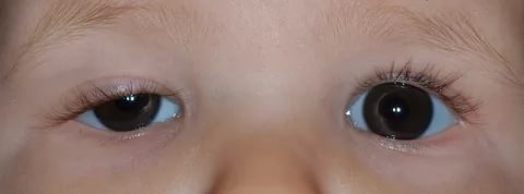
Features of the disease in children
 Ptosis is difficult to diagnose in infancy. This is largely due to the fact that most time the child sleeps and is with eyes closed. You need to carefully monitor the baby's facial expression. Sometimes the disease may manifest as frequent blinking of the affected eye during feeding.
At an older age, ptosis in children can be suspected by the following signs:
- While reading or writing, the child tries to throw back his head. This is due to the limitation of visual fields when the upper eyelid droops.
- Uncontrolled muscle contraction on the affected side. Sometimes this is mistaken for a nervous tic.
- Complaints about rapid fatigue after visual work.
Cases of congenital ptosis may be accompanied by epicanthus(overhanging folds of skin over the eyelid), damage to the cornea and paralysis of the oculomotor muscles. If ptosis in a child is not eliminated, it will lead to development and decreased vision.
Diagnostics
A routine examination is sufficient to diagnose this disease. To determine its degree, it is necessary to calculate the MRD indicator - the distance between the center of the pupil and the edge of the upper eyelid. If the eyelid crosses the middle of the pupil, then the MRD is 0, if higher, then from +1 to +5, if lower, from -1 to -5.A comprehensive examination includes the following studies:
- Determination of visual acuity;
- Determination of visual fields;
- Ophthalmoscopy with examination of the fundus;
- Examination of the cornea;
- Study of tear fluid production;
- Biomicroscopy of the eyes with assessment of the tear film.
It is very important that while determining the extent of the disease, the patient is relaxed and does not frown. Otherwise, the result will be unreliable.
Children are examined especially carefully, since ptosis is often combined with eye amblyopia. Be sure to check visual acuity using Orlova's tables.
Treatment of ptosis
Elimination of ptosis of the upper eyelid can only be done after determining the root cause
Treatment of ptosis of the upper eyelid is possible only after determining the root cause. If it is neurogenic or traumatic in nature, its treatment necessarily includes physical therapy: UHF, galvanization, electrophoresis, paraffin therapy.
Operation
As for cases of congenital ptosis of the upper eyelid, it is necessary to resort to surgical intervention. It is aimed at shortening the muscle that lifts the eyelid.
Main stages of the operation:

The operation is also indicated if the upper eyelid still remains drooping after treatment of the underlying disease.
After the intervention, an aseptic (sterile) bandage is applied to the eye and prescribed antibacterial drugs wide range actions. This is necessary to prevent wound infection.
Medicine
Drooping upper eyelid can be treated conservative method. To restore the functionality of the extraocular muscles, the following therapy methods are used:

If the upper eyelid droops after a botulinum injection, then it is necessary to instill eye drops with alphagan, ipratropium, lopidine, and phenylephrine. Such drugs promote contraction of the extraocular muscles and, as a result, the eyelid rises.
You can speed up the lifting of the eyelid after Botox with the help of medical masks and creams for the skin around the eyelids. Professionals also recommend massaging your eyelids daily and visiting a steam sauna.
Exercises
A special gymnastic complex helps strengthen and tighten the extraocular muscles. This is especially true for involutional ptosis, which occurs as a result of natural aging.
Gymnastics for the eyes with ptosis of the upper eyelid:

Only with regular performance of a set of exercises for ptosis of the upper eyelid will you notice the effect.
Folk remedies
Treatment of ptosis of the upper eyelid, especially on initial stage, perhaps at home. Folk remedies are safe, and side effects practically absent.
Folk recipes for combating ptosis of the upper eyelid:

With regular use folk remedies not only strengthen muscle tissue, but also smooth out fine wrinkles.
Amazing results can be achieved with complex application masks and massage. Massage technique:
- Treat your hands with an antibacterial agent;
- Remove makeup from the skin around the eyes;
- Treat your eyelids with massage oil;
- Perform light stroking movements on the upper eyelid in the direction from the inner corner of the eye to the outer. When treating the lower eyelid, move in the opposite direction;
- After warming up, lightly tap the skin around the eyes for 60 seconds;
- Then continuously press on the skin of the upper eyelid. Do not touch your eyeballs when doing this;
- Cover your eyes with cotton pads soaked in chamomile infusion.
Photo of ptosis of the upper eyelid


Skin of the eyelids very thin and mobile, since their subcutaneous tissue is extremely loose and devoid of fat. This contributes to the easy occurrence and rapid spread of edema with local inflammatory processes, at venous stagnation and some common diseases. The looseness of the subcutaneous tissue also explains the rapid spread of bruising and subcutaneous emphysema of the eyelids.
Sensory nerves of the skin of the eyelids come from trigeminal nerve. The upper eyelid is innervated by terminal branches coming from the first branch of the trigeminal nerve, and the lower eyelid is innervated by the second branch.
Located under the skin orbicularis eyelid muscle(m. orbicularis oculi), innervated by the facial nerve, consists of two parts - palpebral and orbital. When only the palpebral part is contracted, the eyelids slightly close; their complete closure is achieved by contraction of both parts of the muscle. Muscle fibers, running parallel to the edge of the eyelids between the roots of the eyelashes and around the excretory ducts of the meibomian glands, form the Riolan muscle; it presses the edge of the eyelid to the eye and promotes the removal of secretions from the meibomian glands to the surface of the intermarginal edge of the eyelid. Excessive tension of the orbicularis muscle leads to blepharospasm, and often to spastic volvulus, which can also be caused by contraction of the Riolan muscle, especially in the elderly.
It should be noted that with pronounced and prolonged spasm of the muscle, significant swelling of the eyelids also develops, since this greatly compresses the eyelid veins that pass between the fibers of the orbicularis muscle. Facial nerve palsy can lead to inversion of the lower eyelid and non-closure of the palpebral fissure (lagophthalmos).
TO eyelid muscles This also includes the muscle that lifts the upper eyelid (m. levator palpebrae superior), innervated by the oculomotor nerve. Starting deep in the orbit, the levator reaches the cartilage and attaches to its upper edge and anterior surface. Between the two tendon layers of the levator there is a layer of smooth fibers - the Müller muscle, innervated by the sympathetic nerve; it is also attached to the upper edge of the cartilage. In the lower eyelid there is no muscle similar to the levator, but there is a Müller muscle (m. tarsalis inferior). An isolated contraction of the Müller muscle causes only a slight widening of the palpebral fissure, therefore, with sympathetic nerve palsy, slight ptosis is observed, while ptosis with levator palsy is more pronounced and may even be complete.
A solid foundation for the century forms cartilage (tarsus), consisting of dense connective tissue. Physiological significance The cartilage of the eyelids, in addition to its protective function, is due to the presence of meibomian glands in its thickness, the secretion of which lubricates the intermarginal edge of the eyelid, protecting the skin of the eyelids from maceration by tear fluid. The most important feature The structure of the eyelids is their extremely rich blood supply. Numerous arteries anastomosing among themselves originate from two systems - from the ophthalmic artery system and from the facial artery system. Arterial branches running towards each other merge and form arterial arches - arcus tarseus. There are usually two of them on the upper eyelid, and often one on the lower eyelid.
The abundant blood supply to the eyelids is, of course, of great practical importance; in particular, this explains the excellent healing of eyelid wounds both with extensive damage and during plastic surgery.
Veins of the eyelids even more numerous than arteries; outflow from them occurs both in the veins of the face and in the veins of the orbit. It is necessary to emphasize that the orbital veins do not have valves, which are to a certain extent a natural barrier along the way venous blood. Because of this, it is difficult infectious diseases eyelids (abscess, erysipelas, etc.) can spread directly through the venous bed not only into the orbit, but also into the cavernous sinus and cause the development of purulent meningitis.
The eyelids have an anterior and posterior surface and two edges: the orbital (margo orbitalis) and the free (margo liber) - forming the palpebral fissure, the length of which is about 30 mm, height - 10-14 mm. When looking straight ahead, the upper eyelid closes top part cornea, and the lower one does not reach the limbus 1-2 mm. The upper eyelid is limited at the top by the eyebrow. The free (ciliary) edge of the eyelids is arched anteriorly. It distinguishes the anterior and posterior ribs and the intermarginal space lying between them, which has a thickness of up to 2 mm. IN medial section eyelids come together internal adhesion, forming a rounded medial corner of the eye. At the inner corner of the palpebral fissure there is a lacrimal lake (lacus lacrimalis), at the bottom of which there is a lacrimal caruncle (caruncula lacrimalis - anatomically it has the structure of the skin with rudimentary sebaceous glands, hairs and muscle fibers). More laterally, a duplication of the conjunctiva is visible - the semilunar fold. The free edge of the eyelid passes into the anterior and posterior surfaces of the eyelid, separated from them by the anterior and posterior ribs, respectively. At the inner corner, the edge of the upper and lower eyelids, at the level of the outer periphery of the lacrimal caruncle, bears lacrimal papillae with lacrimal puncta. The orbital margin is the point of transition of its skin into the skin of adjacent areas.
Eyelids perform protective function, protecting the eyeball from harmful external influences and the cornea and conjunctiva from drying out. With great mobility, the eyelids have significant strength, thanks to plates that have the consistency of cartilage. The normal blinking frequency is 6-7 times per minute, with tears evenly distributed over the surface of the cornea.
Eyelid layers:
1) skin with subcutaneous tissue– the skin of the eyelids is thin, easily removable, the subcutaneous tissue is poorly expressed, loose, devoid of fat, which is its peculiarity. Beneath the skin is the superficial fascia covering the orbicularis eyelid muscle. The rounded anterior rib has eyelashes. Modified sweat (Moll) and sebaceous (Zeiss) glands open into the hair follicles of the eyelashes.
2) muscle layer - consists of the orbicularis oculi muscle.
The circular muscle of the eye (musculus orbicularis oculi) consists of two parts:
a) palpebral (pars palpebralis) part of the upper and lower eyelids - has a semilunar shape, begins at the internal ligament and, without making a full circle, reaching the outer canthus, connects into a tendon bridge, under which lies the outer ligament of the eyelid. Some of the fibers of the palpebral part begin from the posterior process of the internal ligament and lie behind the lacrimal sac - Horner's muscle (lacrimal muscle), which expands the lacrimal sac. The muscle fibers of the palpebral part at the edge of the eyelids between the roots of the eyelashes and the gland ducts are called the ciliary muscle of Riolan (m. subtarsalis Riolani), which presses the edge of the eyelid to the eyeball and helps remove the secretion of the tarsal glands. This muscle is more pronounced in the lower eyelid and in pathological cases causes entropion of the eyelid.
b) orbital part (pars orbitalis) – begins at the inner corner of the eye from the frontal process upper jaw and, making a full circle, is attached at the place of its origin.
The orbital portion, contracting twice as slowly, has a stronger effect. Contraction of the palpebral part causes blinking movements of the eyelids and slight closure. Tight squinting, both voluntary and reflex, is ensured by contraction of the orbital portion together with the palpebral portion. The facial muscles also participate in the mechanism of closing the eyelids. The orbicularis muscle of the eyelids is innervated by the facial nerve, the fibers of which pass at great depths - almost at the level of the periosteum.
Lifting of the eyelids is carried out by the levator of the upper eyelid and smooth muscles - the superior and inferior tarsal muscles of Müller. The function of raising the lower eyelid is performed by the inferior rectus oculi muscle, which provides an additional tendon to the thickness of the lower eyelid.
The levator (musculus levator palpebrae), or muscle that lifts the upper eyelid, begins at the apex of the orbit, from the tendon ring of Zinn, and goes forward under the upper wall of the orbit. Not far from the upper edge of the orbit, the muscle passes into a broad tendon in the form of three plates, which is located behind the orbicularis muscle and the tarsoorbital fascia. The most anterior part of the tendon is directed to the tarso-orbital fascia, slightly below the upper orbito-palpebral fold, penetrates in thin bundles through this fascia and the fibers of the orbicularis muscle, reaches the anterior surface of the cartilage and spreads under the skin of the upper eyelid, where it is lost. The middle part of the tendon consists of a thin layer of fibers that are woven into the upper edge of the cartilage. The third, posterior portion is directed to the upper fornix of the conjunctiva. Attaching the levator in three places ensures simultaneous elevation of all layers of the eyelid. The levator is innervated by the oculomotor nerve (n. oculomotorius).
On the posterior surface of the levator, approximately 2 mm posterior to the junction with the tendon, the Müller muscle begins, consisting of smooth muscle fibers and attached to the upper edge of the cartilage. Its isolated contraction causes a slight widening of the palpebral fissure. Because The Müller muscle is innervated by sympathetic fibers; with paralysis of the sympathetic nerve, slight ptosis is observed. With paralysis or with transection of the levator, complete ptosis is observed.
The lower eyelid also has a Müller muscle located under the conjunctiva, from the arch to the edge of the cartilage.
The main structures that make up the levator complex include the levator body, aponeurosis, transverse ligament of the upper eyelid (Whitnall ligament), and Müller's muscle.
Whitnall's ligament (Whitnall S. E., 1932) is interesting in the following way - its superficial part, covering the muscle from above, immediately behind the aponeurosis becomes denser, forming the designated cord of the ligament, which extends in the transverse direction and, crossing the orbit, reaches its walls on both sides; the ligament is located parallel to the aponeurosis, but is attached at a higher level; medially, the main place of attachment of the ligament is the trochlea, but behind it some fascicles go to the bone, while at the same time a clearly visible strip extends forward to bridge over the superior orbital notch; Laterally, the ligamentous cord is connected to the stroma of the lacrimal gland, cutting into it like lateral horn aponeurosis, and outside the gland it reaches the outer edge of the orbit; for the most part it lies freely over the aponeurosis, but dense threads of connective tissue can bind them. In front of the ligamentous seal, the leaf suddenly becomes so thin that it forms a free edge, but it can still be traced as it extends forward in a thin layer to the upper orbital edge. This cord is well expressed in the fetus. When force is applied posteriorly to the levator, the cord becomes tense and thus acts as a limiting ligament for the muscle, preventing its excessive action - a function which, by reason of its position and attachment, it performs better than the aponeurosis, the horns of which are fixed at a level below, and which, in in the general understanding, they perform in commonwealth. The action of the levator is thus limited to the attachment of its fascial layers, as is the case with all extraocular muscles.
3) cartilage (however, there are no cartilage elements in it) - a dense fibrous plate (tarsal), which gives the eyelids their shape. Its posterior surface is tightly fused with the conjunctiva, and its anterior surface is loosely connected to the orbicularis muscle. The free edges of the plates are facing each other, the orbital edges are arched. The length of the free edge is about 20 mm, the thickness of the tarsal plate is 0.8-1 mm, the height of the lower cartilage is 5-6 mm, the upper one is 10-12 mm. The orbital margins are fixed at the edge of the orbit by the tarso-orbital fascia (anterior border of the orbit). In the region of the corners of the palpebral fissure, the tarsal plates are connected to each other and fixed to the corresponding bone walls by means of the internal (ligamentum palpebrarum mediale) and external (ligamentum palpebrarum laterale) ligaments of the eyelids. It should be noted here that the internal ligament has three processes: two go anteriorly and merge with the inner ends of the cartilages of the upper and lower eyelids, and the third bends backward and attaches to the posterior crest of the lacrimal bone. Rear end The ligaments, together with the main anterior part and the lacrimal bone, limit the lacrimal fossa. The external ligament is attached to the outer edge of the orbit at the level of the suture between the frontal and zygomatic bones. Dissection of the external commissure of the eyelids with scissors during canthotomy should not reach the bone, since it is here, under the external commissure in the thickness of the orbital part of the orbicularis muscle of the eyelid, that arterial and venous vessels pass in the vertical direction. In the thickness of the cartilage there are meibomian glands (about 30 in each eyelid) - modified sebaceous glands, the excretory ducts of which open in the intermarginal space, closer to the posterior rib.
4) conjunctiva - covers the posterior surface of the cartilage of the eyelids, runs up the posterior surface of the muscles to the levator, and downwards approximately 1 cm above the fascial processes of the inferior rectus muscle and, wrapping further onto the eyeball, forms the conjunctival fornix.
