It is important to know what the lungs are, where they are located in a person, what functions they perform. The respiratory organ is located in humans in the chest. The rib cage is one of the most interesting anatomical systems. There are also bronchi, heart, some other organs and large vessels. This system is formed by the ribs, spine, sternum and muscles. It reliably protects all important internal organs and at the expense pectoral muscles ensures the uninterrupted operation of the respiratory organ, which is almost completely occupied chest cavity... The respiratory organ expands and contracts several thousand times a day.
Where are the lungs of a person?
The lungs are a paired organ. Right and left lung play the main role v respiratory system... It is they who distribute oxygen throughout the circulatory system, where it is absorbed by erythrocytes. The work of the respiratory organ leads to the release of carbon dioxide from the blood, which breaks down into water and carbon dioxide.
Where are the lungs? The lungs of a person are located in the chest and have a very complex connecting structure with the airways, circulatory systems and lymphatic vessels and nerves. All these systems are intertwined in an area that is called the "gate". Here is located pulmonary artery, main bronchus, branches of nerves, bronchial artery. The so-called "root" contains the lymphatic vessels and pulmonary veins.

The lungs look like a vertically dissected cone. They have:
- one convex surface (ribbed, adjacent to the ribs);
- two convex surfaces (diaphragmatic, medial or median, separate the respiratory organ from the heart);
- interlobar surfaces.
The lungs are separated from the liver, spleen, colon, stomach and kidneys. The separation is carried out using a diaphragm. These internal organs border on large vessels and the heart. The back restrains them from behind.
The shape of the respiratory organ in humans depends on the anatomical features of the body. They can be narrow and elongated or short and wide. The shape and size of the organ also depend on the breathing phase.
To better understand where and how the lungs are located in the chest and how they border on other organs and blood vessels, you need to pay attention to the photos that are located in the medical literature.
Covered respiratory organ serous membrane: smooth, shiny, moist. In medicine, it is called the pleura. The pleura in the area of the pulmonary root passes to the surface of the chest cavity and forms the so-called pleural sac.
Lung anatomy
It is important to remember that the right and left lung have their own anatomical features and differ from each other. First of all, they have different amount lobes (separation occurs due to the presence of the so-called cracks located on the surface of the organ).
In the right - there are three lobes: lower; medium; top (in upper lobe there are oblique slit, horizontal slit, lobar right bronchi: upper, lower, middle).
In the left one there are two lobes: the upper one (the lingual bronchus, the tracheal keel, the intermediate bronchus, the main bronchus, the left lobe bronchi - the lower and upper, oblique fissure, the cardiac notch, the tongue of the left lung) and the lower one are located here. The left one differs from the right one in its larger size and the presence of a tongue. Although in terms of such an indicator as the volume of the right lung is greater than the left.
The lungs rest against the diaphragm with their base. The upper part of the respiratory organ is located in the clavicle.

The lungs and bronchi must be closely related. The work of some is impossible without the work of others. Each lung contains the so-called bronchial segments. There are 10 in the right, and 8 in the left. Each segment contains several bronchial lobules. It is believed that there are only 1600 bronchial lobules in the human lungs (800 each in the right and left).
The bronchi branch (the bronchioles form alveolar passages and small alveoli, which form the breathing tissue) and form a complexly woven network or bronchial tree, which provides nutrition circulatory systems oxygen. Alveoli contribute to the fact that when you exhale, the human body releases carbon dioxide, and when you inhale, it is from them that oxygen enters the bloodstream.
Interestingly, when inhaling with oxygen, not all alveoli are filled, but only a small part of them. The other part is a kind of reserve that comes into effect during physical activity or stressful situations. Maximum amount of the air that a person can inhale characterizes the vital capacity of the respiratory organ. It can be from 3.5L to 5L. In one breath, a person absorbs about 500 ml of air. This is called the tidal volume. Vital capacity lungs and tidal volume in women and men are different.
The blood supply to this organ occurs through the pulmonary and bronchial vessels. Some perform the function of gas outlet and gas exchange, others provide nutrition to the organ, these are the vessels of the small and large circle. The physiology of respiration will necessarily be disrupted if the ventilation of the respiratory organ is knocked down or the rate of blood flow decreases or increases.
Lung function
- normalization of blood pH;
- protection of the heart, for example from mechanical stress (when it hits the chest, it is the lungs that suffer);
- protection of the body from various respiratory infections(parts of the lung secrete immunoglobulins and antimicrobial compounds);
- storage of blood (this is a kind of blood reservoir human body, here is about 9% of the total blood volume);
- creating voice sounds;
- thermoregulation.
The lungs are a very vulnerable organ. Its diseases are very common all over the world and there are a lot of them:
- COPD;
- asthma;
- bronchitis different types and types;
- emphysema;
- cystic fibrosis;
- tuberculosis;
- pneumonia;
- sarcoidosis;
- pulmonary hypertension;
- pulmonary embolism, etc.
They can be provoked various pathologies, gene diseases, wrong lifestyle. The lungs are very closely connected with other organs in the human body. It often happens that they suffer even if the main problem is associated with a disease of another organ.
In structure human body quite interesting is such an "anatomical structure" as the chest, where the bronchi and lungs, heart and large vessels, as well as some other organs are located. This part of the body, formed by the ribs, sternum, spine and muscles, is designed to reliably protect the organ structures located inside it from external influence... Also, due to the respiratory muscles, the chest provides breathing, in which the lungs play one of the most important roles.
The human lungs, the anatomy of which will be discussed in this article, are very important organs, because it is thanks to them that the breathing process is carried out. They fill the entire chest cavity, with the exception of the mediastinum, and are the main ones in the entire respiratory system.
In these organs, the oxygen contained in the air is absorbed by special blood cells (erythrocytes), and carbon dioxide is also released from the blood, which then breaks down into two components - carbon dioxide and water.
Where are the lungs in humans (with photo)
When approaching the question of where the lungs are located, it is worth first paying attention to one very interesting fact concerning these organs: the location of the lungs in humans and their structure are presented in such a way that they very organically combine the airways, blood and lymphatic vessels and nerves.
Outwardly considered anatomical structures are quite interesting. In their shape, each of them looks like a vertically dissected cone, in which one convex and two concave surfaces can be distinguished. Convex is called costal, due to its direct attachment to the ribs. One of the concave surfaces is diaphragmatic (adjacent to the diaphragm), the other is medial, in other words the median (i.e., located closer to the median longitudinal plane of the body). In addition, interlobar surfaces are also distinguished in these organs.
With the help of the diaphragm, the right side of the anatomical structure we are considering is separated from the liver, and left side from the spleen, stomach, left kidney and transverse colon. The median surfaces of the organ are bordered by large vessels and the heart.
It is worth noting that the place where the lungs are located in a person also affects their shape. If a person has a narrow and long chest, then the lungs are correspondingly elongated and vice versa, these organs have a short and wide appearance with a similar shape chest.
Also, in the structure of the described organ, there is a base that lies on the dome of the diaphragm (this is the diaphragmatic surface) and an apex protruding into the neck region about 3-4 cm above the clavicle.
To form a clearer idea of what these anatomical formations look like, as well as in order to understand where the lungs are located, the photo below will be perhaps the best visual aid:
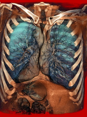

Anatomy of the right and left lung
Remember that the anatomy of the right lung is different from that of the left lung. These differences are, first of all, in the number of shares. There are three of them on the right (the bottom, which is the largest, the top, slightly smaller, and the smallest of the three is the middle), while in the left there are only two (top and bottom). In addition, the left lung has a tongue located on its leading edge, as well as this organ due to the lower position of the left dome of the diaphragm in length slightly longer than the right one.
Before entering the lungs, air first passes through other, equally important parts of the respiratory tract, in particular the bronchi.
The anatomy of the lungs and bronchi overlaps, and so much so that it is difficult to imagine the existence of these organs separately from each other. In particular, each lobe is divided into bronchopulmonary segments, which are parts of the organ, to one degree or another isolated from the same neighboring ones. Each of these sites has a segmental bronchus. In total, there are 18 such segments: 10 on the right and 8 on the left of the organ.
The structure of each segment is represented by several lobules - areas within which the lobular bronchus branches out. It is believed that a person has about 1600 lobules in his main respiratory organ: about 800 each on the right and left.
However, the conjugation of the location of the bronchi and lungs does not end there. The bronchi continue to branch, forming bronchioles of several orders, and already they, in turn, give rise to alveolar passages, dividing from 1 to 4 times and ending, in the end, with alveolar sacs, into the lumen of which the alveoli open.


This branching of the bronchi forms the so-called bronchial tree, otherwise called the airways. In addition to them, there is also an alveolar tree.
Anatomy of the blood supply to the lungs in humans

Anatomy connects the blood supply to the lungs with the pulmonary and bronchial vessels. The first, entering the small circle of the bloodstream, are mainly responsible for the function of gas exchange. Second, belonging big circle, provide nutrition to the lungs.
It should be noted that the provision of the body largely depends on the extent to which the various pulmonary areas are ventilated. It is also influenced by the relationship between the rate of blood flow and ventilation. A significant role is also played by the degree of saturation of the blood with hemoglobin, as well as the rate of passage of gases through the membrane located between the alveoli and capillaries, and some other factors. When even one indicator changes, the physiology of respiration is disturbed, which negatively affects the entire body.
Article read 99,234 times (a).
Outside, the trachea and large bronchi are covered with a loose connective tissue sheath - adventitia. The outer shell (adventitia) consists of loose connective tissue containing in large bronchi fat cells... Blood vessels and nerves pass through it. Adventitia is not clearly demarcated from the peribronchial connective tissue and together with the latter, it provides the possibility of some displacement of the bronchi in relation to the surrounding parts of the lungs.
Further inward are the fibrocartilaginous and partially muscular layers, the submucosal layer and the mucous membrane. In the fibrous layer, in addition to the cartilaginous semirings, there is a network of elastic fibers. The fibrocartilaginous membrane of the trachea is connected with the adjacent organs with the help of loose connective tissue.
The anterior and lateral walls of the trachea and large bronchi are formed by cartilages and annular ligaments located between them. The cartilaginous skeleton of the main bronchi consists of semi-rings of hyaline cartilage, which, as the diameter of the bronchi decreases, decrease in size and acquire the character of elastic cartilage. Thus, only large and medium bronchi consist of hyaline cartilage. Cartilage occupies 2/3 of the circumference, the membranous part - 1/3. They form a fibro-cartilaginous framework, which ensures the preservation of the lumen of the trachea and bronchi.
Muscle bundles are concentrated in the membranous part of the trachea and the main bronchi. Distinguish between the surface, or outer, layer, consisting of rare longitudinal fibers, and deep, or inner, which is a continuous thin shell formed by transverse fibers. Muscle fibers are located not only between the ends of the cartilage, but also enter the inter-annular spaces of the cartilaginous part of the trachea and, to a greater extent, the main bronchi. Thus, in the trachea, bundles of smooth muscles with a transverse and oblique arrangement are located only in the membranous part, that is, the muscle layer as such is absent. In the main bronchi rare groups smooth muscles are present around the entire circumference.
With a decrease in the diameter of the bronchi, the muscle layer becomes more developed, and its fibers go in a somewhat oblique direction. Muscle contraction causes not only a decrease in the lumen of the bronchi, but also some shortening of them, due to which the bronchi participate in exhalation by reducing the capacity of the airways. Muscle contraction allows you to narrow the lumen of the bronchi by 1/4. When inhaling, the bronchus lengthens and expands. The muscles reach the 2nd order respiratory bronchioles.
Inside of the muscle layer is the submucosal layer, which consists of loose connective tissue. It contains vascular and nerve formations, a submucous lymphatic network, lymphoid tissue and a significant part of the bronchial glands, which are of the tubular-acinous type with mixed muco-serous secretion. They consist of end sections and excretory ducts, which open with bulbous extensions on the surface of the mucous membrane. The relatively long length of the ducts contributes to the prolonged course of bronchitis in inflammatory processes in the glands. Atrophy of the glands can lead to drying out of the mucous membrane and inflammatory changes.
The largest number of large glands is located above the tracheal bifurcation and in the area of division of the main bronchi into lobar bronchi. Have healthy person up to 100 ml of secretion is released per day. It consists of 95% water, and 5% contains an equal amount of proteins, salts, lipids and inorganic substances... The secret is dominated by mucins (high molecular weight glycoproteins). To date, there are 14 types of glycoproteins, 8 of which are found in the respiratory system.
The mucous membrane of the bronchi
The mucous membrane consists of integumentary epithelium, the basement membrane, the lamina propria of the mucous membrane and the muscle plate of the mucous membrane.
The bronchial epithelium contains high and low basal cells, each of which is attached to the basement membrane. The basement membrane thickness ranges from 3.7 to 10.6 microns. The epithelium of the trachea and large bronchi is multi-row, cylindrical, ciliated. The thickness of the epithelium at the level of the segmental bronchi ranges from 37 to 47 microns. It consists of 4 main types of ciliates: ciliate, goblet, intermediate and basal. In addition, there are serous, brush, Clara and Kulchitsky cells.
Ciliated cells predominate on the free surface of the epithelial layer (Romanova L.K., 1984). They have an irregular prismatic shape and an oval vesicular nucleus located in the middle of the cell. The electron-optical density of the cytoplasm is low. There are few mitochondria, the endoplasmic granular reticulum is poorly developed. Each cell bears on its surface short microvilli and about 200 ciliated cilia 0.3 µm thick and about 6 µm long. In humans, the density of cilia is 6 μm 2.
Spaces are formed between adjacent cells; the cells are interconnected with the help of finger-like outgrowths of the cytoplasm and desmosomes.
According to the degree of differentiation of their apical surface, the population of ciliated cells is subdivided into the following groups:
- Cells in the phase of formation of basal bodies and axonemes. At this time, cilia are absent on the apical surface. During this period, the accumulation of centrioles occurs, which move to the apical surface of the cells, and the formation of basal bodies, from which axonemes of the cilia begin to form.
- Cells in the phase of moderate ciliogenesis and cilia growth. A small number of cilia appear on the apical surface of such cells, the length of which is 1 / 2-2 / 3 of the length of the cilia of differentiated cells. In this phase, microvilli predominate on the apical surface.
- Cells in the phase of active ciliogenesis and cilia growth. The apical surface of such cells is almost completely covered with cilia, the size of which corresponds to the size of the cilia of cells in the previous phase of ciliogenesis.
- Cells in the phase of complete ciliogenesis and cilia growth. The apical surface of such cells is entirely covered with densely spaced long cilia. The electron diffraction patterns show that the cilia of adjacent cells are oriented in the same direction and are curved. This is an expression of mucociliary transport.
All these groups of cells are clearly distinguishable in photographs obtained using light electron microscopy (SEM).
The cilia are attached to the basal bodies located in the apical part of the cell. The cilium axoneme is formed by microtubules, of which 9 pairs (doublets) are located along the periphery, and 2 single pairs (singlets) are located in the center. Doublets and singlets are connected by nexin fibrils. On each of the doublets, on one side, there are 2 short "handles" that contain ATP-ase, which is involved in the release of ATP energy. Due to this structure, the cilia oscillate rhythmically with a frequency of 16-17 in the direction of the nasopharynx.
They move the mucous membrane covering the epithelium at a speed of about 6 mm / min, thereby ensuring continuous drainage function of the bronchus.
Ciliated epithelial cells, according to most researchers, are at the stage of final differentiation and are not capable of division by mitosis. According to modern concept Basal cells are precursors of intermediate cells that can differentiate into ciliated cells.
Goblet cells, like ciliated cells, reach the free surface of the epithelial layer. In the membranous part of the trachea and large bronchi, ciliated cells account for up to 70-80%, and goblet cells account for no more than 20-30%. In those places where there are cartilaginous half rings along the perimeter of the trachea and bronchi, zones with a different ratio of ciliated and goblet cells are found:
- with a predominance of ciliated cells;
- with an almost equal ratio of ciliated and secretory cells;
- with a predominance of secretory cells;
- with full or almost complete absence ciliated cells ("non-ciliated").
Goblet cells are unicellular glands of the merocrine type that secrete mucous secretions. The shape of the cell and the location of the nucleus depend on the phase of secretion and the filling of the supranuclear part with mucus granules, which merge into larger granules and are characterized by a low electron density. Goblet cells have an elongated shape, which, during the accumulation of secretions, takes the form of a glass with a base located on the basement membrane and intimately connected to it. The wide end of the cell domed protrudes on the free surface and is equipped with microvilli. The cytoplasm is electron dense, the nucleus is rounded, the endoplasmic reticulum is of a rough type, well developed.
Goblet cells are unevenly distributed. Scanning electron microscopy revealed that different zones the epithelial layer contains heterogeneous areas, consisting either only of ciliated epithelial cells, or only of secretory cells. However, continuous accumulations of goblet cells are relatively few in number. Along the perimeter of the section of the segmental bronchus of a healthy person, there are areas where the ratio of ciliated epithelial cells and goblet cells is 4: 1-7: 1, and in other areas this ratio is 1: 1.
The number of goblet cells decreases distally in the bronchi. In the bronchioles, goblet cells are replaced by Clara cells, which are involved in the production of serous components of mucus and alveolar hypophase.
In small bronchi and bronchioles, goblet cells are normally absent, but can appear in pathology.
In 1986, Czech scientists studied the reaction of the epithelium of the airways of rabbits to the oral administration of various mucolytic substances. It turned out that goblet cells are the target cells for the action of mucolytics. After mucus is removed, goblet cells tend to degenerate and are gradually removed from the epithelium. The degree of damage to the goblet cells depends on the injected substance: lasolvan gives the greatest irritating effect. After the administration of broncholysin and bromhexine, massive differentiation of new goblet cells occurs in the epithelium of the airways, resulting in goblet cell hyperplasia.
Basal and intermediate cells are located deep in the epithelial layer and do not reach the free surface. These are the least differentiated cellular forms, due to which physiological regeneration is mainly carried out. The shape of the intermediate cells is elongated, the basal cells are irregularly cubic. Both have a rounded, DNA-rich nucleus and a small amount of cytoplasm, which is dense in basal cells.
Basal cells are capable of giving rise to both ciliated and goblet cells.
Secretory and ciliated cells are united under the name "mucociliary apparatus".
The process of movement of mucus in airways of the lungs is called mucociliary clearance. The functional efficiency of MCC depends on the frequency and synchronism of movement of the cilia of the ciliated epithelium, and also, which is very important, on the characteristics and rheological properties of mucus, i.e., on the normal secretory capacity of the goblet cells.
Serous cells are few in number, reach the free surface of the epithelium and are distinguished by small electron-dense granules of protein secretion. The cytoplasm is also electron dense. Mitochondria and rough reticulum are well developed. The nucleus is round, usually located in the middle of the cell.
Secretory cells, or Clara cells, are most abundant in the small bronchi and bronchioles. They, like serous ones, contain small electron-dense granules, but are distinguished by a low electron density of the cytoplasm and a predominance of a smooth, endoplasmic reticulum. The rounded nucleus is located in the middle of the cell. Clara cells are involved in the formation of phospholipids and, possibly, in the production of a surfactant. Under conditions of increased irritation, they, apparently, can turn into goblet cells.
Brush cells carry microvilli on the free surface, but lack cilia. The cytoplasm of their low electron density, the nucleus is oval, bubble-shaped. In the manual of Ham A. and Cormac D. (1982), they are considered as goblet cells that have secreted their secret. Many functions are attributed to them: absorption, contractile, secretory, chemoreceptor. However, in the human airways, they are practically not studied.
Kulchitsky's cells are found throughout the bronchial tree at the base of the epithelial layer, differing from the basal ones by the low electron density of the cytoplasm and the presence of small granules, which are detected under electron microscope and under light when impregnated with silver. They are referred to as neurosecretory cells of the APUD system.
Under the epithelium is the basement membrane, which consists of collagen and non-collagen glycoproteins; it provides support and attachment of the epithelium, is involved in metabolism and immunological reactions. The condition of the basement membrane and the underlying connective tissue determines the structure and function of the epithelium. The lamina propria is the layer of loose connective tissue between the basement membrane and the muscle layer. It contains fibroblasts, collagen and elastic fibers. The lamina propria contains blood and lymph vessels. The capillaries reach the basement membrane, but do not penetrate into it.
In the mucous membrane of the trachea and bronchi, mainly in the lamina propria and near the glands, free cells are constantly present in the submucosa, which can penetrate through the epithelium into the lumen. Among them, lymphocytes predominate, less often plasma cells, histiocytes, mast cells(mast cells), neutrophilic and eosinophilic leukocytes. The constant presence of lymphoid cells in the bronchial mucosa is designated by the special term "broncho-associated lymphoid tissue" (BALT) and is considered as an immunological defense reaction to antigens that penetrate the air into the respiratory tract.
The structure of the bronchi
Bronchi (which in Greek means breathing tubes) represent the peripheral part of the respiratory tract, through which atmospheric - oxygen-rich - air enters the lungs, and exhausted, oxygen-poor and carbon-dioxide-rich air is removed from the lungs, which is no longer suitable for breathing.
Gas exchange takes place in the lungs between air and blood; oxygen enters the blood, and carbon dioxide is removed from the blood. Thanks to this, the vital activity of the organism is supported. But the bronchi do not just conduct air into the lungs, they change its composition, humidity, and temperature. Passing through the bronchi (and other respiratory tract- nasal cavity, larynx, trachea), the air is heated or cooled to the temperature of the human body, moistened, freed from dust, microbes, etc., which protects the lungs from harmful effects.
Implementation of these complex functions provided by the structure of the bronchi. 2 main bronchi depart from the trachea large diameter(average 14-18 mm) to the right and left lung... From them, in turn, depart smaller - lobar bronchi: 3 on the right and 2 on the left.
The lobar bronchi are divided into segmental (10 each on the left and on the right), and those, gradually decreasing in diameter, into bronchi of the fourth and fifth order, which pass into the bronchioles. This division of the bronchi leads to the fact that not a single functional unit of the lungs (acinus) is left without its own bronchiole, through which air enters it, and all lung tissue can participate in breathing.
The collection of all bronchi is sometimes called the bronchial tree, since, dividing and decreasing in diameter, they very much resemble a tree.
The wall of the bronchi has complex structure, and the most complex is the wall of large bronchi. There are 3 main layers in it: 1) outer (fibrocartilaginous); 2) medium (muscular); 3) internal (mucous membrane).
The fibrocartilaginous layer is formed cartilage, collagen and elastic fibers, smooth muscle bundles. Thanks to this layer, the elasticity of the bronchi is provided, and they do not collapse. With a decrease in the diameter of the bronchi, this layer becomes thinner and gradually fades away.
The muscle layer consists of smooth muscle fibers, combined in circular and oblique beams; with their reduction, the lumen of the airway changes. With a decrease in the caliber of the bronchus, the muscle layer becomes more developed.
The mucous membrane is very complex and plays an important role. It consists of connective tissue, muscle fibers, permeated with a large number of blood and lymphatic vessels... It is covered with a columnar epithelium, equipped with ciliated cilia, and a thin layer of serous-mucous secretion to protect the epithelium from damage. Thanks to this structure, it performs a certain protective role.
The cilia of the columnar epithelium are able to capture the smallest foreign bodies(dust, soot), trapped in the air in the bronchi. By settling on the mucous membrane of the bronchi, dust particles cause irritation, which leads to abundant discharge mucus and appearance cough reflex... Due to this, they, together with mucus, are excreted from the bronchi to the outside. Thus, the lung tissue is protected from damage. Thus, a cough in a healthy person plays a protective role, protecting the lungs from the penetration of the smallest foreign particles.
With a decrease in the diameter of the bronchi, the mucous membrane becomes thinner and the multi-row columnar epithelium turns into a single-row cubic one. It should be noted that the mucous membrane contains goblet cells that secrete mucus, which plays an important role in protecting the bronchi from damage.
Mucus (which in a person forms up to 100 ml during the day) performs another important function... It humidifies the air entering the body (atmospheric humidity is somewhat lower than in the lungs), thereby protecting the lungs from drying out.
The role of the bronchi in the body
Passing through the upper respiratory tract, the air also changes its temperature. As you know, the temperature of the air surrounding a person fluctuates depending on the season in rather significant ranges: from -60-70 ° to + 50-60 °. Contact of such air with the lungs would inevitably cause damage. However, the air passing through the upper respiratory tract is heated or cooled, depending on the need.
The bronchi play the main role in this, since their wall is abundantly supplied with blood, which ensures good heat exchange between blood and air. In addition, the bronchi, by dividing, increase the contact surface between the mucous membrane and air, which also contributes to rapid change air temperature.
The bronchi protect the body from the penetration of various microorganisms (of which there are quite a few in the atmospheric air) due to the presence of villi, the secretion of mucus, which contains antibodies, phagocytes (cells that devour microbes), etc.
Thus, the bronchi in the human body are an important and specific organ that ensures the conduction of air into the lungs, while protecting them from various external stimuli.
Conductor defense mechanisms bronchus is nervous system, which mobilizes and controls all the protective mechanisms of the body (humoral, immunobiological, endocrine, etc.). However, when the protective mechanisms of the bronchi are disturbed, they lose their ability to fully resist the effects of various harmful factors... This leads to the occurrence in the bronchi pathological process- bronchitis develops.
Related materials:
There are no similar materials ...
Everyone needs to know where the bronchi are located. This will help in the event that therapy or diagnosis is needed. In addition, it is the bronchi that are a vital organ, without normal work which man will not live long. Human anatomy is both an interesting and complex area of science that you need to know everything about.
The bronchi are a paired organ that is a natural extension of the trachea. At the level of the fourth (for males) and fifth (for females) vertebra, the tracheal region divides, forming two tubes. Each of them is directed towards the lungs. After penetration into the pulmonary region, they are divided again: into three and two branches, respectively, the right and left parts.
The location shown corresponds to parts of the lung repeating his drawing. It should be noted that:
- the location where the lungs are located in a person has a direct effect on their shape;
- if a person's chest is narrow and long, then the epithelium and lungs will take the indicated shape;
- the presented organs of the human type are characterized by a short and wide appearance with a conjugated form of the chest, which predetermines the functions of the bronchi.
The structure of the bronchial region
All bronchial lobes are subdivided into fragments of the bronchopulmonary type. They are sections of an organ that are isolated from similar adjacent areas. In each of the presented areas, there is a segmental bronchus. There are 18 similar segments: 10 on the right and 8 on the left, which is confirmed by the figure.
The structure of each of the presented segments has several lobules, or sections, inside which the lobular bronchus is divided, which are located on top.
Pulmonologists assure that a person has at least 1600 lobules: 800 each on the right and left sides.
The similarity in the placement of the bronchial and pulmonary regions does not end there. The former, like the epithelium, branch out further, forming bronchioles of the secondary and tertiary order. They give rise to alveolar-type ducts, which divide 1 to 4 times and end with alveolar sacs. Alveoli open into their lumen, which is why human anatomy is logical. It is she who predetermines the functional significance of the presented organ.
Functional features
The function of the bronchi is multifaceted - it is the conduction of air masses through the respiratory system during inhalation and exhalation, protective and drainage functions. Due to the latter two, foreign bodies that have got inside with air masses leave the respiratory system by themselves. Thus, the human anatomy removes harmful microorganisms.
The epithelium of the bronchial region includes goblet-type cells that contain mucus. Foreign bodies and objects adhere to it, and the ciliary part of the epithelium sets the presented mucus in motion and contributes to the removal of the object outside. The presented process provokes a cough in a person, which does not always manifest itself with bronchitis. The functional significance of the bronchi may lie in other actions:

How to maintain bronchial health
The structure of the bronchi should remain complete, without flaws and foreign complications. This will keep your bronchi in perfect health. For this, use medicines(bronchodilators, mucolytics and expectorants), resort to special diet and maintaining healthy way life. The latter excludes the use of alcoholic beverages, nicotine addiction.
Shown high physical activity, that is, daily hiking, hardening, charging.
All this will strengthen the body, which cannot be achieved without constant efforts.
Another condition for bronchial health is exercise breathing exercises and visiting sanatoriums. They strengthen the immune system, optimize the functioning of the pulmonary system, which has a positive effect on the structure of the bronchi and, accordingly, the respiratory process. In this case, the epithelium and respiratory pattern will not be subject to complications in terms of the general condition.
Additional Information
Failure to comply with medical recommendations and maintaining an unhealthy lifestyle provoke the formation of bronchial diseases. The most common are bronchitis, which is caused by inflammation of the bronchial walls. Pathology is formed under the influence of viruses and bacteria, some of which are needed by the body in minimal quantities.
Another complication is bronchial asthma, which is characterized by attacks of asphyxia, forming with a clear cyclicality. Allergic exposure, air pollution, all kinds of infections can become a catalyst for this. To others negative processes refers to:
- bronchial tuberculosis, accompanied by a forced cough with the excretion of a significant ratio of sputum and aggravated breathing;
- candidiasis, which forms when the protective functions of the body are weakened, when the epithelium is weakened, forming a fuzzy pattern;
- cancer, in which the human anatomy changes, and the pathology is accompanied persistent cough with discharge of light pink sputum and edema.
Thus, in order for the bronchi to remain absolutely healthy, it is necessary to know everything about their location, division into certain parts and the nuances of maintaining health. This will allow you to maintain maximum activity, heal the bronchi and lungs, making it possible to live a full life.
