Representing the device and location of the internal organs of a person, you can independently determine the source pain and which specialist should be contacted for help in the first place. Every organ human body occupies a certain place and has its own unique structure.
In this regard, you should know the location of the internal organs of a person in order to independently diagnose the localization of pain and immediately contact the right doctor.
The structure, location and function performed are closely interconnected, the images in the article and the video after will help to facilitate the process of remembering. Conditionally human body It is customary to divide into three cavities, inside which all organs of the human body are located:
- Thoracic cavity - from the neck to the end of the sternum.
- Abdominal cavity - from the end of the sternum to the hip joint.
- Pelvic cavity (small and greater pelvis) - within the boundaries of the hip joints.
The chest cavity is separated from the abdominal cavity by a special muscle - the diaphragm, designed to expand the lungs. Human internal organs: the layout and structural features begin to be studied, as a rule, from top to bottom - from the neck to the pelvic organs. Therefore, the first organ is the thyroid gland, located in the neck, usually under the Adam's apple.
However, its location in adult not always . It can grow in size or get smaller, in some cases, there is a prolapse of this organ of the endocrine system.
Location of organs in the chest cavity
The location of the photo below shows the internal organs of a person more clearly. Here are located the heart, lungs, bronchi and mysterious thymus also called the thymus.
Heart
 Responsible for ensuring the movement of blood in the vessels important element cardiovascular system - the heart. Its location is rib cage above the diaphragmatic muscle. To the right and left of it are the lungs.
In this case, the heart is not located with symmetrical accuracy in the center of our body, but slightly at an angle. Two-thirds of the cardiomuscle is located to the left of the midline, and a third to the right. The shape of the heart is an individual sign and depends on age, gender, body constitution, health status, etc.
Lungs
The location of the internal organs of a person in a picture that schematically reflects the structure chest cavity, continue the most important elements of the respiratory system - the lungs. Their volume is slightly less than the cavity, and the dimensions themselves depend on the phase of the respiratory cycle: they expand on inspiration, and contract on exhalation. The shape of the lungs resembles a truncated cone. The base of this cone rests on the diaphragmatic muscle in the form of a dome, and the apex is directed to the subclavian region.
Bronchi
![]() Anyone who is familiar with the device can show the location of the internal organs of a person in the chest cavity bronchial tree- continuation of the trachea outside the windpipe. Each of the branches of this tree is in a strict hierarchy and has its own name and structural features. Directly from the trachea are two main bronchi, each of which goes to the corresponding lung. The thin, long and not so vertical left main bronchus can be easily distinguished from the right in the figure.
Anyone who is familiar with the device can show the location of the internal organs of a person in the chest cavity bronchial tree- continuation of the trachea outside the windpipe. Each of the branches of this tree is in a strict hierarchy and has its own name and structural features. Directly from the trachea are two main bronchi, each of which goes to the corresponding lung. The thin, long and not so vertical left main bronchus can be easily distinguished from the right in the figure.
In the lungs of the internal organs of a person, the location depends on the place of their localization: on the surface of the lung or inside it. Therefore, the bronchial branches of the first and second order are called extrapulmonary, and all the rest will be intrapulmonary. Each order has its own name: the first is equity, the second is segmental, the third is subsegmental, etc. Branching ends with bronchioles, gradually passing into the alveoli of the lungs.
Thymus
For a long time, why same exactly intended thymus iron for scientists remained a mystery. If you look at the location of the internal organs of a person in the video, you can see that it is located at the very top of the sternum.
To date, its role has also been studied. It is now known that the thymus gland is the most important element immune system. The name is identified with appearance: the shape of the gland resembles a two-pronged fork.
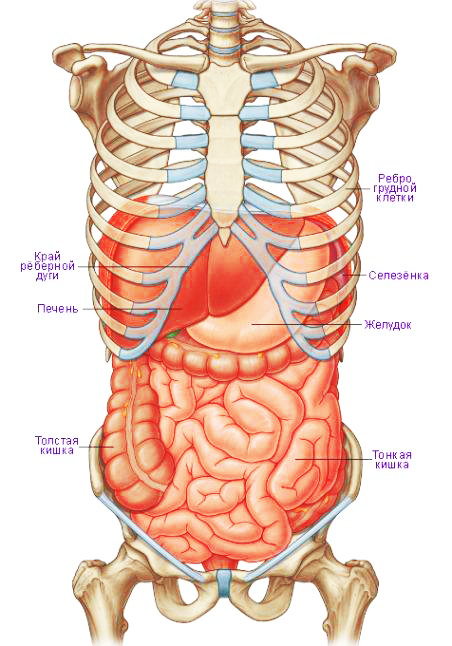
Location of organs in the abdominal cavity
The abdominal cavity is the focus of almost all elements gastrointestinal tract, digestive glands and organs of the excretory system. The incoming "food lump" begins to be digested in the stomach, then it enters the intestines, from where the ducts of the pancreas and gallbladder open, collecting the secret of the liver.
Absorption is completed in the large intestine, while filtration continues in the kidneys and spleen. The adrenal glands are also present here, controlling many processes in our body. A drawing will help to better understand the location of the internal organs of a person.
Stomach
The abdominal cavity is separated from the thoracic cavity by the diaphragm, therefore, immediately below it, to the left of the midline, is the stomach - a sac-like outgrowth of the digestive canal. Its main function is the primary reservoir for food and the first stage in the breakdown of incoming complex nutrients into simpler elements.
The fullness of the stomach determines its size. Food from the esophagus enters the stomach, where gastric juice biological oxidation processes begin.
Pancreas
The location of the internal organs of a person in the peritoneal region is subject to their role in metabolic processes. Therefore, immediately below the stomach, closer to the spine, there is a place of permanent localization of the pancreas. This is one of the largest secretory organs of the human body, performing a dual function.
The pancreatic juice produced by it is saturated with digestive enzymes and is a waste product of the exocrine gland. At the same time, the pancreas secretes a whole complex of hormones that regulate the metabolism of proteins, fats and carbohydrates, as it should be for the endocrine glands.
Liver
Describing internal organs human, the layout of which is limited to the abdominal region, one cannot help but dwell on the liver - one of the vital elements of our body. The liver is located right side from the stomach under the dome of the diaphragm and is an organ of purification.
It consists of two unequal lobes: a small left and a large right, occupying the upper right position under the diaphragm. She is in charge of a whole range of physiological programs, the slightest failure in the implementation of which is detrimental to the body:
- neutralization medicines and other unsafe substances into substances that are less toxic in composition;
- excretion of excess hormones and other substances;
- satiety and blood glucose control;
- regulation of fat metabolism, production of cholesterol and lipids;
- participation in the hematopoietic processes of the embryo, etc.
gallbladder
The main function of this organ is the accumulation of bile (a greenish viscous liquid) synthesized by the liver, and its excretion in 12 duodenum through the bile and cystic ducts. It is located in the lower part of the liver, on the border of its lobes. By shape gallbladder resembles a pouch of a longitudinal shape, having very thin walls.
In this kind of sac, the bile produced by the liver is collected, which is then portioned out into the area duodenum to regulate the digestion of dietary fats. Some internal organs, the location of the photo will only help to find out, for example, the gallbladder is localized in the lower third of the liver can be seen in the pictures at the end of the article.
Spleen
Considering the location of the internal organs of a person in the pictures in the peritoneum, at the top left you can see the spleen in shape resembling a flattened hemisphere. In shape, it is represented by a hemisphere, which is flattened. In both a child and an adult, this organ performs hematopoietic and immune function through the formation of lymphocytes.
Also, the spleen filters the flow of platelets and red blood cells if their structure is damaged. If we continue talking about filtration, it can be noted that the spleen also does not allow protozoa, foreign particles and bacteria to pass. It is also worth noting that this body takes an active part in the exchange of food.
Functions of the spleen
The spleen is actively involved in both the hematopoietic and immune processes of our organism, namely:
- formation of lymphocytes and filtration of microorganisms;
- participation in metabolic reactions and filtration of damaged blood cells;
- platelet accumulation and platelet organ and hematopoietic organ at the initial stage of embryonic development.
Intestines
It is quite simple to imagine the location of the internal organs of a person below the stomach, since all this space is occupied by a compactly laid intestine. A long tangled tube starting immediately from the stomach is small intestine, which on the right is transformed into colon. The latter describes a kind of circle, the end point of which is the anus.
The smooth functioning of the intestines is the key to the health of the human body. Two-thirds of all cells that provide immunity are localized in this area of the human internal organs: the location of such a large number of immunocytes is the best proof of the importance of this organ. Starts peristalsis drunk on an empty stomach glass warm water while cleansing the entire body of accumulated toxins.
kidneys
One of the few organs represented by a pair. One of the few organs represented by a pair. The kidneys are paired bean-shaped elements of the urinary system. They are to the right and left of spinal column in the area of lumbar. Their sizes do not exceed 10-12 cm, while right kidney slightly smaller than the left. By studying the location of the internal organs of a person on video, you can understand the main function kidneys, which is maintaining invariability in the internal environment and in urination. It is the main organ of the urinary system.
The localization of the kidneys in the body is the lumbar region, behind the intestines, and, accordingly, the parietal abdominal sheet. Without pathology, this organ has a weight of 110 to 190 grams. The main functions of the kidneys are the secretion and filtration of urine, the regulation of chemical homeostasis.
The kidneys are divided into cortex and medulla. On its side is the renal pelvis, in which there is an opening for the renal vein, artery, and also for the ureter. From above, this organ is covered with a fibrous membrane.
adrenal glands
Localized at the top of the cortical substance of the kidney from the outside. These are paired, like the kidneys, glands internal secretion. Like the kidneys, they consist of a cortical (outer) substance and a medulla (internal). The activity of the adrenal glands is regulated by the parasympathetic and sympathetic nervous systems.
They, in turn, regulate metabolism, and also help the body adapt to changes. external environment. The latter function is due to the fact that the synthesis of norepinephrine, adrenaline, corticosteroid hormones and androgens, which are the main hormones of the human reproductive system, is carried out in the adrenal glands.
On the upper parts kidneys are localized paired endocrine glands - the adrenal glands, consisting of the medulla and cortical substance. Their main function is to regulate metabolic processes, especially during stressful situations and adaptation period.
How are the organs of the large and
Between hip joints the location of the internal organs of a person in the figure fits into the following sequence: if the body is female, then the ovaries and uterus are localized here, if the male is the testicles and prostate. This is the location of the bladder.
ovaries
These are the glands of the reproductive system, represented by a pair and performing endocrine function. They synthesize female sex hormones (steroid, estrogen, partially androgens), as well as the maturation and excretion of the cells of the reproductive system.
Localized on both sides of the walls of the uterus. As already mentioned, a pair of female sex glands - the ovaries, not only produces hormones (estrogen, steroids and weak androgens), but also serves as a place for the development and maturation of eggs.
Uterus
The uterus is a hollow organ made of smooth muscles, the purpose of which is to carry the fetus during pregnancy. The pelvis is a relatively small cavity, so the location of the internal organs of a person in this area is described relative to each other. So, the uterus is located in front of the rectum directly behind bladder.
The rounded lower part ends with the cervix. The size of this organ depends on the presence / absence of pregnancy and the stage of embryonic development. During gestation, the uterus increases along with the fetal egg, and after childbirth returns to its usual size, not exceeding 10 cm.
Bladder
In the lower third of the small pelvis is a hollow smooth muscle element of the excretory system - bladder. Its functionality is associated with the reservation of urine secreted by the kidneys and its subsequent excretion during urination. V male body below it is the prostate gland, and in the female behind it is the vagina.
By imagining the location of the internal organs in your body, you can quickly identify the suffering organ and build a constructive conversation with the doctor. And this, in turn, will lead to a more accurate diagnosis and the appointment of a timely and effective treatment which will have a positive effect on the speed of recovery.
Location of internal organs: table and figures
 |
 |
 |
Video: Anatomy through human eyes
The structure of the human body has long worried leading scientists and researchers. Over the course of a thousand years, a wide variety of theories have been put forward that have tried to explain the simple, and at the same time complex issue How exactly does the human body function?
It is important to understand the fact that most of the work of ancient researchers was carried out in secret from everyone. This is due to the fact that the religious canons of that time did not allow a detailed study of the human body.

Avicenna and Paracelsus
Scientists such as Avicenna and Paracelsus drew their first conclusions from the dissection of cadavers. It sounds a little unpleasant, but only such material could answer a lot of questions.
Human anatomy is needed for several purposes: first of all, we need to know what a healthy and strong organism is.
And treatment can be carried out only if the changes that occur inside organs and systems under the influence of diseases and pathogens are known.
In simple terms, it is impossible to treat diseases without understanding those pathological processes and changes that occur at the level of organ systems, organs, tissues and cells.
The primitive tools of the first scientists were rather scarce: only a lancet (the world's first scalpel), a bright head for understanding what they saw, and paper with a pen were present. The data obtained were recorded in diaries, and the first detailed diagrams and drawings appeared several centuries before our era.
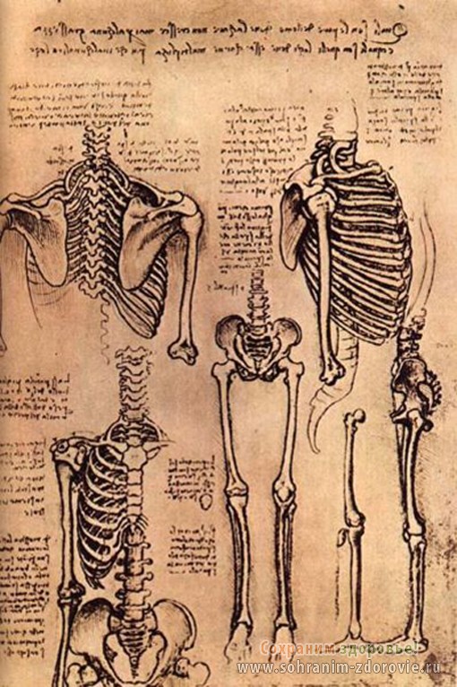
Rice. 1. Human skeleton painted by Leonardo da Vinci
Research methods were gradually improved, the information obtained acquired a clearer structure. And the communication of leading doctors among themselves made it possible to create single system terms and concepts in medicine.
A new era began in the 18th century scientific discoveries: scientists not only actively study anatomy, but also reveal the secrets of new sciences. Among them is histology, which actively studies the structure and functions of tissues, embryology, the science of the development of the fetus in the mother's womb.

After Harvey studied the properties of blood in detail, scientists had many unresolved questions.
One of the main secrets concerned the transfer of blood from arteries to veins.
Marcello Malpighi fully confirmed the guesses of his colleagues who spoke about the presence of transitional bridges or anastomoses.
With the advent of the microscope, all theories were confirmed. Capillaries were discovered, which turned out to be the missing link in anatomical structure circulatory system.
New tools have allowed scientists to dive deeper into this fascinating science.
Organs as an object of study
As you know, in our body is a large number of specialized bodies, each with its own function. The stomach is part digestive system and is responsible for the digestion of food eaten, the heart is part of the circulatory system that saturates nutrients every cell in the body.
A detailed study of each organ allows you to get a deeper picture of the functioning of the whole organism as a whole. The main task of anatomical research is to understand the functions that this or that object of research performs.
As modern methods studies apply examination of the body and its autopsy after death. The days of old prejudice are long gone, so scientists can go about their work without too much fear. All the results obtained during the study are recorded in detail and recorded.
On the basis of the data obtained, it is possible to draw conclusions about the functions of the organ, its state at the time of a person's life, and to establish the exact cause of death.
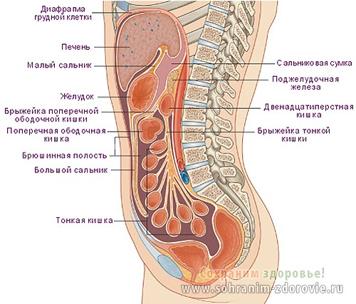
Rice. 2. Sagittal (longitudinal) section of a woman's body.
Unfortunately, a dead human body cannot provide answers to all questions. You will not be able to see how the cells are saturated with nutrients and absorb oxygen, you cannot see the growth processes in the tissues.
Therefore, as an object for research, living and healthy person. In such cases, the main tools for research are not a scalpel with knives, but X-ray machines, scanning systems and magnetic resonance imaging.
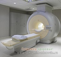
This amazing device allows you to conduct research in several planes and make the so-called "slices".
The field of study can be very diverse: starting from the study of the organ as a whole, and uploading it to a functional unit - a cell. For example, for muscles, the basic structural unit is a cell called a myocyte, a neuron is the main building block from which our nervous system is built.
If we consider the lungs, then the process of gas exchange takes place in a small sac called the alveolus.
Suitable for her blood vessels of two types: arterioles carry blood enriched with oxygen, and venules (microscopic veins) bring carbon dioxide to the lungs, which was formed during the life of our body.
Human organs as an important element of the whole organism
Based on the data obtained on the structure of each cell, conclusions can be drawn about the structure of the entire human body as a whole. To obtain accurate data, there are five main sections in anatomy. Each of them has a clearly defined range of tasks:
- systematic anatomy deals with the introductory part of this science. There is a study of basic terms and concepts that are unknown to beginners. This section deals with the study of all existing systems in the human body.
- topographic anatomy is a separate field of science that studies the location of internal organs. This section is studied by all future doctors, but this knowledge is especially useful for surgeons. Can't be held surgical intervention on the stomach, if the doctor does not know exactly where it is located. In addition, there are pathological options for the placement of internal organs, up to a mirror image (for such people, the heart is on the right, the stomach is shifted to the left, the kidneys also change places, etc.)
- plastic anatomy deals with such important issues, as features of the structure of the body and external features of a person. This section is studied in detail by practitioners and future plastic surgeons. As you know, these doctors specialize in the correction various pathologies. For example, correction of the shape of the nose after an injury, breast augmentation, removal of wrinkles and folds on the body. This section studies the internal organs in terms of changes in our appearance under the influence of internal factors. An example is thyroid gland: with an excess amount of hormones produced, a person significantly loses weight, facial features are sharpened, and a huge goiter appears on the neck.
With regard to operations on the mammary glands of women, they cannot be performed without gaps in the field of anatomical knowledge.
- comparative anatomy studies such important point as the development of the human body under the influence of evolution. According to Darwin's theory, over the course of millions of years, man has changed significantly. The first species were not "homo sapiens" or Homo sapiens. In the early stages of evolution, our ancestors learned to walk upright, used primitive tools and learned the basics of hunting. And some organs are vestigial, or obsolete. They came to us from the ancestors of animals.
An example is the appendix - this process is not involved in the processes of digestion of the human body. But in animals, it plays one of the key functions. The same applies to the coccyx - a person does not need it, and in animals a tail is attached to this part of the spine.
- anthropological anatomy deals with the study of such issues as racial characteristics, gender and age differences in individuals. The study of this section allows you to understand evolutionary changes, features of the development of people of a particular race. For example, Africans do not just have black skin. This reliable protection from scorching sunlight, which, if exposed for a long time, can lead to burns and sunstroke. The inhabitants of the far north have a characteristic, narrow section of the eyes. This is due to the impact of powerful winds and snowstorms, which are often found in those areas.
If you are interested in human anatomy, internal organs, pictures will help to more clearly present the structure of the human body.
Without a visual representation, it is very difficult to understand what exactly is being discussed in a particular case. For these purposes, there are various scientific manuals that demonstrate the structure of internal organs, their location.
Atlases are the most popular: such publications are published every year, constantly filled with new elements and images. The information received will be of interest to everyone: as students medical universities, and ordinary people who are interested in science.
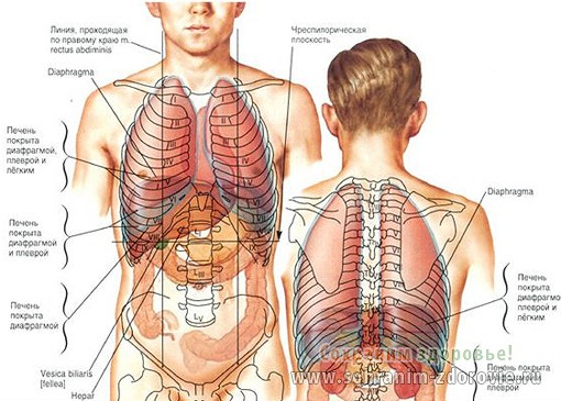
Rice. 3. The location of the internal organs in a man.
As for doctors and medical staff, then such atlases have long turned into a reference book. Even the acquired knowledge must be periodically updated and consolidated. Therefore, it will be useful to open the atlas and flip through a few pages.
There is one more interesting direction in the field of studying the structure of the human body, it is ...
Pathological anatomy
It is a scientific discipline that studies all pathological processes in organs, tissues and cells that occur during various diseases. Many scientists believe that the founder of this direction is Rudolf Virchow, the famous German scientist.
He was the first to suggest that all changes occur at the cellular level. Up to this point, there were many theories, but none of them considered the cell as the main place of struggle between the body and pathogens.
Modern pathological anatomy studies the causes of the disease (the so-called etiology), the main mechanisms of its development (in science, the term is known as pathogenesis) and various forms diseases (among scientists this phenomenon known as pathomorphosis.)
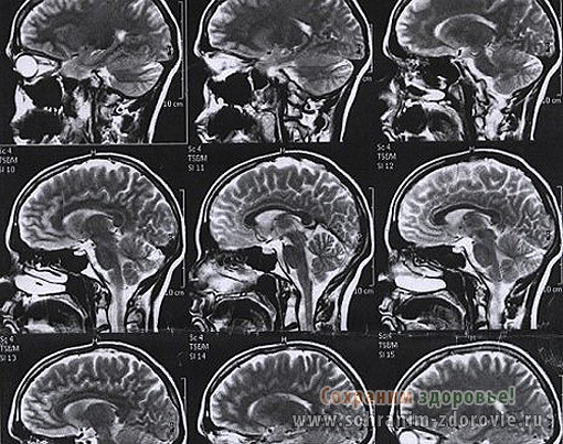
Rice. 4. Results of magnetic resonance imaging of the brain.
The main tasks of modern pathological anatomy look like this:
- Identification of the causes of pathological changes and conditions for the development of the disease. The causative agent is called a pathogen and is a leading factor in the development of changes in cells and organs.
- The study of the mechanism of development of certain pathogenic processes. At the same time, in the human body there are characteristic changes, which allow you to more accurately determine the type of disease.
- The study of such a phenomenon as the general picture of the disease (identification of the main signs both at the macro and at the micro level.)
- Pathological anatomy actively explores the outcome of each disease and possible complications for the human body.
- Studying the changes that occur in human body after taking medicines or procedures performed.
- A detailed study of the diagnosis based on the totality of the data obtained.
- With help various methods all pathological processes are studied. Research is carried out both during the life of the patient and after his death.
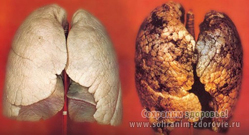
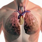
Rice. 5. Pathological changes in the lungs of a smoker.
Human anatomy, internal organs and the structure of the human body is a fascinating science.
Its study is carried out as part of school curriculum, as well as in specialized medical institutions. But it is not at all necessary to go to universities to get the information you are interested in.
To do this, it is enough to acquire the relevant literature and an anatomical atlas. And we invite you to watch a video about how alcohol affects the internal organs of a person. What happens to them and much more. Take care of yourself and your health!
Sincerely, the team of the project “Save Health!
