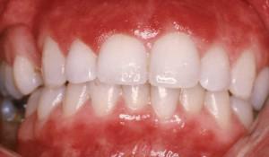
Oral diseases are largely determined by lifestyle, nutrition, environmental influences and household habits. At all times, the problem was reasonably associated with hygiene and the general condition of the body, as well as the absence of pathologies of teeth and gums. It is known that the oral mucosa is closely connected with a number of internal organs. That is why her health is the key to good health, high performance and an active lifestyle.
Each of us has at least once encountered such a pathology and probably tried to independently determine how serious it is and whether it is worth seeing a doctor. This article will help you decide on the type of disease, find out which ailment can be cured on your own, and when you should visit a doctor.
Types of diseases
To date, lesions in the oral cavity do not have any clear structure. The classification of diseases is quite extensive and in various scientific works it is generalized according to different characteristics. This situation makes it much more difficult to understand the material. Therefore, we will try to systematize the information and consider the most common pathologies.
By their nature, lesions in the oral cavity can be infectious, fungal, viral, inflammatory or oncological in nature. For this reason, you should not engage self-diagnosis and treatment. It is prudent to seek professional medical help.
Infectious nature of the disease
This group includes all pathological conditions that arise as a result of bacterial damage and are accompanied by a necrotic process in tissues.
Stomatitis begins with the appearance of erosive ulcerations on the mucous membrane
Infectious and inflammatory lesions of the oral cavity traditionally include:
- all kinds of stomatitis (catarrhal, ulcerative, erosive, traumatic;
- diseases of teeth and gums;
- tongue damage (glossitis);
- sore throat.
All of them are the result of non-compliance with hygiene measures or illiterate care of teeth and gums. Often, inflammation of the mucous membrane occurs against the background of certain gastrointestinal diseases - gastritis, enterocolitis, duodenitis, helminthic infestations.
Stomatitis
The lion's share of oral cavity pathologies is made up of infectious stomatitis. They are diagnosed in adults and children equally often. In some cases, the disease goes away on its own after a few days, but more often the patient needs health care. The type of inflammation should be determined by a therapist or a dentist at a dental clinic.
At mild degree For oral lesions in adults, no specific treatment is required. In general, it is enough to rinse your mouth several times a day with pharmaceutical antiseptics or infusions of medicinal herbs and adhere to a gentle diet. To reduce discomfort, use Kamistad ointment and baking soda.
Diseases of teeth and gums
Dental problems are often to blame for oral lesions. In this case, not only the mucous membrane suffers. The gums begin to bleed and ulcerate, the shape of the teeth changes, and the roots become exposed.

Often the cause of damage to the oral mucosa and gums is diseased teeth.
The following diseases cause such manifestations:
- periodontal disease;
- periodontitis;
- gingivitis.
In terms of damage to the oral cavity, dentistry is in second place after stomatitis. This disease requires specialist help and thorough treatment. With late or illiterate therapy, the patient runs the risk of being left without teeth.
Often the cause of the pathological condition is surgical operations (implantations) on the upper or lower jaw. This complex procedure requires highly qualified specialists and long-term treatment in the future.
Diseases of the larynx
Lesions of the oral cavity and pharynx are the most common reasons for visiting medical specialists. People of working age and children most often suffer from the disease.
Among the diseases of this group are pharyngitis and acute pharyngitis, sore throat, chronic tonsillitis and laryngitis. Ailments are manifested by dry mouth, sore throat, sore throat, and fever.
An inflammatory process that disrupts the structure and color of the tongue. Develops under the influence of infections that have entered the oral cavity. Activate pathological condition May be a burn or other injury to the mucous membrane.
The risk group includes people who neglect the rules of oral hygiene, lovers of hot drinks and spicy foods.

Glossitis is also a disease of the oral cavity.
The fight against the inflammatory process consists of rinsing the mouth with antiseptic drugs.
Blame the virus
The main difference between viral diseases and infectious-inflammatory diseases is their ability to be transmitted by air, sexual or contact. What unites these ailments is a similar symptomatology - the appearance of a small vesicle, gradually turning into an erosive lesion.
Viral diseases of the oral mucosa include:
- candidiasis;
- herpes lesions;
- ulcerative-necrotic form of stomatitis;
- papillomas;
- venereal manifestations;
- vesicular pharyngitis (herpes sore throat).
In some cases, other pathologies of a viral nature may develop on the oral mucosa. This process is most often short-term and does not cause much trouble to the patient.
Herpes
Medical statistics show that more than 90% of the entire population of the planet is infected with the herpes virus. In most cases, he is in a dormant state, occasionally reminding of himself with a pimple on his lip. After 8–10 days, the bubble dries out safely.

A severe form of herpes appears multiple foci inflammation
In patients with weakened immune systems, the virus is much more aggressive and manifests itself in many formations on the surface of the lips and inside the mouth. When the gums are damaged, catarrhal gingivitis develops.
When the pimples burst, they merge into a large ulcer that does not heal for a long time. The disease is recurrent in nature, worsening at the slightest disruption in the body. At the first signs of herpes on the lips, it is recommended to use moisturizing gels and ointments. Fenistil Pencivir cream will remove inflammation and speed up recovery.
Candidiasis lesions
Fungal diseases of the oral cavity are no less common than herpes. Under normal conditions, mycoses are passive and do not bother the host. They are activated only under the influence of unfavorable factors:
- hypothermia of the body;
- inflammatory processes;
- decreased immunity;
- frequent stressful situations, physical stress.
IN medical practice The most common types of mycoses are:
- atrophic candidiasis. Manifested by drying and redness of the mucous membrane;
- pseudomembrane candidiasis. Registered most frequently. It occurs acutely, the oral cavity becomes covered with a cheesy coating, itching and burning are felt;
- hyperplastic candidiasis. It is characterized by the appearance of many plaques and the appearance of a white rash on the tongue. With self-treatment, it quickly becomes chronic.

A fungal infection of the mucous membrane is characterized by a white coating on the tongue.
To choose the right treatment regimen, it is necessary to accurately determine the type of candidiasis. This can only be done by a specialist after a visual inspection and receipt of analysis data.
Other viral diseases
Most sexually transmitted infections can enter the body through oral contact. At the site of entry of a pathogen, for example, syphilis, a superficial ulcer forms on a solid base, the so-called chancre. It does not react to irritants and does not cause discomfort.
Oral diseases of a venereal nature easily spread to other parts of the body and are transmitted to others through close contact.
Papillomas caused by a virus are also very contagious. They are localized in the mouth and throat, resembling cauliflower. It is impossible to completely get rid of this disease. Specific therapy can only eliminate the signs of pathology.
Neoplasms
Separately, we should talk about cancer alertness. Oncological diseases are diagnosed very often today and are becoming epidemic. The oral mucosa is especially vulnerable. She is regularly exposed to all sorts of irritants - cigarette smoke, spicy, salty and hot foods, mechanical stress from uncomfortable dentures.
Unfavorable factors provoke the appearance of non-healing microtraumas, which, with constant irritation, turn into oncology.

The appearance of signs of cancer requires immediate contact with a therapist or specialist
Require special attention precancerous conditions. Despite the fact that this is not yet a cancerous process, but only a background for its development, it is necessary to treat the situation very responsibly. With proper treatment and timely diagnosis, it is possible to localize the pathological condition and achieve complete recovery.
Childhood diseases
Oral lesions in children are in many ways similar to pathologies in adults. They are also systematized according to common reasons and signs. Below we will consider what pediatricians and pediatric dentists most often encounter.
Stomatitis
Children often develop all kinds of diseases of the oral mucosa. This is explained by the imperfection of the immune system and children's restlessness. Young children put anything that attracts their attention into their mouths, and the thing may turn out to be far from harmless in terms of transmitting bacterial and viral infections.
With aphthous (erosive) stomatitis, which is diagnosed especially often, ulcers with a white coating appear in the mouth. They are very painful and greatly disturb the child.
Herpes stomatitis is no less often found. Herpes itself is very contagious and is easily transmitted from a sick adult through kissing, through toys and other things that end up in the baby’s mouth. IN childhood the infection develops against the background elevated temperature, irritation and inflammation of the oral mucosa, the appearance of blisters.
With a weakened immune system and excessive intake antibacterial drugs Catarrhal stomatitis often develops.
Pyoderma
This disease usually occurs in weakened and frequently ill children. It manifests itself as cracks in the corners of the lips and on the mucous membrane. It may occur as a result of vitamin deficiency or the introduction of dirt into the oral cavity.
Injuries
A very common cause of illness in childhood. Toys, cutlery, toothbrushes and many other items that children do not know how to use become a traumatic factor.
Thrush
The disease most often occurs in children infancy. The causative agent becomes fungal infection, which weak immunity is not yet able to resist.
Sometimes the cause of damage to the oral cavity is diseases of the teeth and gums, but this happens much less frequently than in adults. Children more often suffer from infectious and traumatic diseases than from dental diseases.

Thrush is a common occurrence in infants
General manifestations of pathologies
Diseases of the oral mucosa do not go unnoticed. They make themselves known through a mass of unpleasant sensations and significantly reduce the patient’s quality of life.
In general, when the mucous membrane is damaged, the following symptoms develop:
- pain and dry mouth;
- discomfort while eating, talking, drinking;
- localization of irritation in the area of infection, the appearance of cracks, wounds, erosions;
- loss of performance;
- weakness, lethargy.
Complex inflammation of the mouth and tongue often leads to loss of taste, swelling and dryness of the tissues, a feeling of swelling and numbness of the organ.
With stomatitis, the pain can be quite severe. The patient's sleep and usual daily routine are disrupted. The mucous membrane becomes loose, bleeds and is easily damaged by hot drinks, toothbrushes, and dentures.
Some diseases are accompanied by the appearance of a cheesy coating or whitish film on the mucous membrane, inside the cheeks, pharynx and tongue. Increased salivation often occurs, and the submandibular lymph nodes become swollen and inflamed. May appear bad taste or bad breath.
Treatment tactics
Pathological processes in the oral cavity imply complex therapy. Treatment is selected individually in each case and depends on the nature of the pathogen, the severity of symptoms and the presence of concomitant pathologies. The age of the patient also matters.
Local help
Diseases of the oral mucosa require regular treatment of foci of inflammation - ulcers, erosions, cracks, wounds and herpetic blisters. For this purpose, pharmaceutical antiseptics or infusions of medicinal herbs are used:
- Furacilin;
- Miramistin;
- Stomatidin;
- Chlorhexidine;
- Malavit;
- Octenisept
- hydrogen peroxide solution;
- boric alcohol;
- sage, calendula, chamomile.

Pharmacy antiseptics used for mouth rinsing
Pathological ulcerations can be washed with ordinary soda dissolved in a glass of water. For spot treatment of foci of inflammation, blue or brilliant green is used. However, this generally effective method has a drawback - the mouth and tongue will turn bright.
A gauze swab is used for contact with ulcers and erosions. In this case, cotton wool cannot be used. The slightest lint stuck on the surface of the ulcer will cause an aggravation.
After disinfection, ulcers and wounds are lubricated with Solcoseryl, sea buckthorn or almond oil. In case of severe pain, medications with anesthetics are prescribed - Kamistad gel, Lidocaine or Novocaine solution. At viral nature The patient is prescribed Acyclovir, Valtrex, Famvir, Valacyclovir for the disease.
Local treatment of oral diseases also includes dental sanitation of inflammation areas. Diseased teeth are removed or filled, lost teeth are restored.
Diet
Oral diseases require compliance special diet. Dishes should not be hot, spicy or sour. In order not to aggravate the pathological condition, patients are advised to avoid the following products:
- tomato, apple and other juices;
- marinades, spices;
- hot and sour dressings, sauces;
- alcohol;
- citrus;
- cookies, crackers, chips;
- seeds.
The food should be pleasantly warm, soft and tender. The patient is prescribed porridges, slimy soups, dairy products, and boiled vegetables. After eating, it is recommended to thoroughly rinse your mouth with an antiseptic or an infusion of anti-inflammatory herbs. If this is not done, food particles will cause active growth of bacteria.
In case of severe damage to the mucous membrane, when any food causes discomfort, the use of dry nutritional mixtures can be recommended.
Medications
At pathological development disease, patients are prescribed general therapy aimed at destroying the pathogen and eliminating the symptoms of the disease. For this purpose, the following groups of medications are used:
- antibiotics - Amoxiclav, Sumamed, Metronidazole, Flemoklav Solutab, Augmentin, Ciprofloxacin;
- drugs that improve microcirculation - Agapurin, Vazonit, Latren, Pentilin, Pentoxifarm, Pentoxifylline NAS, Trental;
- vitamin and mineral complexes in capsules and injections;
- antihistamines - Suprastin, Tavegil, Claritin;
- immunostimulants - Viferon, echinacea tincture, Amixil, Viferon, Neovir, Arbidol.

For moderate and severe forms of mucosal damage, medications are prescribed
Oncologists treat cancerous tumors. In this case, in addition to medications, the patient is prescribed chemotherapy.
How to protect yourself
Prevention of mucosal diseases is based on hygiene. It is necessary to brush your teeth twice a day and rinse your mouth after each meal. In addition, dentists advise using dental floss daily.

To prevent oral diseases, it is necessary to undergo a dental examination every six months.
It is very important to visit the dental office regularly and not only for treatment. Preventive examinations play a huge role. It will be much easier to get rid of a disease detected early than from an advanced disease that has already caused a lot of complications.
Immunity plays a huge role in preventing oral diseases. In most cases, lesions of the mucous membrane occur precisely at the moment when the body’s defenses are weakened.
Of course, being sick is very difficult. Unfortunately, it is not always possible to protect yourself from the disease. If trouble has already occurred and you feel that the infection has already entered the body, do not delay in contacting a doctor.
Doctors have conventionally divided all pathological processes into diseases of the teeth, gums, and oral mucosa. It will be useful for each person to learn brief information about the most common diseases. After all, early diagnosis is most often the key to successful and rapid treatment.
Causes of oral diseases
In everyone's mouth healthy person there are a huge number of opportunistic microorganisms. As long as the protective functions work correctly, they do not pose a particular threat. Let's consider the factors that provoke the unhindered development of bacteria:
- Unsatisfactory.
- Weakening of the immune system after taking antibiotics or other strong drugs.
- Diseases or malfunctions of internal systems.
- Oncology, HIV, AIDS.
- Inflammatory or infectious diseases.
- Availability bad habits.
- Poor nutrition.
- Hypothermia or overheating.
- Dehydration.
- Hormonal imbalances.
- Genetic predisposition.
Some pathologies occur more often in the oral cavity in children or in old age. This fact is explained by the fact that in the former the protective functions have not yet been formed, and in the latter they have already weakened.
Symptoms
Disease of the oral cavity and tongue is not difficult to notice on your own if you are attentive to changes. There are several signs indicating the presence of a pathological process:
- sensation of pain, itching, burning;
- swelling of the oral mucosa;
- redness of soft tissues;
- the appearance of wounds, ulcers, blisters;
- formation of purulent abscesses;
- violation of the integrity of the enamel;
- accession;
- general malaise.
It is worth noting that there are ailments that last for a long period without visible symptoms. Usually they can be discovered by chance during radiographic studies or at later stages of development.
Infectious and inflammatory diseases
This group of diseases includes different kinds. The pathology is characterized by the appearance of small ulcers covered with plaque on the mucous membranes. Stomatitis is classified based on the causes of manifestation and the causative agent. Localization of the pathology is the inner sides of the lips, cheeks, tongue, palate, larynx. In severe cases, the esophagus or other internal organs are even affected.
Let's consider the types of pathology:
- Catarrhal type – the disease manifests itself as swelling and rashes covered with a white or grayish coating.
- Aphthous appearance – the mucous membrane becomes covered with bubbles. After a while they burst. Then aphthae (erosions) form, causing great discomfort to the person.
- Ulcerative appearance mainly develops against the background of catarrhal type.
Viral diseases of the oral cavity
This group also includes several types of stomatitis (ulcerative-necrotic, herpetic, specific). These diseases are more difficult to treat. For example, specific stomatitis is diagnosed as a secondary phenomenon of an underlying disease (syphilis, tuberculosis, etc.).
The most common pathology is infection caused by the herpes virus. The disease is mainly localized around the lips. But when weakening protective functions the body also spreads to the mucous membranes inside the mouth.
Fungal pathologies
Mucosal lesions in this type of stomatitis are caused by Candida fungi. Oral candidiasis in adults and children is quite common. The fact is that a certain amount of yeast-like fungi is always present in the microflora of a healthy person. And when the immune system fails, their number increases, provoking pathological proliferation of spores and damage to the mucous membranes.
Major diseases of teeth and gums
Let us consider the most common pathological processes affecting teeth and periodontal tissues.
- Caries – Every person encounters this disease sooner or later. On initial stage on the enamel layer you can see light or dark spots. Then, due to the activity of microorganisms, hard tissues are destroyed, affecting ever deeper layers.
- Gingivitis is an inflammatory process in which the integrity of the dentogingival junction remains unaffected. It manifests itself as swelling, bleeding and tenderness of soft tissues. Lack of treatment leads to aggravation of the situation.
- Periodontitis - considered the most common and insidious disease. Gradual development is almost asymptomatic. Only after damage to the bone and soft tissues of the supporting apparatus of the unit does a person experience pain, itching and discomfort.
- Periodontal disease occurs quite often. The disease is expressed in systemic damage to the periodontium. Symptoms of the disease are the formation of hard sub- and supragingival deposits, exposure of the necks of teeth, bad smell. In the acute stage, pockets may form from which purulent contents are separated. leads to tooth loss, so treatment should begin as soon as the first signs appear.
Diagnostic principles
Determination of disease of the oral mucosa or dentition should be carried out first. Without proper diagnosis, treatment cannot be prescribed. Let's look at how the examination is carried out:
- The doctor visually examines the oral cavity using a probe and a mirror.
- Percussion (tapping) is performed.
- A thermal test is performed (a stream of cold or hot air is directed at the causative tooth).
- If it is necessary to confirm a preliminary diagnosis, an x-ray examination is prescribed.
- In case of gum disease, additional histological determination of the type of causative agent of the disease may be prescribed.
What kind of doctor deals with oral diseases?

If discomfort or any symptoms of the onset of pathological processes appear, it is important to quickly diagnose the disease. The treatment will depend on the stage at which this is done.
Any problems that arise in the oral cavity should be addressed to a dentist. After examination, he will determine the cause and provide treatment. If necessary, the patient will be referred to a specialized specialist, for example, a periodontist. If oral diseases are concomitant pathologies of any underlying illness, consultation and treatment with other doctors (gastroenterologist, allergist, immunologist, infectious disease specialist) is recommended.
Preventive measures for oral diseases
- Proper and regular hygiene involves cleaning twice a day, using,.
- A balanced diet has a positive effect on the entire body, and especially on the dental system.
- A healthy lifestyle will lead to strengthening of protective functions. Having strong immunity, people are less likely to end up in the doctor's office.
- Getting rid of bad habits. Alcohol, nicotine and other combustion products during smoking negatively affect the mucous membranes of the oral cavity.
- Parents should teach their child to properly care for their teeth and gums from childhood.
- The older a person gets, the more malfunctions in the functioning of the body's systems appear. Therefore, it is imperative to monitor your oral health in old age.
- Visiting the dentist at least once every six months is a good preventive measure for the development of many diseases and complications.
Experts point out that many diseases can occur with virtually no symptoms. That is why it is important to visit a doctor in a timely manner, professional cleaning and eliminate pathologies at the very beginning of their development.
Useful video about the main diseases of the oral cavity
Diseases that develop in the oral cavity often bring discomfort to the sick person and interfere with his full life. They appear at any age, but more often in weakened people. Diseases that occur in the mouth can be viral and infectious, not dangerous to health and precancerous, but they all require high-quality diagnosis and treatment.
Types of oral diseases with photos
When an infection enters the oral cavity, the mucous membrane is the first to suffer. It becomes inflamed, becomes thinner and becomes a breeding ground for infections. The disease can affect the tongue, gums, inner cheeks and tonsils. All diseases of the oral cavity are conventionally called stomatitis, but stomatitis is not the only ailment that affects the oral mucosa.
Let's look at the most common diseases in the mouth and mucous membranes, their symptoms and causes. The general classification and statistics of oral diseases in adults can be seen in the photo with the names of the diseases:

Stomatitis and thrush
Stomatitis is an inflammatory reaction in the oral mucosa. People with reduced immunity and thinned mucous membranes (infants and the elderly) are susceptible to it.
Stomatitis causes discomfort in the patient, can signal the presence of a pathological process in the body and be a harbinger of oncology. There are many varieties of this disease. More details about the types of stomatitis, possible causes of the disease and symptoms can be found in the table.
| Types of stomatitis | Symptoms | Causes of the disease |
| Infectious | Various rashes developing into ulcers | Occurs against the background of the underlying infectious disease |
| Traumatic | Starts with a wound and its redness, progresses to rashes and ulcers | Occurs after damage to the mucous membrane (scratches, burns from hot food or drinks) |
| Bacterial | Yellowish crust on the lips, plaque and blisters with pus in the mouth | Getting germs and dirt onto the mucous membrane |
| Fungal (candidiasis, thrush) | Thick, cheesy white coating covering the oral cavity | Low immunity, long-term use antibiotics, infection from mother to child during childbirth |
| Allergic | Swelling and dryness of the mucous membrane, burning and itching, bright spots white or red | Individual reaction to food, medicine and hygiene products |
| Herpetic | Blistering rashes inside and on the lips turning into ulcers. Increased body temperature, possible vomiting and diarrhea | Airborne herpes virus infection |
| Aphthous | Small round or oval rashes covered with a gray-yellow coating with a red border (we recommend reading: why is there a yellow coating on the tongue and what could it be?). Can be single or multiple | Occurs more often in adults with reduced immunity and vitamin deficiency |
| Nicotinic | It begins with irritation of the soft or hard palate, progresses to hardening of the palate, and multiple ulcers appear. | Occurs in smokers due to irritating effect tobacco smoke on the mucous membrane. May develop into cancer |
Glossitis or inflammation of the tongue
The tongue is called the mirror of human health, because by its condition one can determine the presence of diseases in the body. Inflammatory lesions of the tongue in medicine are called glossitis; it can be acute or chronic.
Based on the causes of the disease, glossitis is divided into primary (an independent disease) and secondary (joined against the background of other diseases). Depending on the form of the lesion, glossitis can be deep or superficial. Glossitis often appears with stomatitis.
General symptoms of glossitis:

In the chronic form, papillomas and warts may appear. Types of glossitis, its symptoms and causes are described in the table. You can see what kind of rashes and ulcers there are in the photo.
| Kinds | Signs | Causes |
| Desquamative | Uneven desquamation of the epithelium (light spots) in the form of a geographical pattern | Viral and infectious diseases, illnesses gastrointestinal tract |
| Gunterovsky | Lacquer finish and bright red tongue | Lack of vitamins and folic acid |
| Catarrhal | Superficial inflammation and increased sensitivity language in the initial stage | Stomatitis, teething in children |
| Candida | Curd-like coating with brown specks, swelling and burning, unpleasant odor | Yeast fungus, thrush |
| Aphthous | Ulcerative lesions in the form of aphthae ( purulent pimples with red border) | Aphthous stomatitis |
| Allergic | Swelling, itching and burning | Individual reaction to food or hygiene products |
| Atrophic | Dying of the papillae and muscles of the tongue, decreased sensitivity | Lack of vitamins A and E, infections |
| Diamond-shaped | Pathology of the basal part of the tongue in the form of a diamond, does not cause pain or discomfort | Abnormal development, diseases of the gastrointestinal tract, have a chronic course |
| folded | The appearance of multidirectional stripes and folds | Abnormal language development |
| Interstitial | Increased density and limited mobility of the tongue | Syphilis |
Herpes virus
The well-known “cold rashes” on the lips can also appear in the oral cavity. The cause of such rashes is infection with the herpes virus, which can occur in acute and chronic forms.
The most common type of herpetic rash in the oral cavity is acute herpetic stomatitis. Characterized by rapid spread and sudden development of symptoms. It is most often transmitted by airborne droplets, but there are cases of infection through blood and from mother to child during childbirth.
In the initial stage, herpes infection manifests itself as pain, burning and swelling of the mucous membrane. Light form The disease does not manifest itself with clear symptoms. A severe form of acute herpetic stomatitis manifests itself with severe symptoms:

The main symptoms of the disease are rashes in the form of blisters with a yellowish-white coating, which when ruptured form ulcers. The rash can affect the tongue, gums, cheeks and even tonsils.
Herpetic stomatitis is not dangerous disease, but brings great discomfort to the sick person. With proper and timely treatment, the prognosis for recovery is favorable.
Gingivitis or gingivostomatitis
When we are talking about an inflammatory process localized primarily on the gums, without affecting the periodontal junction, gingivitis can be diagnosed. If the gums are damaged and ulcers appear on the inner surface of the cheeks, gingivostomatitis is diagnosed (more often it affects children).

Gingitis is often a consequence of poor dental care, occurs predominantly in men and depends on lifestyle and the general condition of the body. In the absence of proper treatment, the disease progresses and turns into periodontitis, which threatens tooth loss.
With neglectful care of teeth and oral cavity, microorganisms accumulate, as a result of which dental plaques form and the inflammatory process begins. Gingivitis can be acute, chronic and recurrent. There are several types of gingivitis:
- Ulcerative - the initial acute form. Characterized by swelling of the gums, their redness and appearance foreign odor from mouth.
- Catarrhal. Severe swelling, pain in the gums and slight bleeding appear. Gum pockets are not affected in this form.
- Hypertrophic - advanced stage of the disease. At this stage, the gums and gingival papillae thicken and enlarge, and the gum pocket turns red. There are two forms of hypertrophic gingivitis - edematous, characterized by swollen, smooth-red, bleeding gums, and fibrous - with this form the gums are very dense, there is no pain or bleeding (cannot be treated with medication, surgical treatment is used).
Other types of diseases
There are also less common diseases of the oral lining, such as cheilitis, leukoplakia, xerostomia, lichen planus, glossalgia (more details in the article: red gums and other diseases of the oral cavity). Some of them are diagnosed only by experienced doctors.

Diagnosis and symptoms
If you experience any unpleasant symptoms of oral disease, you should contact your dentist. It will not be difficult for an experienced doctor to diagnose the disease by examining the oral mucosa. This may be enough to establish the correct diagnosis.
In some cases, examinations may be prescribed:
- scraping from the site of the lesion for examination under a microscope;
- bacterial culture to determine the sensitivity of the fungus to the drug;
- allergy tests;
- general examination of the body to identify a systemic disease.
When should you see a dentist? If any common symptoms diseases of the mucous membrane and mouth:
- pain, swelling and burning;
- change in color of the mucous membrane or the appearance of spots on it;
- increased or decreased work salivary glands;
- the appearance of any rashes, ulcers and wounds.
Treatment of oral diseases in adults
Due to the wide variety of diseases, there is no single treatment regimen. First of all, the cause of the disease and associated diseases are identified and treated. The treatment regimen is drawn up individually for each patient.
Treatment is prescribed comprehensively and includes medications for internal and local application. Recovery can be long.
Medicines

Folk remedies
- Traditional treatment is effectively supplemented with folk remedies. For these purposes, herbal decoctions are used, soda rinses and applications with natural oils.
- Rosehip, sea buckthorn or St. John's wort oil is used as an application to the affected areas. Effectively heal wounds and injuries. Use a gauze swab soaked in oil to remove plaque from thrush.
- Calendula and chamomile flowers, oak bark, and eucalyptus leaves are used in the form of decoctions for rinsing. They have antibacterial and wound-healing effects.
- A weak soda solution is used to treat oral candidiasis. Can be used for rinsing and as a means to remove plaque in the mouth.
Prevention of oral diseases
Contact your dentist not only when symptoms of the disease appear, but also twice a year for a preventive examination. To prevent diseases in the mouth, you need to know the main factors influencing their occurrence and try to eliminate them:

Oral diseases are very common. Suffice it to say that they occur in almost all adults and 70 percent of children. Most are characterized by pathological processes of an inflammatory nature.
The oral mucosa is reliable protection body and has a close connection with a number of internal organs and systems. Diseases can be caused by a variety of reasons. This includes various injuries, for example, hot food burns, dehydration, reduced immunity, lack of important vitamins and microelements, allergies, infectious components, tartar, poor oral care.
Of all the numerous diseases that can form in the mouth, infectious-inflammatory, fungal and viral, and dental diseases should be distinguished, dividing them into appropriate groups. In addition, there are a number of manifestations that are difficult to attribute to any of the listed groups. They will also be discussed below.
As you know, any disease has its own characteristic symptoms. This helps to decide not only on the choice of a specialist, but also to find out which pathology can be cured yourself, and which only with a specialist. Below are some of the most known diseases oral cavity.
Human papilloma and herpes viruses can cause the formation of these pathologies in the patient’s oral cavity
Herpes. It is considered one of the most common diseases. Research shows that about ninety percent of people are infected with this virus. It is often localized in the body, taking latent form. If the patient’s immune defense functions well, then herpes in most cases manifests itself as a small pimple that resolves without outside intervention in a short time, which is usually no more than seven to ten days. When the immune system is weakened, the disease takes on more serious forms.
While at rest, the disease begins to take an active form with the following provoking factors:
- stress;
- cold;
- non-compliance with sleep patterns, lack of sleep,
- climatic: in cold, windy weather;
- menstruation,
- surgical interventions,
- immunosuppression.
The process of disease development occurs in stages and usually includes six stages. The first signs that make themselves felt are characterized by itching on the lips and slight tingling in this area. Then the lips become swollen, reddish and pain appears, which interferes with eating and talking. The next stage is characterized by the appearance of separate small bubbles or entire groups of them. After some time, the blisters begin to burst, turning into tiny ulcers. They have a hard crust coating that often cracks. The final stage is the gradual passage of ulcers and the disappearance of redness, as well as pain.
If the first symptoms of herpes are detected, you need to apply something cold, preferably a piece of ice, to your lips. And also treat with a high-quality balm to moisturize. The small bubbles that subsequently form must be lubricated with an ointment intended for this, which can always be purchased at any point in the pharmacy chain. For example, penciclovir helps. If the patient is frequently susceptible to exacerbations of this disease (on the recommendation of a doctor), antiviral drugs can be taken.
Papilloma. This known formation can, caused by the papilloma virus, form on different areas bodies. There are several types of viruses, but one of them (human papillomas) can cause the manifestation of the disease in the oral cavity. There, these formations resemble white plaques in appearance. Papillomas may present serious danger, especially when it spreads to the throat area, when the patient has problems not only with hoarseness, but also with breathing. Besides this type The virus cannot be completely cured, you can only eliminate the clinical manifestations using one of the following methods:
- surgical intervention;
- chemically;
- cytostatic;
- the use of immunotherapy.
However, the possibility of combining the above methods is not excluded.
Infectious and inflammatory diseases
Diseases of this nature are a very common reason for patients to turn to various types of specialists - therapists, dentists or otolaryngologists. The most common infectious and inflammatory diseases will be discussed next.
Pharyngitis. Characterized by inflammation of the pharyngeal mucosa and, in particular, the throat. The disease is usually accompanied by soreness, a feeling of discomfort when eating and acute pain in the throat area.
Acute pharyngitis. There are many reasons for its manifestation. The development of the process can be provoked by cold or polluted air that the patient inhales, tobacco smoke, and various types of chemical substances. In addition, the development of pharyngitis can also be affected by the presence of pneumococcal infection. In addition to the above-mentioned general signs of pharyngitis, one can also name such as general weakness, an increase in body temperature, while its increase is noted only in acute cases. This symptom is not typical for the chronic form.
As a rule, the diagnosis of this disease is carried out by general examination by a specialist and taking a throat swab. Experts prescribe treatment of pharyngitis with antibiotics only in exceptional cases when the disease has a complex course. Basically, it is sufficient for the patient to follow a certain diet and take hot foot baths. Warm compresses on the neck, inhalations and warm milk with honey are excellent in overcoming the disease.
Glossitis. This disease is characterized by an inflammatory process, as a result of which the structure and color of the tongue changes. The provoking factors in most cases are oral infections. Various other causes can also affect the manifestation of the disease, for example, tongue burn, overindulgence alcohol, oral injuries, since they are a kind of port of infection. In addition, often among the people who become ill with this disease are patients who abuse spicy foods and low-quality mouth fresheners. Of course, the risk of developing glossitis increases significantly in patients who do not follow basic hygiene rules, for example, using someone else’s teeth cleaning equipment.
Signs of the disease include a burning sensation and a feeling of discomfort. Then, after some time, the patient’s tongue takes on a pronounced red color, and the process of salivation is activated. After some time, signs of the disease will become dullness or complete absence taste. At the same time, in some cases, taste sensations do not disappear, but become perverted.
In the absence of timely and high-quality treatment, the consequences can be the most severe, because the tongue can swell so much that it will be difficult for the patient not only to eat, but also to speak. In addition, growths can cover the entire tongue. As a rule, treatment for glossitis consists of taking medicines, as well as following a special diet that is based on liquid food.
Stomatitis. When people talk about infectious diseases of the oral cavity, the first thing they mean is stomatitis, which is the most common among them. Infection of the patient's body occurs in different ways. This can happen, for example, as a result of a mechanical injury. When the infection enters the patient’s body, ulcers begin to form, which have signs unique to stomatitis. In most cases, they begin to form inside the cheeks and lips, spreading to the root of the tongue and under it. The characteristic of ulcers may be that they are shallow. They can be single and round with smooth edges, covered in the center with a film. In most cases, these formations are very painful.
When treating stomatitis, the patient must use special toothpastes and rinses. They should not contain sodium lauryl sulfate. For severe pain, it is recommended to use anesthetics. In addition there are special means, which help remove the film from the ulcers, which significantly reduces the treatment time. This disease also involves the use of anti-infection medications prescribed by the doctor.
Herpes and stomatitis: what is the difference
Often, even doctors cannot immediately distinguish stomatitis from viral herpes; as a result of the infection, painful ulcers form in the patient’s mouth. Currently, the distinction is made based on some essential features characteristic of this particular pathology:
- if stomatitis is constantly localized in new areas, then herpes does not tend to choose other affected areas, appearing in the same place;
- stomatitis in most cases is localized on soft tissues, while herpes, as a rule, affects tissue areas adjacent to bone structures;
- with herpes, the manifestation of ulcers begins after the bursting of small blisters; with stomatitis, the inflammatory process begins with the formation of ulcers.
Gingivitis. This pathology is an inflammation of the mucous membrane of the gums. Happens quite often. This is a disease that affects women during pregnancy and adolescent patients to a greater extent.
Currently, gingivitis is divided into four types:
- catarrhal. The most characteristic signs are bleeding with swelling and redness of the gums, itching;
- ulcerative-necrotic. It is characterized by the appearance of ulcers and necrosis on the gums. Also, the distinctive signs inherent in this type of gingivitis are such as the presence of bad breath, acute pain, and enlarged lymph nodes. In addition, the patient experiences general malaise and a rise in temperature;
- hypertrophic. With this type of disease, you can observe an increase in the gum papillae, which cover some area of the tooth, and there may also be mild bleeding of the gums and slight pain;
- atrophic. It is characterized, as a rule, by the fact that the patient has an acute reaction to cold and hot food, while there is a decrease in the level of the gums and exposure of part of the tooth.
Treatment of gingivitis early stages can be painless and quick if the patient follows all the doctor’s oral hygiene instructions. Sometimes curettage is required. The doctor will remove plaque either closed, using special instruments, or open, cutting the gums.
In case of gingivitis of an infectious-inflammatory nature, the first step is to eliminate the causative agent of the infection. Antibacterial or antifungal agents. The chronic form of the disease requires treatment of the oral cavity with sodium bicarbonate solution, as well as anti-inflammatory drugs.
The presence of gingivitis often indicates a weakened immune system in the patient. In this regard, it is necessary to treat not only the inflammatory process, but also to increase the body’s defenses. Drugs from the group of immunocorrectors will help with this.
Fungal diseases
Research has found that almost fifty percent of the world's population are passive carriers of Candida fungus. It can become active under favorable conditions, especially when the patient’s immune system is weakened. There are several types of candidiasis:
Thrush. The most common type. It is characterized, as a rule, by a white coating and dryness of the cheeks and lips from the inside, as well as on the back of the tongue and palate. These symptoms may be accompanied by a feeling of discomfort and burning, while young patients cope with thrush much easier than adults.
Atrophic. This type is considered to be the most painful. As the disease develops, the mucous membrane becomes dry and the oral cavity becomes bright red. Hyperplastic candidiasis is characterized by a large layer of plaque; if an attempt is made to remove it, it ends in bleeding of the surface. In most cases, this type of fungal disease is caused by wearing a plate prosthesis for a long time, resulting in drying out of the mucous membrane of the tongue, palate and corners of the mouth with the further development of the inflammatory process. For proper treatment of candidiasis, it is necessary to accurately determine its type, and this is only possible by conducting a whole range of tests.
Lichen planus. Also a fairly common type of fungal disease. It is characterized by the formation of ulcers, plaques and blisters and redness in the patient’s mouth. Quite often, lichen can be combined with gastric and liver disorders, diabetes.
Other diseases
In addition to the diseases that were mentioned above, there are a number of such diseases that hardly belong to one or another group. An example is the disease of the so-called geographic tongue. This, as a rule, is not a dangerous pathology of the tongue, and it is difficult to call it a disease. It is characterized by grooves covering the tongue, which are somewhat similar to a geographical map.
However, among the diseases that cannot be classified, there are also those that need attention. One of these ailments is oral dysbiosis. The factors that cause the development of the disease in most cases are the use of antiseptics and antibiotics to eliminate other causes during treatment.
The presence of dysbacteriosis may be indicated by minor cracks in the corners of the lips, as well as loose teeth with damage to the enamel. To combat dysbiosis, you need to restore the microflora of the oral mucosa.
Xerostomia. This disease is characterized by dryness, burning and inflammation of the oral mucosa. The reason for this is a disruption in the functioning of the mucous and salivary glands, as a result of which the secretion of mucus and saliva is reduced. In most cases, the disease manifests itself in patients who suffer from allergies, diabetes, and some other pathologies. Treatment is based on eliminating provoking factors. In addition, there is necessary rehabilitation oral cavity.
Dental diseases
Periodontal disease. As a result of the inflammatory process, dystrophy of all periodontal tissues is formed. Characteristic signs are anemic gums; you can notice not only the necks, but the roots of the teeth. Tooth displacement and mobility are noted. In this case, patients do not experience pain. All this affects timely contact with a specialist, since the disease often has an unnoticeable course. Which ultimately leads to the development of the following disease, which is discussed below.
Periodontitis. It is characterized by inflammation of the tissues that surround the teeth, mainly the gums and bones. Patients over forty years of age suffer less from caries than from this disease. And among those who have reached the age of sixty-five, according to studies, almost 80 percent develop this disease. It has been noted that men are less likely to suffer from periodontitis than women. The first symptom of the disease is gum bleeding, which can be observed both during brushing and eating. Further symptoms may be accompanied by gum swelling, as well as sensitivity to both cold and hot foods.
The development of periodontitis in most cases is influenced by malocclusion, as a result of which a separate group of teeth experiences overload. By the way, more than 60 percent of the population suffers from this pathology. As the patient ages, they may act as provoking factors. systemic diseases, for example, in men it is ischemia and diabetes. The weaker half has hormonal imbalances during menopause and various problems associated with the thyroid gland.
During treatment, first of all, it is necessary to take measures to remove plaque and dental deposits. Then treat the gums with medications that relieve inflammation. In almost one hundred percent of cases, antibiotics are used to treat this disease. Patients are often prescribed physical procedures, for example, involving a laser, which affects the inflammatory focus.
It is also practiced surgical intervention, especially in cases where tooth mobility occurs and there is severe inflammation and tooth mobility. Thanks to them, it becomes possible to clean dental deposits in hard-to-reach areas under the gums. For the purpose of prevention, it is necessary to take measures to correct the bite and try to avoid complications of chronic diseases that can provoke the development of periodontitis.
As mentioned at the beginning, these are only the most common diseases. There are quite a lot of them. Some inflammations are not very complex in nature, while others, on the contrary, pose a serious threat. The main thing is to identify the disease in time and take measures to treat it.
Finally
Oral disease is a kind of mirror that reflects problems occurring within the entire body. The development of the disease can be triggered by gastrointestinal disorders, various types of anemia and lack of vitamins in the body, weakened immunity, endocrine problems, allergies, and often more serious diseases. In this regard, experts advise not to delay visiting a doctor if you suddenly discover the presence of ulcers, blisters or redness in the oral cavity, regardless of whether they are painful or not. This is the only way to hope for quick and effective treatment.
More

DISEASES OF THE ORAL MUCOSA
According to their manifestations, diseases of the mucous membranes of the oral cavity can mainly be divided into three groups: 1) inflammatory lesions - stomatitis; 2) lesions similar to a number of dermatoses, dermatostomatitis, or stomatosis; 3) diseases of a tumor nature. Recognizing all these diseases requires first of all knowledge normal anatomy and physiology of the oral mucosa, the ability to study it taking into account the state of the whole organism, directly connected in its existence with the external environment.
RESEARCH METHODS. GENERAL SYMPTOMATOLOGY
The structure of the oral mucosa. The oral mucosa consists of three layers: 1) epithelium (epithelium); 2) the mucous membrane itself (mucosa propria); 3) submucosa (submucosa).
Epithelial layer formed by stratified squamous epithelium. The epithelial layer contains cells of various shapes - from a cylindrical, cubic layer to a completely flat surface epithelium. As in the skin, the epithelial cover can be divided depending on the characteristics and function of its individual rows into four layers: 1) horny (stratum corneum), 2) transparent (stratum lucidum), 3) granular (stratum granulosum), 4) germinative (srtatum germinativum).
The germinal layer makes up a significant part of the epithelium of the mucous membrane. Its lower row consists of cylindrical, densely colored cells, with their narrow side facing their own membrane. These cells are considered to be the germinal layer of the germinal layer. It is followed by several rows of flatter cells, which are also well painted and connected to each other by jumpers. Then there are layers of cells that are in various stages of keratinization: 1) granular layer - the initial degree of keratinization, 2) transparent layer - a more pronounced degree of keratinization, which is the transition to the last, clearly defined stratum corneum. The transparent layer of epithelium on the oral mucosa is mainly observed in those places where keratinization manifests itself with greater intensity.
Actually mucous membrane formed by dense connective tissue with a fibrillar structure. IN connective tissue the shells themselves are small blood vessels such as capillaries and nerves. The membrane at the border with the epithelium forms papillary outgrowths. These papillae come in different sizes. Each papilla has its own feeding vessel.
Submucosa also of a connective tissue structure, but it is looser than the shell itself and contains fat and glands; it contains larger vascular and nerve branches.
The mucous membrane of the oral cavity is supplied with nerve fibers - sensory and motor. The innervation of the mouth involves the cranial and spinal nerves, as well as the cervical sympathetic nerve. The following cranial nerves approach the walls of the oral cavity: trigeminal, facial, glossopharyngeal, sublingual, and partly vagus.
To study the oral mucosa, we use a number of techniques, which, depending on the characteristics of the case, are used in varying numbers and combinations. The main examination of the oral cavity consists of the following points: 1) -questioning, 2) examination, 3) feeling - palpation, 4) microscopic studies. In addition, a study of the general condition of the body and individual systems and organs, and often additional serological, hematological and other laboratory tests.
Onpoс. As always, in case of diseases of the mouth, general indicative questions are asked first, and then questions of a specific nature. When interviewing patients suffering from oral lesions, the doctor often immediately detects a number of objective symptoms that are associated with speech disorder (dyslalia). They appear as a result of damage to oral tissues by inflammatory processes or the presence of congenital or acquired defects of the oral cavity. Disorders manifest themselves in changes in the sonority of speech and the nature of the pronunciation of individual sounds - letters.
Inflammatory processes on the lips, which reduce mobility or swelling of the lips due to pain, often distort pronunciation for the most part labial sounds: “m”, “f”, “b”, “p”, “v” (dyslalia labialis).
Inflammatory processes in the tongue, especially ulcerative or other diseases leading to limited mobility of this organ, make it difficult to pronounce almost all consonant sounds, which leads to a lisp (dyslalia labialis). When the posterior part of the tongue is affected, the pronunciation of the sounds “g” and “k” is especially affected.
When the integrity of the hard palate is damaged (syphilis, congenital cleft defects, trauma) and when the soft palate is damaged, even slightly, speech takes on a nasal tone: all consonants are pronounced through the nose. The pronunciation of the so-called closed consonants is especially impaired: “p”, “b”, “t”, “d”, “s”. This speech disorder is called rhinolalia aperta in contrast to rhinolalia clausa (dull sound). The latter disorder is observed during infiltrating processes of the palatine velum.
The doctor draws attention to all these disorders at the beginning of the conversation with the patient, thus introducing elements into the survey functional research mouth
Particular attention should be paid to complaints of difficulty and pain during eating, mainly when the soft palate is affected. Swelling of the palate and pain interfere with the normal act of active swallowing. If the integrity of the palatine vault is damaged, liquid food flows into the nose. Small abrasions on the hard palate often cause severe pain when eating solid food. Painful lesions of the tongue also cause difficulty in eating solid food; liquid food passes more easily. Complaints of painful eating may also occur if the vestibule of the oral cavity is affected. With stomatitis and ulcerative processes in the mouth, patients complain of bad breath (foetor ex ore).
It is important to establish a connection between mucosal lesions and some other diseases. In the presence of stomatitis and stomatosis, it is necessary to pay special attention to general infectious diseases, diseases of the digestive system, and metabolism.
In acute cases, it is important to determine the presence of any acute general infection, such as influenza. Often, influenza infection can precede stomatitis. In some acute diseases, damage to the mucous membrane provides very valuable diagnostic signs, for example, Filatov's spots in measles. Often stomatitis complicates some general debilitating disease or follows a disease, especially often after the flu. Acute as well as chronic lesions of the mucous membrane can be associated with skin diseases, general poisoning (drug, occupational, etc.), diseases of the gastrointestinal tract (anid and anacid gastritis, membranous colitis, etc.), helminthic infestation, nutritional disorders ( vitamin deficiencies - scurvy, pellagra, etc.), blood diseases (anemia, leukemia, etc.). Specific infections - tuberculosis and syphilis - should be highlighted. Diseases of the glands internal secretion, such as thyroid dysfunction, should also be noted during the interview.
Examination of the oral mucosa. The most valuable method of examining the mouth is examination. Regardless of the intended diagnosis, all parts of the mouth should be examined. It is necessary to examine the mouth in very good lighting, preferably daylight. Not only the affected area is subject to examination, but the entire mucous membrane of the oral cavity and the affected areas of the mucous membrane of the pharynx, skin, perioral area and face.
Lips and cheeks. The oral mucosa mainly differs from the skin in the presence of a thin epithelial layer, very slight keratinization of the surface layers, abundant blood supply due to the presence of a dense vascular network, and the absence hair follicles and sweat glands, a small amount sebaceous glands, which are predominantly located in the area of the mucous membrane of the lips from the corners of the mouth to the free edge of the teeth. The skin, located at the junction with the mucous membrane in the area of the red border of the lips, is also similar in structure to the mucous membrane. These features of the latter, as well as the presence of bacteria and a moist, warm environment in the form of oral fluid, determine different manifestation lesions on the mucous membrane and skin of the same origin.
The examination begins from the vestibule of the mouth. Using a mirror, spatula or hook, first pull back the lip, then the cheek. On the inner surface of the lip, thin superficial veins are visible from under the mucous membrane and intertwined strands of loose connective tissue and the orbicularis oris muscle protrude. Upon closer examination, sparsely scattered small yellowish-white nodules can be seen. These are the sebaceous glands. In people suffering from seborrhea, the number of sebaceous glands in the oral cavity is often increased. On the lateral parts of the lips, especially the upper, small nodular protrusions are visible - mucous glands. On the mucous membrane of the cheek, sebaceous glands are sometimes found in significant numbers in the form of a scattering of yellowish-white or grayish tubercles, which are usually located along the bite line in the area of molars and premolars. Acinous glands are also found on the mucous membrane of the cheeks. There are fewer of them here than on the lip, but they are larger in size. A particularly large gland is located opposite the third upper molar (gianduia molaris). It should not be confused with a pathological formation. At inflammatory processes mucous membrane, the number of visible glands usually increases.
On the buccal mucosa at the level of the second upper molar, if you pull back the cheek, you can see a small protrusion like a papilla, at the top of which the stenon duct opens - the excretory duct of the parotid gland. To determine the patency of the Stenon's duct, the examination can be supplemented with probing. The direction of the Stenon's duct in the thickness of the cheek is determined by a line drawn from the earlobe to the red border upper lip. Probing is done using a thin, blunt probe; the cheek should be pulled outward as much as possible. The probe, however, cannot be passed into the gland. Usually the probe gets stuck in the place where the stenope duct passes through m. buccinator. Unless absolutely necessary, probing is not recommended to avoid infection and injury. Is it easier and safer to examine gland function through massage? massage the area of the parotid gland from the outside; the doctor observes the opening of the duct; saliva flows normally. When the gland becomes inflamed or the duct is blocked, saliva is not released, but pus appears.
In the transitional fold, mainly at the point of transition of the mucous membrane of the cheek to the gum, in the area of the upper molars, blood vessels, especially veins, are sometimes clearly visible. They should not be mistaken for pathological formations.
The normal mucous membrane of the lips and cheeks is mobile, especially on the lower lip; it is less mobile on the cheeks, where it is fixed by fibers of the buccal muscle (m. buccinator). In the presence of inflammatory processes, deeply penetrating ulcers, the mucous membrane takes on an edematous, swollen appearance, sometimes tooth marks are visible on it, and its mobility is sharply limited.
In addition to inflammatory processes, swelling of the mucous membrane is observed in heart and kidney suffering, in some diseases associated with dysfunction of the endocrine glands (myxedema, acromegaly).
After examining the vestibule of the mouth (lips and cheeks), the oral cavity is examined (Fig. 175).
Mucous membrane of the hard palate in appearance it differs significantly from that on the cheeks. It is paler, denser, motionless and has a different relief. In the anterior part there are symmetrical, transverse elevations of the mucous membrane (plicae palatinae transversae), which smooth out with age. The relief of the mucous membrane of the palate is significantly distorted under the influence of wearing plastic prostheses. In the midline of the central incisors there is a pear-shaped prominence called the palatine papilla (papilla palatina). In some subjects it may be pronounced, but it should not be mistaken for a pathological formation. The area of the palatine papilla corresponds to the location of the incisive canal of the upper jaw (sapalis incivus). Sometimes in the middle of the hard palate there is a rather sharply protruding longitudinally located elevation (torus palatinus). This formation represents a thickening of the palatine suture (raphe palatini), and it also cannot be considered pathological. Numerous glands are embedded in the thickness of the mucous membrane covering the palate. They are located mainly in the mucous membrane of the posterior third of the hard palate, closer to the soft palate. The excretory ducts of these glands open in the form of pinholes - depressions on the mucous membrane of the palate (foveae palatinae, fossae eribrosae).
The glands located under the mucous membrane of the hard palate extend to the soft palate. The mucous membrane of the palate rarely looks like a uniformly colored cover. In smokers, it is almost always inflamed and colored deep red. With lesions of the liver and biliary tract, the color of the soft palate sometimes takes on a yellowish tint, and with heart defects - bluish.
Language. When examining the tongue, a very complex picture is revealed. Its surface has a villous appearance due to the presence of various papillae. Usually the back of the tongue is colored pink with a matte tint. However, the tongue is often coated or coated, most often gray-brown in color. Any plaque should be regarded as a pathological phenomenon. Sometimes the tongue, even in its normal state, may appear coated with a white coating, which depends on the length of the filiform papillae (papillae filiformes) scattered along its upper surface - the back and root. This plaque may disappear with age, and sometimes change during the day (more pronounced in the morning, less pronounced by the middle of the day, after meals).
The tongue, as a rule, becomes coated in cases where, due to inflammatory processes and pain in the oral cavity or other reasons, its normal mobility is disrupted or speech, chewing, swallowing is difficult, or there is a disease of the stomach or intestines. In such cases, plaque appears not only on the back and root of the tongue, but also on the tip and side surfaces. Plaque can also cover the palate and gums. Plaque, or deposits, is usually formed due to increased desquamation of the epithelium and mixing of desquamation products with bacteria, leukocytes, food debris and oral mucus. The presence of plaque on only one side of the tongue depends, for the most part, on the limitation of the activity of this side of the tongue, which is observed with hemiplegia, trigeminal neuralgia, hysterical anesthesia, and unilateral localization of ulcers. I.P. Pavlov believes that the basis for the occurrence of plaque is the neuroreflex mechanism.
Around the angle formed by the large papillae, at the apex of which there is a blind opening (foramen coecum), the posterior part of the tongue begins, devoid of papillae. The follicular apparatus of the tongue is located here and, thanks to the presence large quantity crypt (bays), this part in appearance resembles an amygdala. Some people call it the “lingual tonsil.” The follicular apparatus often enlarges during inflammatory processes in the oral cavity and pharynx. An increase can be observed in the normal state of these departments, with changes in lymphatic system body.
When examining the lateral surface of the tongue at the root, rather thick venous plexuses are visible, which can sometimes mistakenly appear abnormally enlarged (Fig. 176).

In the lower part of the tongue, the mucous membrane becomes more mobile in the middle, passes into the frenulum of the tongue and into the covering of the floor of the oral cavity on the sides. Two sublingual folds (plicae sublinguales) extend from the frenulum on both sides, under which the sublingual glands are located. Closer to the middle, lateral to the intersection of the sublingual fold and the frenulum of the tongue, there is the so-called sublingual caruncle (caruncula sublingualis), in which the excretory openings of the sublingual and submandibular salivary glands are located. Inward from the sublingual fold, closer to the tip of the tongue, a thin, uneven, fringed process of the mucous membrane (plica fimbriata) is usually visible. In this fold there is an opening for the anterior lingual gland of Blandin-Nun (gl. Iingualis anterior), which is located at the tip of the tongue or at the site of the transition of the mucous membrane from the bottom to the lower surface of the tongue. During inflammatory processes that spread to the floor of the mouth, the caruncle swells, rises, the mobility of the tongue is limited, and the tongue itself moves upward.
Symptoms of inflammation. When examining the mucous membranes of the oral cavity, you should pay attention to a number of symptoms and take into account the degree and nature of their deviation from the normal appearance. The following features should be fixed first.
Firstly, type of mucous membrane: a) color, b) shine, c) surface character.
Inflammatory processes cause a change in color a. At acute inflammation due to hyperemia, the mucous membrane takes on a bright pink color (gingivitis and stomatitis). The intensity of the color depends not only on the degree of congestion of the superficial vessels, but also on the tenderness of the mucous membrane. For example, on the lips, cheeks and soft palate the color is brighter than on the tongue and gums. At chronic inflammation(congestive hyperemia) the mucous membrane takes on a dark red color, a bluish tint, and a purplish color.
Changes in the normal luster of the mucous membrane depend on damage to the epithelial cover: keratinization or disruption of integrity (inflammatory and blastomatous processes), or the appearance of fibrinous or other layers (aphtha).
Surface character may vary depending on changes in the level of the mucous membrane. Based on the depth of destruction of the latter, one should distinguish: 1) abrasions (erosions) - violation of the integrity of the surface layer of the epithelium (there is no scar during healing); 2) excoriation - violation of the integrity of the papillary layer (during healing, a scar is formed); 3) ulcers - a violation of the integrity of all layers of the mucous membrane (deep scars form during healing). Violation of the integrity of the mucous membrane during abrasions and ulcers causes changes in the level of the mucous membrane - a decrease in it. Scars, on the contrary, for the most part produce a limited increase in levels on the mucosal surface. However, atrophic scars are known (with lupus), causing a decrease in the level of the mucous membrane. A decrease is also observed in retracted scars after deep destruction of the mucous membrane.
Hypertrophic productive forms of inflammation of the mucous membrane also noticeably change it appearance.
Changes the surface relief of the mucous membranes and the presence of nodular and tuberculate rashes. A nodule, or papule, is a small (from a pinhead to a pea) elevation of the mucous membrane in a limited area. The color of the mucous membrane over the papule is usually changed, since the papule is based on the proliferation of cellular elements in the papillary and subpapillary layers, accompanied by dilation of the superficial vessels. Papular rashes on the mucous membrane are observed mainly during inflammatory processes [syphilis, lichen ruber planus]. Large papules (plaques) are observed with aphthous stomatitis, and sometimes with syphilis.
Tubercle in appearance it resembles a papule, differing from it only anatomically. It covers all layers of the mucous membrane. Due to this, the tubercle, unlike the papule, leaves a mark in the form of an atrophic scar during reverse development. Typical manifestations of tubercular lesions on the mucous membrane are lupus and tubercular syphilide. The difference between the tubercular rashes in these two diseases is that with syphilis the tubercle is sharply limited, and with lupus, on the contrary, the tubercle does not have a clear outline. Sometimes, as, for example, happens with lupus, the presence of tubercular lesions of the mucous membrane is masked by secondary inflammatory phenomena. IN in this case To identify the tubercles, it is necessary to squeeze out blood from the hyperemic tissue. This is achieved using diascopy: a glass slide is pressed onto the area of mucosa being examined until it turns pale, then the lupus tubercle, if present, is indicated as a small yellowish-brown formation.
A gross change in the surface level of the mucous membrane is caused by the presence of neoplasms (tumors).
Thus, studying the appearance of the mucosa can be valuable for diagnosis. Determination of color, gloss, level must also be supplemented with data on the extent of the lesion and the location of its elements.
Banal stomatitis and gingivitis usually give diffuse lesions, some specific gingivitis, such as lupus, are mostly limited and strictly localized in the anterior area. upper teeth. Lupus erythematodes has a favorite localization on the oral mucosa - it is mainly the red border of the lips and inner surface cheeks in the molar area. Lichen planus is located mainly on the mucous membrane of the cheek according to the bite line.
Next, one should distinguish a confluent lesion from a focal one, when the elements are located separately. In the oral cavity, the focal arrangement of elements produces predominantly syphilis. In tuberculous and common inflammatory processes, a confluent arrangement of elements is observed. Almost always, when examining the oral cavity, the outer coverings should also be examined.
Below is an inspection diagram.
Inspection scheme
1. Statement of damage to the mucous membrane.
2. Nature of appearance and course.
3. The main elements of the lesion.
4. Grouping elements
5. Growth of elements.
6. Stages of development of elements.
For a spot
1. Size.
3. Coloring.
4. Durability.
5. Topography.
6. Current.
7. Availability of other elements.
For papule and tubercle
1. Size.
3. Coloring.
4 Stages of development.
5. Topography.
For ulcers
1. Size.
5. Depth.
6. Secret.
7. Density.
8. Soreness.
9. Surrounding tissue
10. Development.
11. Current.
12. Topography.
For scars
1. Size.
4. Depth.
5. Coloring.
Having completed the morphological analysis of the lesion, the doctor supplements it, if necessary, with palpation and palpation. This cannot be neglected.
Examination of the outer integument is aimed mainly at establishing changes in the color and appearance of the skin, and the presence of swelling. Such an examination usually does not provide solid indicative signs, since the appearance of the swelling often says little about its nature and origin. Swelling of the cheek and chin can be caused by the presence of collateral edema, which is very often caused by either phlegmonous inflammation of the subcutaneous tissue, or a tumor process. To establish the nature of the swelling, it is necessary to perform a palpation examination.
TO palpation examination mouth lesions have to be resorted to quite often. Palpation must be performed when examining oral tumors, some ulcers, and in all cases of lesions of an unknown nature.
When palpating a tumor, in addition to its consistency, one should determine the depth of its location, the mobility of the tumor itself and the mucous membrane above it, and its connection with surrounding tissues and organs. When palpating an ulcer, the doctor should be interested in its density, edges and the nature of infiltration around the ulcer. These data often provide valuable auxiliary information in the differential diagnosis between cancer, tuberculosis, syphilis and nonspecific ulcers on the tongue, cheek, and lip.
A cancerous ulcer is characterized by the presence of a very dense cartilage rim around the ulceration. Feeling a cancerous ulcer is painless. On the contrary, palpation of a tuberculous ulcer often causes pain. The edges of a tuberculous ulcer are slightly compacted and do not give the sensation of a cartilaginous ring when palpated, which is so characteristic of cancer. Sometimes a hard chancre or syphilitic ulcer on the lip or tongue, cheek, due to the presence of a dense painless infiltrate, can be difficult to distinguish by touch from a cancerous ulcer.
Nonspecific ulcers of the oral mucosa, when palpated, for the most part differ significantly from those described above due to their superficial location. Here, however, one should keep in mind chronic ulcers of traumatic origin, especially those located on the lateral surface of the tongue, at its root. These ulcers, due to trauma constantly caused by a carious tooth or poorly fitted prosthesis, are surrounded by a rather dense infiltrate. And yet they remain more superficial and less dense than with cancer.
Often, in order to examine dental patients, it is necessary to use palpation of the external tissues of the face and neck. This study is carried out in search of inflammatory infiltrates, neoplasms, and when examining the lymphatic system. It is recommended to feel the soft tissues of the face with the head well fixed.
Visible diffuse swelling of the soft tissues of the face, which is observed during inflammatory processes in the jaws, mostly occurs due to collateral edema. Palpation examination usually reveals in the doughy mass of edematous tissue the presence (or absence) of a compacted area, infiltrated tissue or a fluctuating area of abscess.
The lymph nodes. Especially often it is necessary to examine the lymph nodes. As is known, the study of nodes is of great importance for the clinical assessment of inflammatory and blastomatous processes. Lymph from the soft and hard tissues of the mouth is drained through the following system of nodes. The first stage is the submandibular, chin, lingual and facial lymph nodes; second - superficial and upper deep cervical nodes; third - lower deep cervical nodes. From the lower deep cervical nodes, lymph enters the truncus lymphaticus jugularis.
Individual areas of the mouth and dental system are connected to the first stage lymph nodes as follows. All teeth, with the exception of the lower incisors, give lymph directly to the group of submandibular nodes, the lower incisors - to the chin and then to the submandibular nodes. The floor of the mouth, cheeks (directly and through the superficial facial nodes), as well as the lips are connected to the submandibular lymph nodes, with the exception of the middle part of the lower lip, which gives lymph first to the mental nodes. The back part of the gums of the lower jaw gives lymph to the submandibular nodes and deep cervical nodes, and the front part - to the mental nodes; the gums of the upper jaw - only in the deep buccal, the tongue - in the lingual and directly in the upper deep cervical. The palate is connected directly to the deep facial lymph nodes (Fig. 177, 178).


Palpation of the chin and submandibular lymph nodes is carried out as follows. The doctor stands to the side and slightly behind the patient. The patient relaxes the neck muscles by slightly tilting his head forward. Using the tips of the three-middle fingers of both hands, the doctor penetrates right and left into submandibular region, pressing soft tissues. Thumbs, while resting on the lower jaw, fixing the head. The submandibular nodes are located inward from the edge of the lower jaw in the following order. In front of the submandibular salivary gland there are two groups of lymph nodes: 1) in front of the external maxillary artery and 2) behind the artery; behind the salivary gland is the third group of submandibular lymph nodes. The mental nodes are located along the midline of the chin between the geniohyoid muscles (Fig. 177).
To palpate the facial lymph nodes, it is more convenient to use a two-handed examination: one hand fixes and lifts the cheek from the inside, the other palpates the glands from the outside. Sometimes it is useful to use a two-handed examination when palpating the submandibular and mental lymph nodes, for example, in very obese subjects with inflammatory infiltration of soft tissues, etc. The facial lymph nodes are located mainly on the buccal muscle in the space between the masseter and orbicularis oris muscles. The cervical nodes run along the internal jugular vein.
When palpating the lymph nodes, it is important to determine their size, consistency, mobility and pain. Normally, the lymph nodes are not palpable at all or are vaguely palpable. Acute inflammatory processes in the mouth cause an increase in the corresponding nodes; the lymph nodes become painful when touched. In these cases, acute perilymphadenitis may also appear; the nodes are palpated in a continuous package. In banal chronic inflammatory processes, the nodes are usually enlarged, mobile and slightly painful. The glands are especially dense in cancer and syphilis; they can also be palpated in separate packets. With cancer in further stages of its existence, limited mobility of nodes may be observed due to metastases. Chronic perilymphadenitis is considered characteristic of tuberculous lesions of the lymph nodes.
